NIH funding Archives - Page 6 of 7 - Sanford Burnham Prebys
Institute News

SBP researcher receives NIH Outstanding Investigator Award to study deadly pathogens

AuthorSusan Gammon
Date

June 7, 2016

Francesca Marassi, PhD, professor in SBP’s NCI-designated Cancer Center, has been awarded an Outstanding Investigator Award from the National Institute of General Medical Sciences (NIGMS). The $4 million grant is to study how proteins on the surface of pathogens promote virulence by mediating the first-line interactions with human host cells. The project has important implications for biology and medicine.

“Our initial focus is on a protein called Ail (attachment invasion locus) that is expressed on the outer membrane of Yersinia pestis, the causative agent of plague,” said Marassi. “The Y. pestis bacterium is highly pathogenic, spreads rapidly and causes an extremely high rate of mortality. Ail is critical for suppressing the human immune defenses and for promoting bacterial invasion”

Although it is sensitive to some antibiotics, the potential use of Y. pestis as a biological weapon has led to its classification as a Tier 1 Biothreat Agent – a designation used by the U.S. Department of Health and Human Services to identify pathogens and toxins that can be misused to threaten public health or national security.

“The emerging threat of bacterial drug resistance makes our work particularly important,” added Marassi. “We will be using a technology called NMR (nuclear magnetic resonance) to determine the three-dimensional structure of Ail and examine how it associates with its human protein partners. Visualizing these biomolecular complexes helps us understand how pathogens engage their human host, and advances our ability to design effective drugs and vaccines for bacteria and viruses,” added Marassi.

Institute News

Soft bone disease research — from bench to bedside and back

Authorjmoore
Date

April 7, 2016

Funding for the laboratory of José Luis Millán, PhD, professor in the Human Genetics Program, has been renewed by the NIH to the tune of $3 million over the next five years. The grant ensures that they can continue to advance understanding of and develop treatments for the rare disease called hypophosphatasia (HPP). This disease—also known as soft bones—can cause skeletal deformities of the limbs and chest and result in frequent fractures and premature loss of teeth. HPP is estimated to affect approximately one per 100,000 live births.

The Millán lab’s decades of research have already led to the first-ever treatment, asfotase alfa, an enzyme that’s injected three to six times per week to help harden bones. This drug is a functional version of the enzyme that’s defective or missing in HPP patients, tissue-nonspecific alkaline phosphatase, that is modified to help it reach mineralizing tissues such as bone. Asfotase alfa is manufactured by Alexion Pharmaceuticals and was approved by the FDA last year.

The new therapy has transformed the lives of patients who received it as part of the clinical trial. For example, San Diego’s own Morgan Fischer couldn’t walk before receiving it, but she has since started skiing, riding ponies, and joined a kids’ baseball team.

“It is a rare privilege for a basic research scientist like myself to be able to translate our laboratory work to the clinic and impact patients’ lives,” said Millán. “It’s been extremely rewarding to see how much this treatment has helped these children, and now also adults, afflicted by this genetic disorder.”

“But our work is not done.”

With their current NIH support, Millán’s research group will address asfotase alfa’s limitations. For one, it doesn’t prevent premature fusion of skull bones, which means that many patients still need surgery to allow the brain to grow properly. Investigating other factors regulating skull development could identify targets for future drugs that could be used in combination with enzyme replacement.

Another potential problem with asfotase alfa is that it may contribute to hardening of the arteries in adults. This may result from the way the enzyme is modified, which could also lead to accumulation in arteries. The researchers will examine whether alternative forms of the enzyme can lessen this effect.

“While our earlier research went from the bench to the bedside, now we’re taking what we’ve learned from patients back to the bench. Hopefully, our studies will lead to insights that allow hypophosphatasia patients to live long, healthy lives,” Millán added.

Institute News

Ze’ev Ronai receives Outstanding Investigator grant

Authorjmoore
Date

February 3, 2016

A renowned cancer researcher at Sanford Burnham Prebys Medical Discovery Institute (SBP) has received a National Cancer Institute (NCI) Outstanding Investigator Award (OIA) for cancer research with breakthrough potential. Ze’ev Ronai, PhD, is scientific director of SBP’s La Jolla campus and professor of its NCI-designated Cancer Center. He will receive $7.9 million over a seven-year period to advance his cancer research. Continue reading “Ze’ev Ronai receives Outstanding Investigator grant”

Institute News

21st Century Cures Act will benefit SBP in Lake Nona, according to Orlando Medical News

Authorjmoore
Date

January 27, 2016

A recent article highlighted how the federal 21st Century Cures Act will benefit Orlando-area research institutes, including SBP. The legislation, which was passed by the House of Representatives in July, would promote medical research and accelerate the translation of discoveries into new drugs and medical devices by increasing funding for the National Institute of Health (NIH) and making research and healthcare policy changes.

The 21st Century Cures Act, which remains to be passed by the Senate, calls for annual increases in the stagnating budget for the NIH amounting to about 3% per year for 3 years when adjusted for inflation, as well as an additional $2 billion per year for 5 years to create an “NIH Innovation Fund.” NIH funding was recently increased by $2 billion (6.7%) in December as part of the 2016 budget.

The article quotes Stephen Gardell, PhD, senior director of Scientific Resources at SBP, on the importance of NIH funding: “The NIH is making an investment in the work of researchers and looking for a return on that investment—discoveries that will provide the foundation for new therapies and new devices that will improve human health and combat disease.”

Gardell’s research focus involves the profiling of metabolites in blood, urine and tissues to discover novel biomarkers. Large-scale profiling of metabolites enabled by remarkable advances in mass spectrometry has created a new area of research called metabolomics. Hundreds of different metabolites (“biomarker candidates”) can now be measured in a single drop of blood. The metabolite profile provides a signature of health, disease and drug action that can help to recognize a disease early and guide the care provider to select the right drug.

Gardell also emphasized that SBP is well equipped to carry out the translation of discoveries from bench to bedside that the act is intended to promote. He described the SBP drug discovery program as “a very capable and powerful resource that is modeled after the infrastructure in the world-leading pharmaceutical companies.”

Institute News

NIH funding gets biggest increase in 12 years

Authorsgammon
Date

December 28, 2015

The National Institute of Health is getting a $2 billion funding increase, giving biomedical research institutions a good reason to celebrate the new year. The increase represents a big turnaround for the agency that has been working with a stagnant budget since 2003. The funding boost promises to ignite the science—and the scientists—that rely on government funding to find new ways to prevent disease and improve health.

“NIH funding fuels some of the most important, influential research that expands our understanding of diseases, and funds new approaches to prevent, diagnose, treat, and in some cases even cure illnesses that impact world health,” said Kristiina Vuori, MD, PhD, president of SBP. “This increase in NIH funding will help SBP—and similar biomedical medical research institutions—to continue to make groundbreaking scientific discoveries and translate our findings into applied medicine for the benefit of patients.  We couldn’t be more pleased.”

SBP, which ranks in the top four of NIH awards to independent research organizations, has a big reason to celebrate.  Almost 50% of the funding for our primary research areas—cancer, neuroscience, immunity, and disorders of the metabolism—comes from NIH grants. Moreover, the money helps support the more than 800 scientific staff at SBP that are directly making and advancing our discoveries.

Included in the $2 billion are $200 million for precision medicine, an additional $350 million for Alzheimer’s disease research, and $85 million for the BRAIN Initiative—the project to map the human brain.

The approval of the spending bill is a significant bipartisan achievement by a Congress that became convinced that investing in medical science is a good use of taxpayer money.

Congratulations to all involved, especially NIH Director Francis Collin for his ongoing efforts to bring this to a successful outcome.

Institute News

San Diego is serious about science

Authorsgammon
Date

October 14, 2015

“San Diego’s non-profit research institutions are the backbone of scientific innovation. They are the birthplace of groundbreaking advances in biosciences and translational research that yield life-changing discoveries and therapies.”

2015 Economic Impact of San Diego’s Research Institutions Report San Diego Regional Economic Development Corporation

On Tuesday, October 13, the San Diego Regional Economic Development Corporation released their annual report on the contribution that non-profit research institutions make to the local economy. And what it revealed is what many of us already knew—that San Diego is the most concentrated scientific R&D market in the United States and a global leader in innovation.

The report was generated with the guidance of numerous research institutions, including SBP, Salk Institute for Biological Studies, Scripps Research Institute, J. Craig Venter Institute, and West Health Institute, and is the most comprehensive analysis on San Diego’s research institutions to date.

Here are some of the highlights from the report’s findings that reinforce San Diego as an epicenter of life science research and innovation:

  • Independent research institutions in San Diego receive more NIH research funding and generate more patents than any other metro area of the United States.
  • Research institutions impact 37,000 jobs and have a combined $4.6 billion impact on the region’s gross regional product (GRP) every year.
  • The $4.6 billion economic impact of research institutions equates to 33 U.S. Open Golf Championships, 4 San Diego Convention Centers, 34 San Diego Comic-Cons, or 6 aircraft carriers.
  • Many of the world’s best and brightest scientists perform cutting edge research in San Diego; there are 111 living National Academy of Science members and more than 2,600 postdocs within research institutions alone.

The information in the report will be used to build supporting coalitions with industry leaders, attract investment in the life science sector, and increase funding to make sure that science, technology, engineering and math (STEM) students stay in the area to address the workforce issues that this dynamic sector requires.


To read the report click here

Institute News

A new approach to treating osteoarthritis

AuthorSusan Gammon
Date

January 20, 2015

In a recent collaborative research study between two brothers—one a rheumatologist and the other a medical engineer—novel shaped nanoparticles were able to deliver anti-osteoarthritis drugs directly to the cells that drive the onset and progression of osteoarthritis (OA). The findings show promise to improve the treatment options for the nearly 21 million Americans, 25 years of age and older, that suffer from this chronic, often debilitating disease.

“We are excited to have developed nanoparticles which can efficiently and safely bring anti-OA drugs into the cells called chondrocytes that cause OA,” said Massimo Bottini, Ph.D., adjunct assistant professor in the Bioinformatics and Structural Biology Program at Sanford-Burnham. “Our method not only delivered the drug effectively, but stayed in the joint for a prolonged time without causing side effects. This is a significant improvement over previous attempts to deliver anti-OA drugs to affected joints.”

About osteoarthritis Under normal conditions, the extracellular matrix of the joint is maintained through a continual remodeling process in which low levels of different enzymes that produce and degrade cartilage are maintained. However, with increasing age and general wear and tear on joints, OA can occur when the enzymes that degrade cartilage are overproduced, creating an imbalance that leans toward the loss of collagen and joint impairment.

“For advanced OA, joint-replacement surgery is the only option for patients to regain comfortable and normal joint functions. For less severe cases, there is currently no medical therapy that can slow down or halt progression of the disease. This makes OA one of the largest unmet clinical needs in the field of rheumatology,” said Nunzio Bottini, MD, Ph.D., an associate professor in the Division of Cellular Biology at the La Jolla Institute for Allergy and Immunology (LIAI), who is also a practicing rheumatologist—and Massimo’s brother.

“The goal of treating OA is to restore the balance of the enzymes that control the matrix environment. Since there is no blood supply to the joint, drugs to treat the disease must be injected directly into the joint,” said Nunzio.

“Until now, scientists have tried using spherical nanoparticles to deliver anti-OA drugs. But the physical shape and size of the spheres predisposes them to diffuse into the synovial fluid and be flushed out of the joint before they can be effective,” said Massimo. “We have designed a one-dimensional linear nanoparticle made of graphite that is 100,000 times thinner than a human hair. This unique nanoparticle is engineered to travel through the negatively charged extracellular matrix and carry molecules to the nucleus of chondrocytes to turn off the genes that cause the disease.”

The study Using a mouse model for OA, the brothers injected the novel nanoparticle loaded with a gene inhibitor into the knees of affected mice. The nanoparticle delivered large amounts of the gene inhibitor to the cytoplasm and the nucleus of chondrocytes. Importantly, particles remained in the joint for two weeks compared to only few days for spherical nanoparticles.

“This is a significant improvement over previous attempts to deliver drugs to OA joints,” said Massimo. “Our next step is to further optimize the nanoparticle, see how long it remains in the body, and move to clinical studies in humans,” said Massimo.

Arthritis is a complex disease and integrated work between technologists—such as my brother Massimo—and biologists like me significantly increases the chance to make major treatment advances. Our next objective is to secure NIH funding to continue applying our complementary expertise to the quest to improve the lives of those suffering from arthritis,” added Nunzio.

The collaborative study was published in ACS Nano and performed at both Sanford-Burnham and LIAI. Cristiano Sacchetti, PhD, a shared postdoctoral fellow in Massimo and Nunzio Bottini’s laboratories was lead author on the paper.

A link to the paper can be found at: http://pubs.acs.org/doi/abs/10.1021/nn504537b.

Institute News

Newly discovered cell stress pathway could hold therapeutic promise for diverse diseases

AuthorGuest Blogger
Date

January 5, 2015

This post was written by Janelle Weaver, PhD, a freelance writer.

When cells are faced with unfavorable environmental conditions, such as limited nutrient availability, the activation of adaptive stress responses can help protect them against damage or death. For example, stressed cells can maintain sufficient energy levels for survival by degrading and recycling unnecessary or dysfunctional cellular components. This survival mechanism, known as autophagy (literally, ‘self-digestion’), also plays key roles in a variety of biological processes such as development and aging, and is often perturbed in various diseases. Even though tight control of autophagy is key to survival, relatively little is known about the signaling molecules that regulate this essential process.

Sanford-Burnham researchers have made important progress in addressing this gap in knowledge by discovering that proteins called STK3 and STK4 regulate autophagy across diverse species. As reported recently in Molecular Cell, the newly identified mode of autophagy regulation could potentially have important clinical implications for the treatment of a broad range of diseases, including cancer, diabetes, Alzheimer’s disease, cardiac dysfunction, and immune-related diseases.

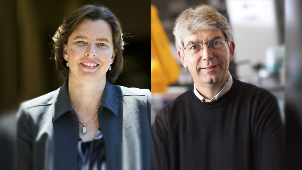
“Our discovery is fundamental to our molecular understanding of how autophagy is regulated,” said senior study author Malene Hansen, PhD, associate professor of the Development, Aging, and Regeneration Program at Sanford-Burnham. “Because impairment in the autophagy process has been linked to many disorders in humans, we believe that pharmacological agents targeting this novel regulatory circuit may hold great therapeutic potential.”

Critical kinases

Autophagy is a cellular recycling process involving a highly intricate and complex series of events. Cellular components such as abnormal molecules or damaged organelles are first sequestered within vesicles known as autophagosomes. These vesicles then fuse with organelles called lysosomes, which contain enzymes that break down various molecules. This fusion process results in the formation of hybrid organelles called autolysosomes, where the defective cellular components are enzymatically degraded and recycled. A protein called LC3 plays crucial roles in the formation of autophagosomes and the recruitment of dysfunctional cellular components to these vesicles. The signaling events that coordinate LC3’s various functions in autophagy have not been clear, but new research from the Hansen lab now proposes a novel and essential role for the mammalian Hippo kinases STK3 and STK4 in regulating autophagy by targeting LC3 for phosphorylation.

In their study, Hansen and her team describe that deficiency in both STK3 and STK4 impairs autophagy not just in mammalian cells, but also in nematodes and yeast. When exploring how the kinases regulate autophagy in mammalian cells, the researchers discovered that phosphorylation of LC3 by STK3 and STK4, specifically on the amino acid threonine 50, is critical for fusion between autophagosomes and lysosomes—an essential step in the autophagy process. “Collectively, the results of this study strongly support a critical and evolutionarily conserved role for STK3 and STK4 in regulating autophagy, by phosphorylating the key autophagy protein LC3, at least in mammalian cells,” Hansen said.

Killing bacteria

Previous studies have shown that STK4 also plays a role in regulating antibacterial and antiviral immunity in mammals, including humans. Moreover, autophagy is known to play a role in the clearance of intracellular pathogens. “These findings, taken together with our discovery that deficiency in STK3 and STK4 severely compromises autophagy, led us to test whether STK4 also plays a role in antimicrobial immunity through its function in autophagy,” said lead study author Deepti Wilkinson, Ph.D., a postdoctoral fellow in Hansen’s lab.

To test this notion, the researchers collaborated with Victor Nizet MD, professor of Pediatrics and Pharmacy  at UC San Diego and found that indeed mouse embryonic cells deficient in both STK3 and STK4 were unable to efficiently kill intracellular group A streptococci—bacteria known to be cleared by autophagy. However, an LC3 mutation that resulted in constant phosphorylation at threonine 50 restored the ability of the STK3/STK4-deficient cells to kill the bacteria. “This finding suggests that the same STK4-LC3 signaling pathway involved in autophagy also contributes to the response of mammalian cells to infection with intracellular pathogens and could play a role in human immune-related disease,” Wilkinson said.

Correcting defects

Moving forward, the researchers plan to further probe the molecular mechanisms by which STK3 and STK4 regulate autophagy. They will also investigate the therapeutic implications of the STK3/STK4 signaling pathway for tumor suppression as well as immune-related disorders such as bacterial and viral infections. “Understanding how autophagy works and why it sometimes stops to function optimally is essential for fighting diseases such as cancer, diabetes and neurodegeneration,” Hansen said.

“We have made a major contribution towards this endeavor by showing that STK3 and STK4 play an essential role in keeping the process of autophagy running smoothly by directly phosphorylating the key autophagy protein LC3. We hope our discoveries will lead to the development of effective drugs that can help correct autophagy defects that commonly occur in these diseases,” added Hansen.

A copy of the paper can be found at: http://www.ncbi.nlm.nih.gov/pubmed/25544559

Institute News

Exercise following bariatric surgery provides health benefits

Authorsgammon
Date

December 1, 2014

A new study by researchers at the Florida Hospital – Sanford-Burnham Translational Research Institute for Metabolism and Diabetes (TRI-MD) in Orlando, Fla., shows that patients who moderately exercise after bariatric surgery (weight-loss surgery) gain additional health improvements in glucose metabolism and cardiorespiratory fitness compared to patients who lead a sedentary lifestyle after surgery. The findings confirm the physiological and potential clinical benefits of adding an exercise regime following weight-loss surgery. Continue reading “Exercise following bariatric surgery provides health benefits”