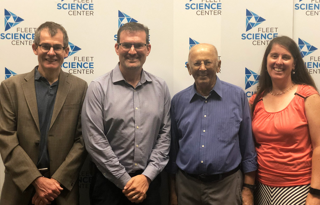
Professors Peter Adams, PhD, and Malene Hansen, PhD, of Sanford Burnham Prebys will lead key research and development cores
Sanford Burnham Prebys researchers are joining forces with University of California San Diego (UC San Diego) and the Salk Institute to form a world-class San Diego Nathan Shock Center (SD-NSC), a consortium established to study cellular and tissue aging in humans. The center will be funded by the National Institute on Aging (NIA), part of the National Institutes of Health, and is expected to receive $5 million over the next two years.
Professors Peter Adams, Ph.D., and Malene Hansen, PhD, of Sanford Burnham Prebys will lead key research and development cores, along with Professors Rusty Gage, PhD, Martin Hetzer, PhD, and Tatyana Sharpee, PhD, of Salk; and Anthony Molina, PhD, of UC San Diego. Salk Professor Gerald Shadel, PhD, will be the director of the SD-NSC.
“This is a special opportunity for San Diego’s aging research community to share our ideas, skills and technologies to drive innovative research in the basic biology of aging,” says Adams. “We are grateful for this support and will work to create the strongest environment possible to achieve meaningful breakthroughs that will benefit human health.”
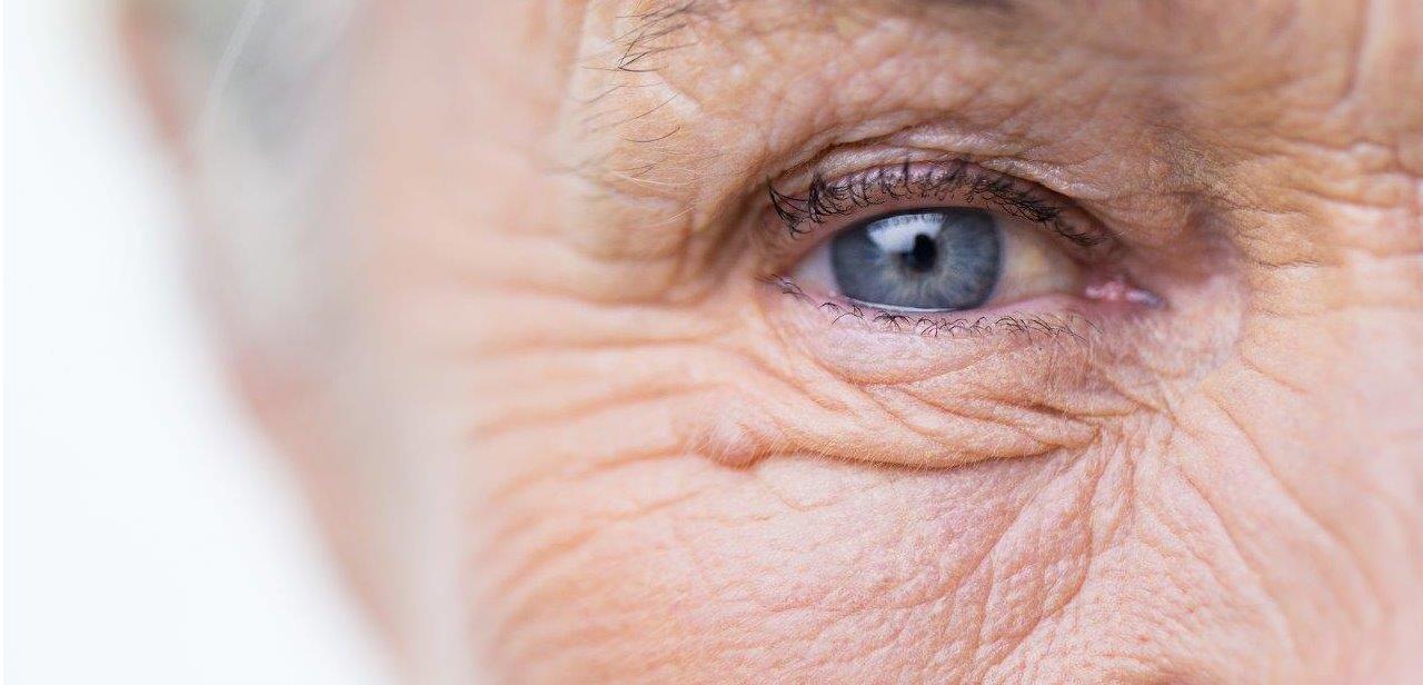
Aging is the most significant risk factor for human disease. Human cells and tissues age at different rates depending on their intrinsic properties, where they are in the body and environment exposures. Yet, scientists do not fully understand this variability (“heterogeneity”) and how it contributes to overall human aging, risk for disease or therapeutic responses.
To explore the complex heterogeneity of human aging, the SD-NSC will deploy three cutting-edge Research Resource Cores, including the Human Cell Models of Aging Core, to be led by Gage and Molina; the Heterogeneity of Aging Core, to be led by Hetzer and Adams; and the Integrative Models of Aging Core, to be led by Sharpee.
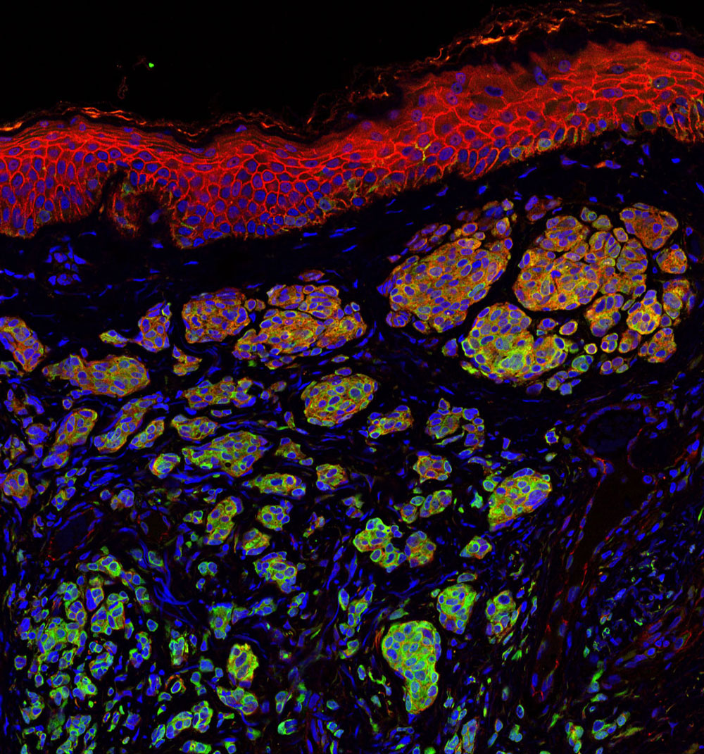
The cores will allow detailed analysis of human cells and organoids (artificially grown clusters of cells that model tissues), derived from a unique aging cohort at UC San Diego that is annotated for multiple measures of the actual biological age of individuals. In addition, the cores will provide scientific services to the Nathan Shock Centers network and the aging research community at large, including the dissemination of samples, protocols and computational tools to facilitate the study of heterogeneity in aging.
A Research Development Core headed by Hansen will also be established to encourage and support new investigators to enter the field of aging research. Through this core, the SD-NSC will provide pilot research grants, workshops and customized mentoring programs to promote the research and development of young investigators, as well as in-person and virtual trainings to spur collaboration and the sharing of knowledge related to the basic biology of aging.
“For years, my colleagues and I have been organizing successful symposia such as the annual La Jolla Aging Meeting (LJAM), where we share new aging research and discuss opportunities for collaboration,” says Hansen, who also hold the positions of associate dean for Student Affairs and faculty adviser for Postdoctoral Training at Sanford Burnham Prebys. “The San Diego Nathan Shock Center will enable us to broaden the reach and impact of LJAM, as well as take training and mentoring of the next generation of researchers to a new level.”
The SD-NSC builds on Sanford Burnham Prebys’ strengths in fundamental aging research, renowned for its use of model organisms to unravel cell changes associated with normal development and aging. By building, analyzing and probing models of disease, scientists in the Institute’s Aging, Development and Regeneration Program are providing new tools to uncover novel therapeutic targets for heart disease, neurodegeneration, muscle disorders, diabetes, cancer and other debilitating diseases.
The SD-NSC will be one of a network of eight Nathan Shock Centers nationwide, and is funded by NIA grant number P30AG068635.