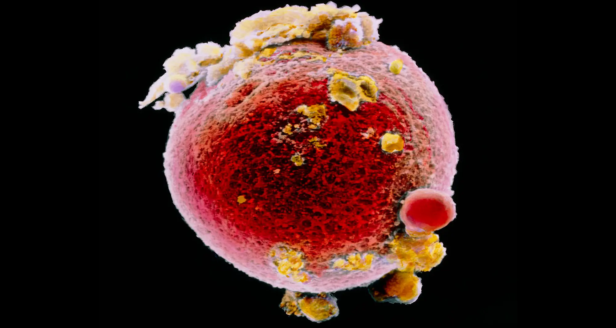
False color scanning electron micrograph of a single human ovum or egg cell. Human ova are among the largest cells in the human body and are visible to the naked eye, roughly the size of a single, small grain of sand.
Image courtesy of SPG.
False color scanning electron micrograph of a single human ovum or egg cell. Human ova are among the largest cells in the human body and are visible to the naked eye, roughly the size of a single, small grain of sand.
Image courtesy of SPG.
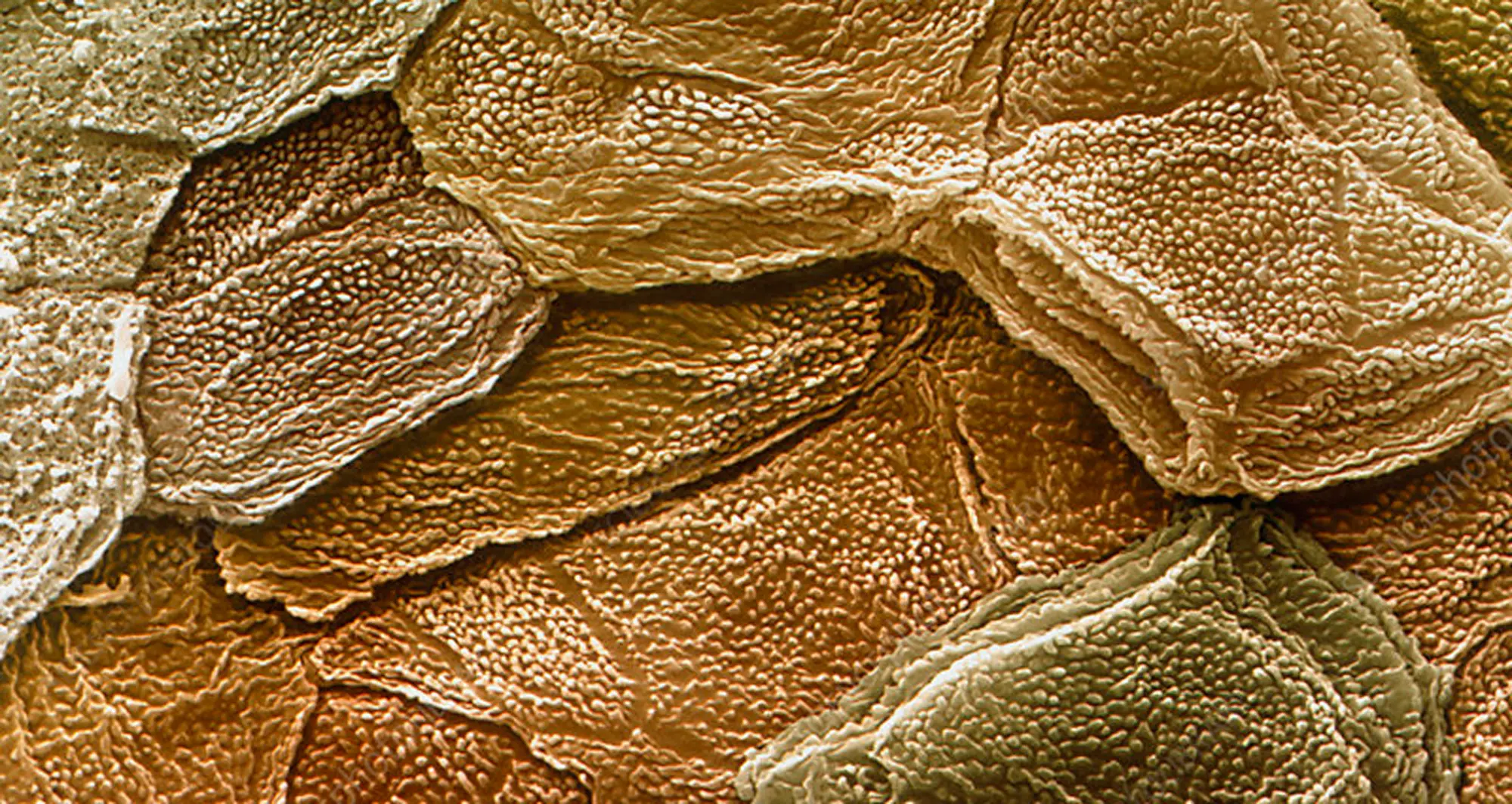
Colorized scanning electron micrograph of surface of human skin. The outermost layer of skin is called the stratum corneum and is composed primarily of keratinocytes, living and dead. Dead cells form horny scales, and release peptides called defensins, which are part of the first line of immune defense mechanisms.
Image courtesy of Eye of Science/SPL.
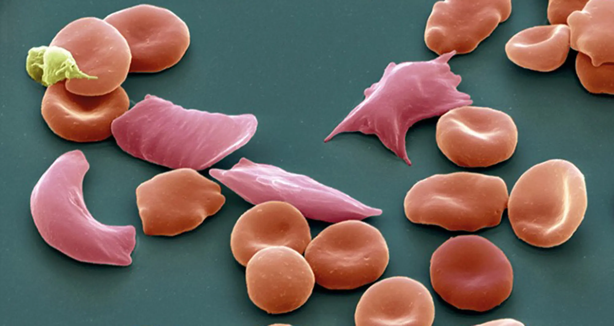
Colorized scanning electron micrograph depicts normal red blood cells (red, concave) with elongated blood cells indicative of sickle-cell disease (pink).
Image courtesy of Eye of Science/SPL.
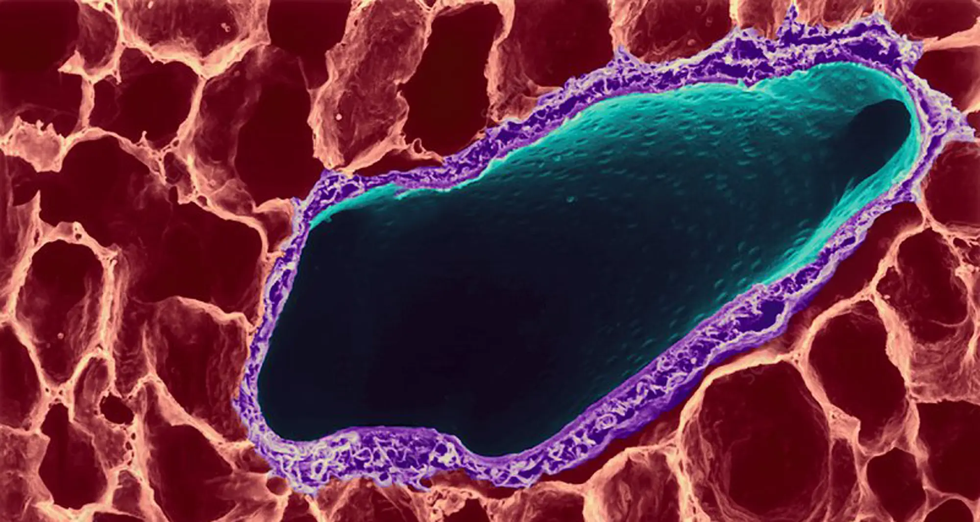
Colorized scanning electron micrograph of human lung tissue, depicting alveoli and bronchus. Lung alveoli are tiny, grape-like sacs, numbering in the millions, where essential gas exchange (oxygen/carbon dioxide) occurs. The bronchus is a major airway that branches from the trachea to conduct air into the lungs.
Image courtesy of Dennis Kunkel/SPL.
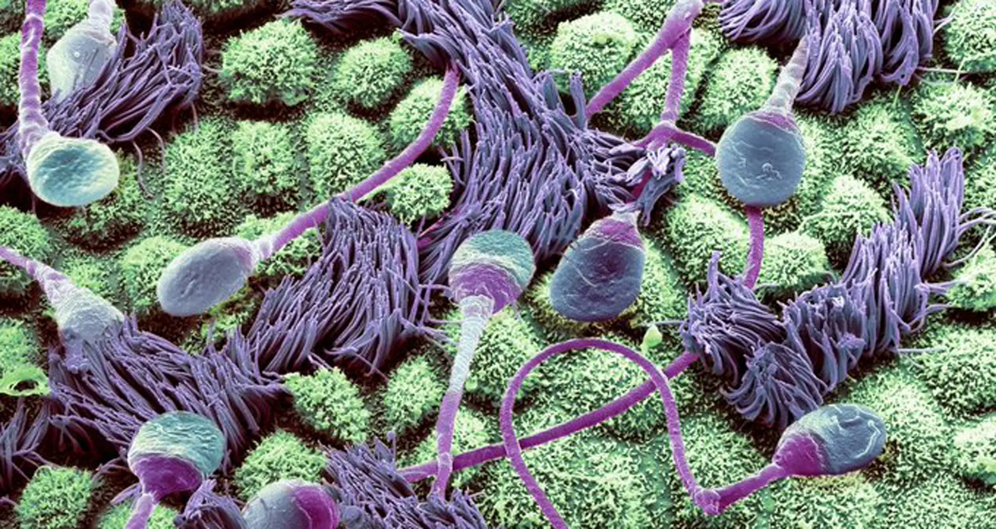
Colorized scanning electron microscope composition of human sperm traveling through a fallopian tube.
Image courtesy of Steve Gschmeissner/Getty.
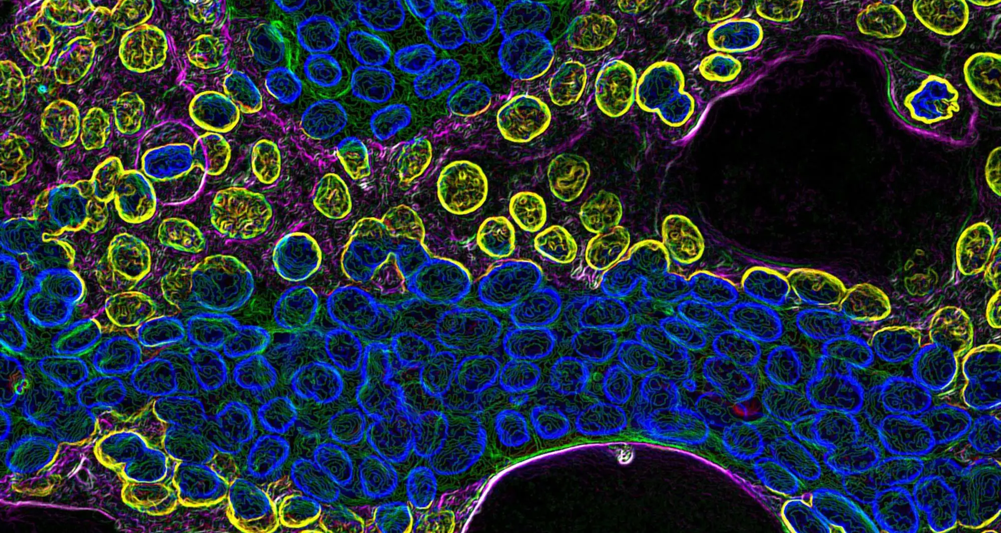
Carcinoma cells via fluorescence microscopy.
Image courtesy of Frederick Keeney, The Wistar Institute, Philadelphia.
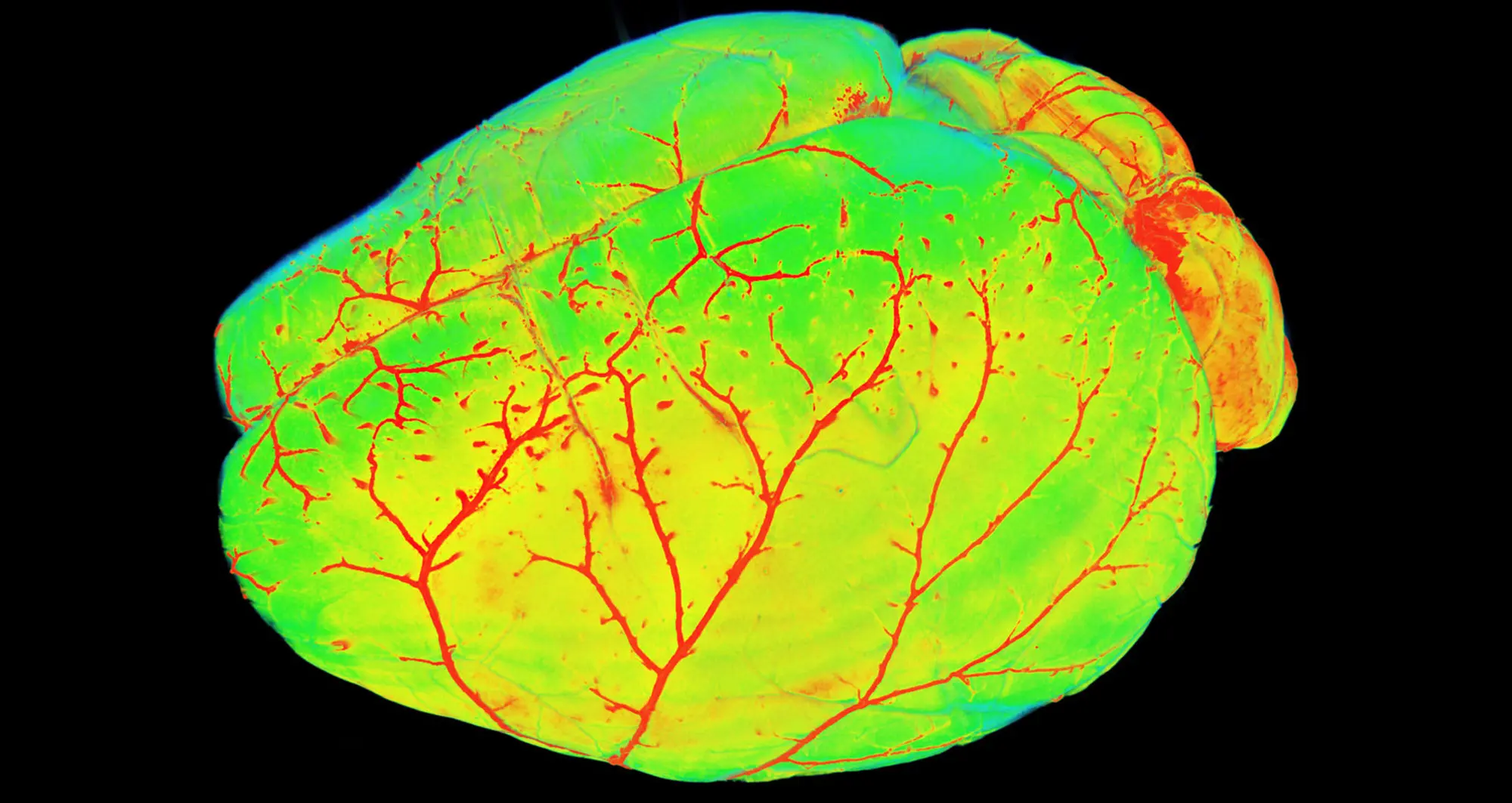
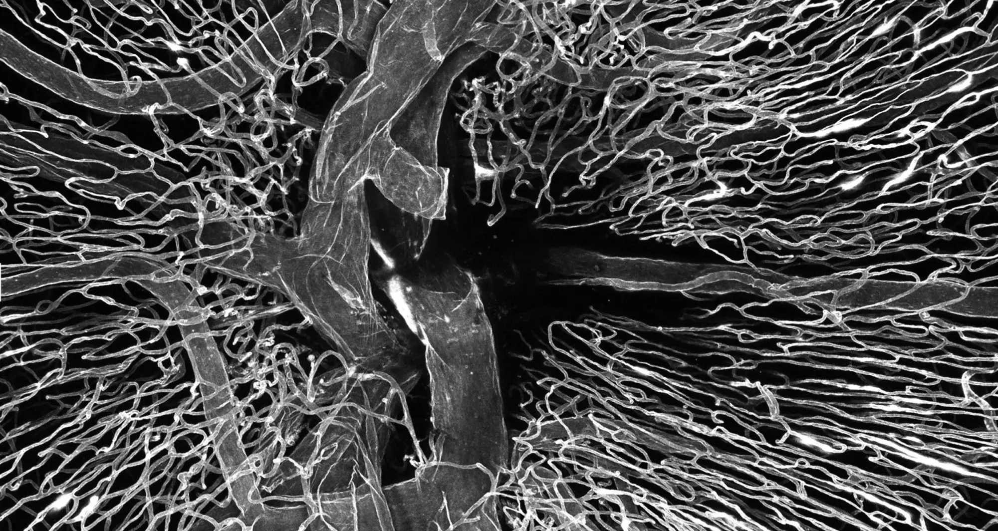
Mouse brain vasculature, as imaged by light sheet fluorescence microscopy.
Image courtesy of Ali Erturk, Munich, Germany.
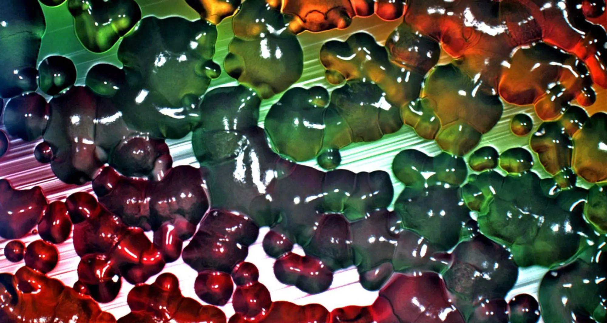
Stereomicrography of bacterial growth in a petri dish.
Image courtesy of Neil James Egan, PPG Industries, Cleveland, Ohio.
A human donor central retinal artery was cannulated (intubated) and perfused with Lectin FITC, a specific protein chemically linked to a fluorescent dye. The microvasculature of the optic nerve head area at the center is visible.
Image courtesy of Dong An, Lions Eye Institute, Australia.
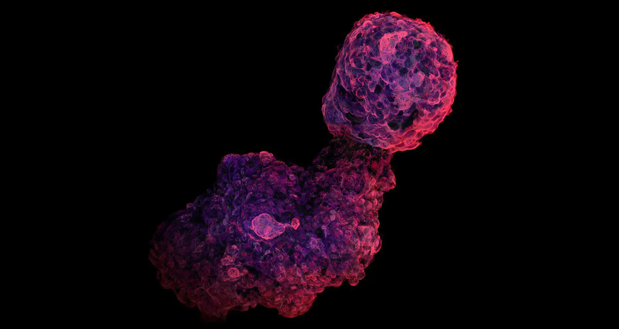
Using confocal, deconvolution and image stacking techniques, a micrograph of a human I pluripotent stem cell-derived cardiac organoid.
Image courtesy of Syed Ashraf, Divya Sridharan and Salvia Zafar, Ohio State University.