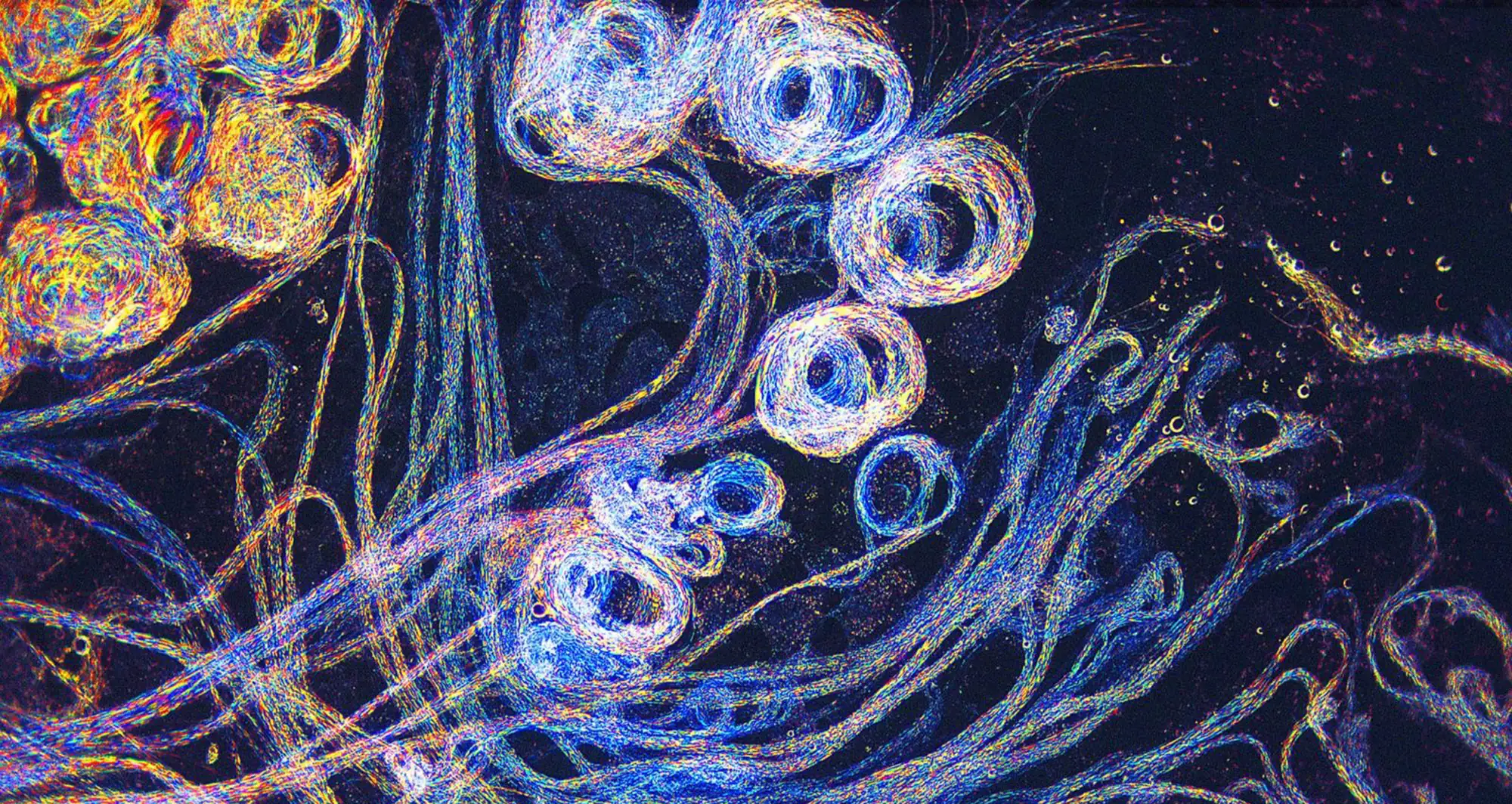
Confocal microscopy reveals human hair follicles growing from a stem cell-derived organoid (cyan) with nerves (red).
Image courtesy of Karl R. Koehler and Jiyoon Lee.

Confocal microscopy reveals human hair follicles growing from a stem cell-derived organoid (cyan) with nerves (red).
Image courtesy of Karl R. Koehler and Jiyoon Lee.

Depicted in this confocal/deconvolution micrograph are the actin cytoskeleton (cyan) and the endoplasmic reticulum (red) of a mouse brain cancer cell.
Image courtesy of Halli Lindamood and Eric Vitriol.

Darkfield microscopy captures the flowing grace of sperm from Drosophila virilus, a species of fruit fly.
Image courtesy of Earl Nishiguchi.

A human fibroblast—the most common cells of connective tissue—undergoes mitosis or cell division. Cell components include actin (gray/brown), myosin II (green) and DNA (magenta).
Image courtesy of Nilay Taneja.

A micrograph depicts the hippocampal region of the human brain. Located in the medial temporal lobe, it acts as a crucial component of the limbic system for learning, memory and spatial navigation.
Image courtesy of Sarah Moghadam and Ahmad Salehi.

Developing mouse nerve cells displaying nuclei (yellow) surrounded by cell bodies, with long extensions called axons and thin branching structures called dendrites. Electrical signals travel from the axon of one cell to the dendrites of another.
Image courtesy of Torsten Wittman, UC San Francisco.

Proteins in the neural tissues of a zebrafish embryo direct cells to line up and form the neural tube, which will become the spinal cord and brain. Zebrafish embryonic development research may help pinpoint the underlying cause of common neural tube defects, such as spina bifida, which occur in about 1 in 1,000 newborn children.
Image courtesy of Alexander Schier, Harvard University.

Colorized scanning electron micrograph of Vibrio bacteria. Some species of the rod-shaped bacteria cause cholera in humans.
Image courtesy of Tina Weatherby Carvalho, University of Hawaii at Manoa.

Colorized scanning electron micrograph of a blood clot, with red blood cells tangled in fibrin, a tough, insoluble fibrous protein that acts like a molecular glue and is essential for clotting and wound healing.
Image courtesy of Thomas Deerinck, UC San Diego.

False color scanning electron micrograph of a single human ovum or egg cell. Human ova are among the largest cells in the human body and are visible to the naked eye, roughly the size of a single, small grain of sand.
Image courtesy of SPG.