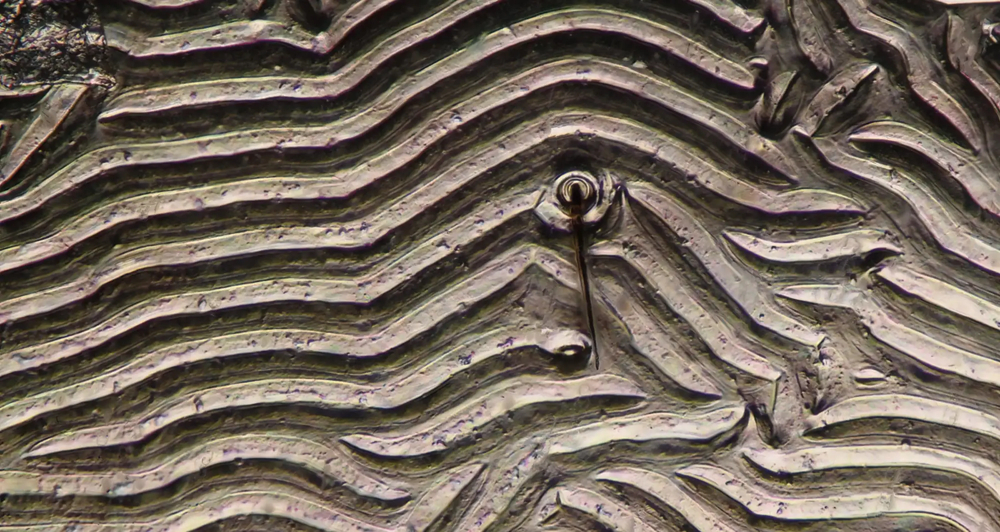
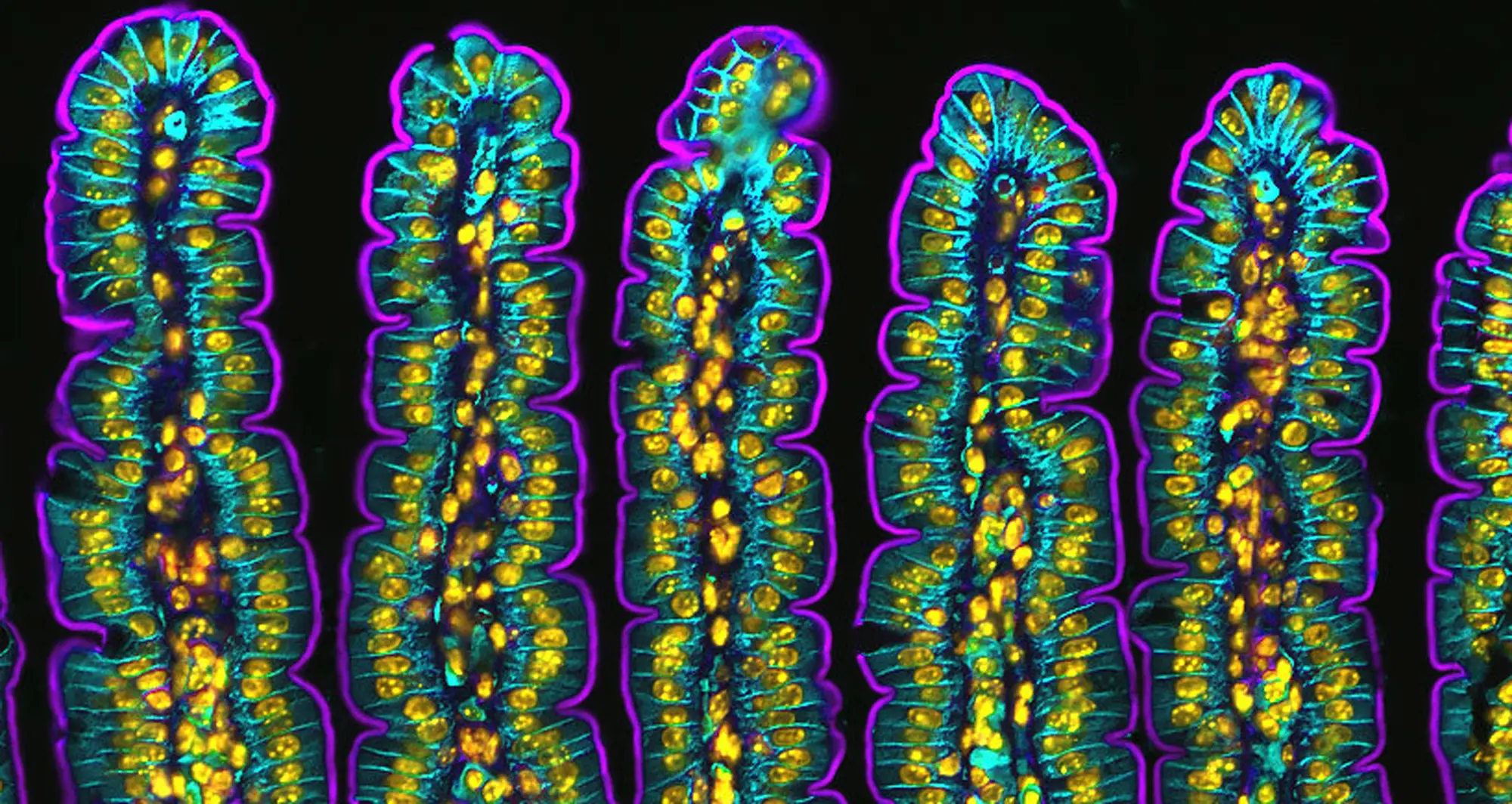
Science in Pictures Archives - Page 3 of 7 - Sanford Burnham Prebys
Institute News

Science in Pictures

AuthorScott LaFee
Date

December 16, 2024

A 3D-structured illumination reveals a synaptonemal complex, a protein structure that forms between homologous chromosomes during cell division. It’s believed SCs function primarily as scaffolds to allow interacting chromatids to complete their crossover activities.

Image courtesy of Chung-Ju-Rachel Wang and Bioscapes.

Institute News

Science in Pictures

AuthorScott LaFee
Date

December 9, 2024

This image of the hippocampus in a rat brain was taken using an ultra-widefield high-speed multiphoton laser microscope. Tissue was stained to reveal the organization of glial cells (cyan), neurofilaments (green) and DNA (yellow).

Image courtesy of Thomas Deerinck, NCMIR and NIH.

Institute News

Science in Pictures

AuthorScott LaFee
Date

December 2, 2024

t’s as lovely as a snowflake in winter, but something entirely different: a crystal of sildenafil, the active ingredient in Viagra tablets.

Image courtesy of Annie Cavanagh, Wellcome Collection.

Institute News

Science in Pictures

AuthorScott LaFee
Date

November 25, 2024

Confocal micrograph of bacterial biofilm on a human tongue cell. The oral cavity harbors more than 700 species of bacteria, second only to the gut.

Image courtesy of Tagide deCarvalho, University of Maryland, Baltimore County.

Institute News

Science in Pictures

AuthorScott LaFee
Date

November 4, 2024

A chromosome from Drosophila melanogaster (fruit fly) salivary glands, using Brightfield microscopy. Fruit flies are model organisms, sharing 75% of the genes that cause disease in humans.

Image courtesy of Earl Nishiguchi, Kauai Community College and Nikon Small World.

Institute News

Science in Pictures

AuthorScott LaFee
Date

October 28, 2024

A colorized scanning electron micrograph of a human brain cancer stem cell. Cell bodies are  in orange; nuclei in green. Cancer stem cells possess characteristics of normal stem cells, specifically the ability to give rise to all cell types found in a particular cancer sample. That makes them a particularly enticing target for developing cancer therapies, especially those prone to metastasis or recurrence.

Image courtesy of Izzat Suffian, Pedro Costa, Stephen Pollard, David McCarthy & Khuloud T. Al-Jamal.

Institute News

Science in Pictures

AuthorScott LaFee
Date

October 21, 2024

A colorized scanning electron micrograph of a natural killer cell from a human donor. NK cells are a type of white blood cell critical to the innate immune system. They  provide rapid responses to virus-infected cells, stressed cells, tumor cells and other intracellular pathogens based on signals from several activating and inhibitory receptors.

Image courtesy NIAID.