Science in Pictures Archives - Page 6 of 7 - Sanford Burnham Prebys
Institute News

Science in Pictures

AuthorScott LaFee
Date

May 20, 2024

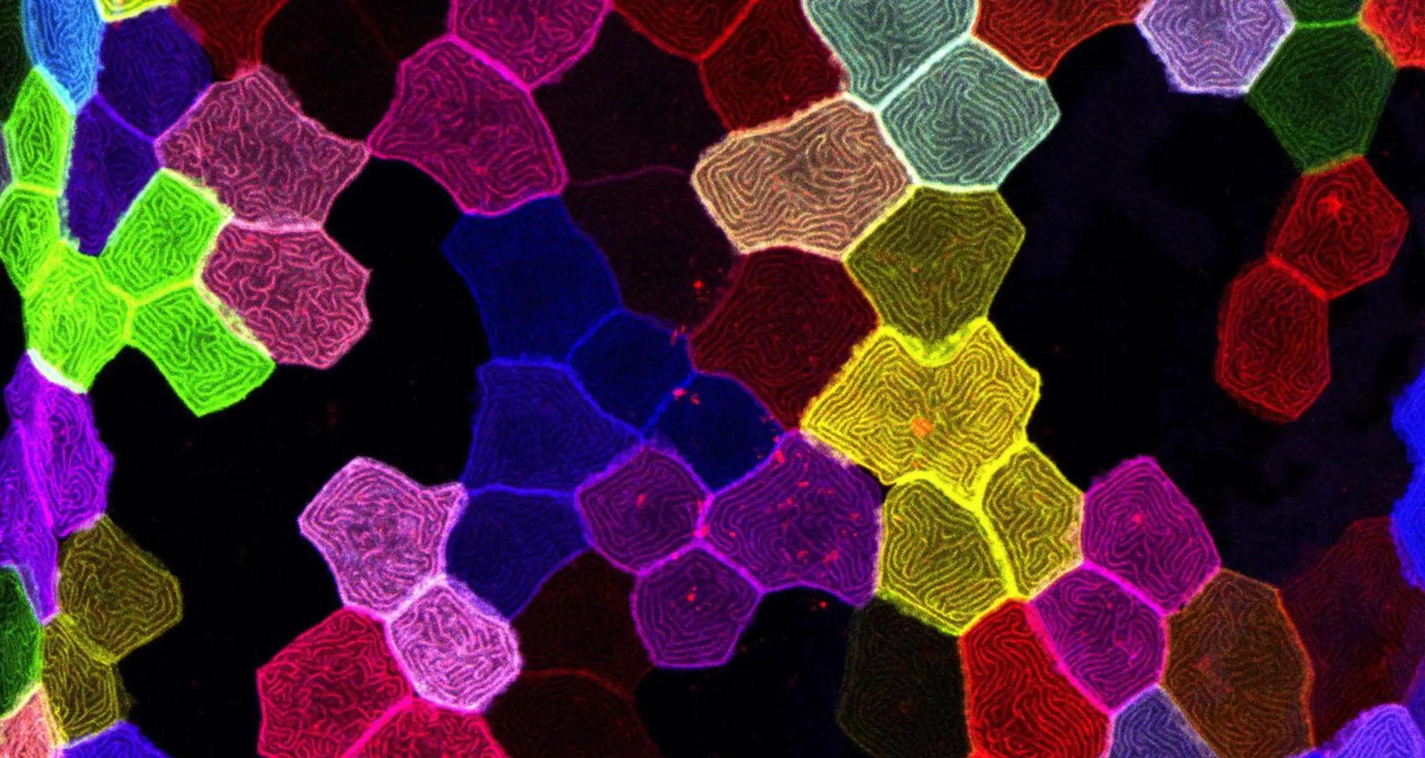
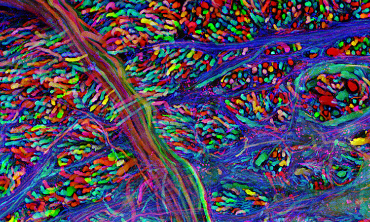
A confocal micrograph of a “brainbow” in the brain stem of a mouse. Brainbows are the result of an imaging technique in which neurons can be distinguished from each other using multiple fluorescent proteins.

Image courtesy of Jeff Lichtman, Harvard University.

Institute News

Science in Pictures

AuthorScott LaFee
Date

May 13, 2024

In this fluorescent micrograph, filamentous actin and microtubules (structural proteins) are revealed in mouse fibroblasts, a type of cell that contributes to the formation of connective tissue.

Image courtesy of Torsten Wittman, Scripps Research.

Institute News

Science in Pictures

AuthorScott LaFee
Date

May 6, 2024

A colorized scanning electron micrograph of a cultured human neuron from an induced pluripotent stem cell.

Image courtesy of Mark Ellisman and Thomas Deerinck, National Center for Microscopy and Imaging Research, UC San Diego.

Institute News

Science in Pictures

AuthorScott LaFee
Date

April 29, 2024

In this image stacking photomicrograph, sunflower pollen cling to an acupuncture needle.

Image courtesy of John-Oliver Dum, Medienbunker Produktion.

Institute News

Science in Pictures

AuthorScott LaFee
Date

April 22, 2024

A polarized light micrograph (magnified 25 times) depicts caffeine crystals.

Image courtesy of Stefan Eberhard, University of Georgia.

Institute News

Science in Pictures

AuthorScott LaFee
Date

April 15, 2024

A quadruple-fluorescence confocal image of a mouse retina, showing the optic nerve layer. Glial cells are colored green and retinal ganglion cells are orange. Optic nerve fibers are red and blood vessels are blue. 

Image courtesy of Thomas Deerinck, National Center for Microscopy and Imaging Research, UC San Diego.

Institute News

Science in Pictures

AuthorScott LaFee
Date

April 8, 2024

As colorful as their consequence, this brightfield reflected light micrograph displays a variety of wildflower seeds

Image courtesy of Yanping Wang, Beijing Planetarium.

Institute News

Science in Pictures

AuthorScott LaFee
Date

April 1, 2024

In this confocal micrograph (magnified 25 times), the epithelial cells of a palmskin zebrafish larva are revealed in colorful detail. Striking similarities between human skin and zebrafish skin make it an important model organism

Image courtesy of Chen-Hui Chen, Institute of Cellular and Organismic Biology in Taiwan.

Institute News

Science in Pictures

AuthorScott LaFee
Date

March 25, 2024

A confocal micrograph captures endothelial cells in the intestine of an 18.5-day-old mouse embryo. Blood vessels and intestinal lining in blue; nerves in red and green.

Image courtesy of Nathan Burns, National Heart, Lung and Blood Institute.

Institute News

Science in Pictures

AuthorScott LaFee
Date

March 18, 2024

In this reflected light micrograph, life teems in a single drop of seawater below the eye of a needle.

Image courtesy of Peter Parks.