Science in Pictures Archives - Page 7 of 13 - Sanford Burnham Prebys
Institute News

Science in Pictures

AuthorScott LaFee
Date

March 10, 2025

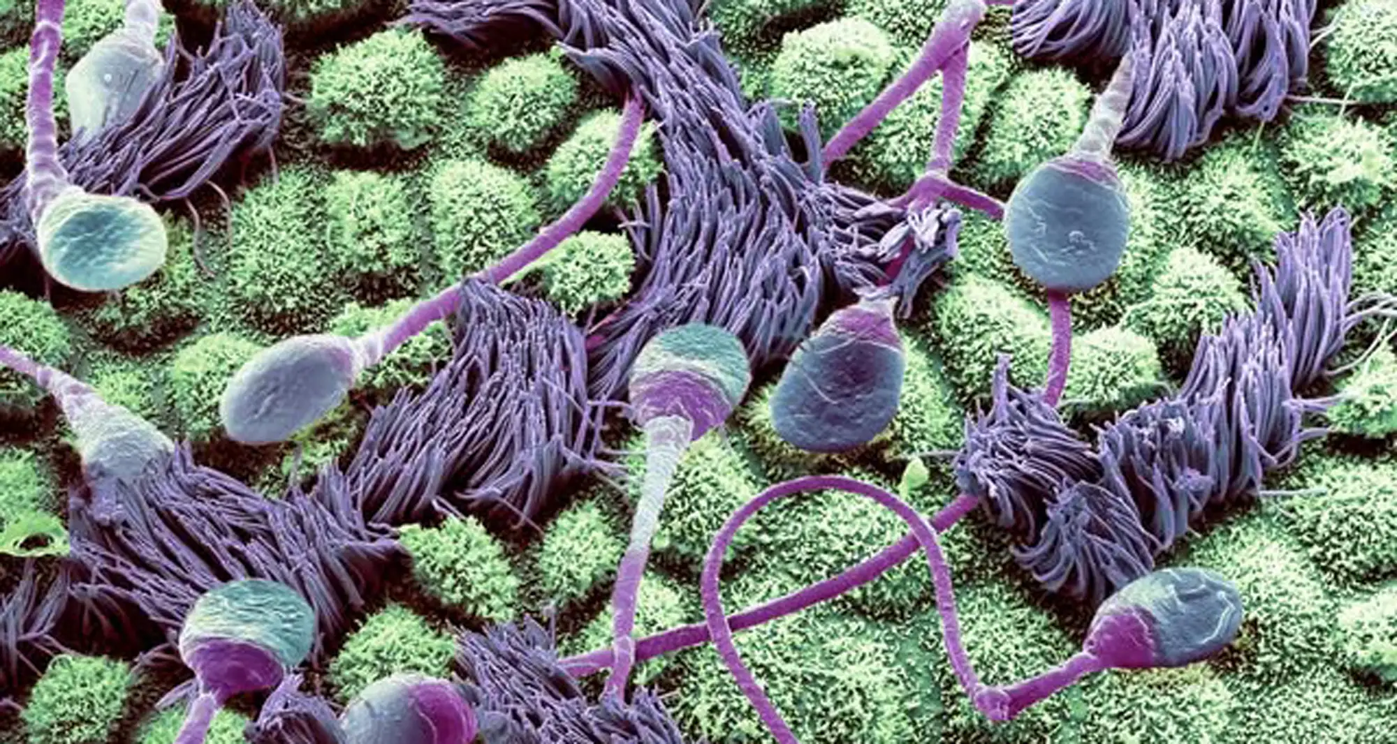
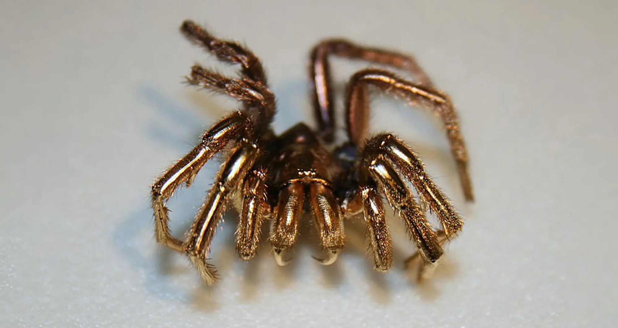
A spider is coated in gold to prepare it as a specimen for scanning electron microscopy. Gold is used because of its high electrical conductivity, which enables the electron beam to interact with a specimen more effectively.

Image courtesy of Steve Gschmeissner.

Institute News

Science in Pictures

AuthorScott LaFee
Date

March 3, 2025

A colorized scanning electron micrograph reveals in fine detail human sperm traveling through a fallopian tube. Ciliated cells (purple with hairlike extensions) help move eggs from the ovary to the uterus and regulate flow of fluid. Secretory cells (green) produce a nutrient-rich fluid that bathes sperm and egg.

Image courtesy of Steve Gschmeissner.

Institute News

Science in Pictures

AuthorScott LaFee
Date

February 24, 2025

A differential interference contrast image of an amoeba magnified 60 times. Amoebas are a type of cell or unicellular organism with the ability to alter their shape, primarily by extending and retracting pseudopods or “false feet.”

Image courtesy of Håkan Kvarnström, Stockholm, Sweden and Nikon.

Institute News

Science in Pictures

AuthorScott LaFee
Date

February 17, 2025

Melanoma or melanocyte skin cancer cells (orange) under microscope. Skin cancer is the most common type of cancer, with rising prevalence. One in five Americans will develop skin cancer in their lifetime. Because melanomas can develop anywhere in the body, they pose particular challenges for diagnoses and treatment.

Institute News

Science in Pictures

AuthorScott LaFee
Date

February 10, 2025

A confocal micrograph depicts an embryonic mouse kidney showing the collecting duct (blue) and nephron progenitor (yellow) cells.

Image courtesy of Lori O’Brien, University of North Carolina at Chapel Hill and Nikon.

Institute News

Science in Pictures

AuthorScott LaFee
Date

February 3, 2025

Using confocal microscopy, this image depicts breast tissue showing contractile myoepithelial cells wrapped around milk-producing alveoli.

Image courtesy of Caleb Dawson at Walter and Eliza Hall Institute of Medical Research in Australia and Nikon.

Institute News

Science in Pictures

AuthorScott LaFee
Date

January 27, 2025

A network of dopaminergic neurons generated from human stem cells. Dopaminergic neurons regulate many brain functions, including voluntary movement, learning and reward and working memory.

Image courtesy of Nick Gatford, University of Oxford and Nikon Small World.

Institute News

Science in Pictures

AuthorScott LaFee
Date

January 13, 2025

Healthy arteriole in eye, with tough, flexible elastin wall (pink), red blood cells (red) and supporting collagen fibers (web-like netting) surrounded by yellow and green areas.

Image courtesy of Donald Pottle, The Schepens Eye Research Institute, Boston and Bioscapes.

Institute News

Science in Pictures

AuthorScott LaFee
Date

January 6, 2025

Beta-tubulin expression of a Drosophila (fruit fly) third instar larval brain, with attached eye imaginal discs.

Image courtesy of Christian Klambt and Imke Schmidt, University of Munster, Germany and Bioscapes.