Rare Disease Day gathers scientists, doctors and families
AuthorMiles Martin
Date
March 3, 2022
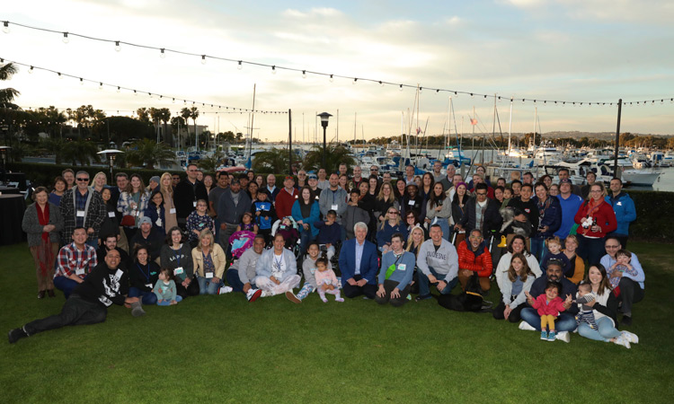
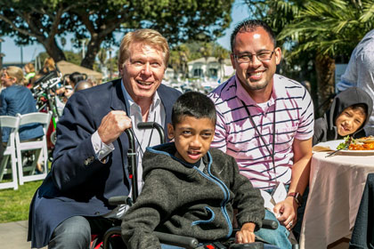
The 2022 Rare Disease Day Symposium took place last weekend at the Dana On Mission Bay Resort in San Diego. The event, sponsored by Sanford Burnham Prebys and CDG CARE, brought together researchers, clinicians and families from around the world to discuss new medical breakthroughs and meet other families living with rare diseases.
Rare Disease Day is celebrated on the last day of February to raise awareness for rare diseases, defined by the United States government as those that affect fewer than 20,000 people. Although there are more than 7,000 individual types of rare diseases that affect more than 30 million people in the United States, this year’s conference gathered more than 200 people focused on CDG, an extremely rare group of genetic disorders that affect children.
CDG, which stands for congenital disorders of glycosylation, occurs when sugar molecules on many of our proteins are absent or incomplete. CDG causes serious, often fatal, malfunctions in various organ systems throughout the body.
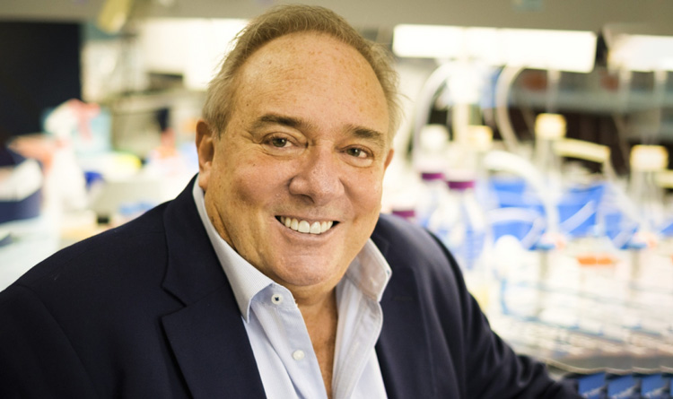
“This is a chance for the global CDG community to come together, support one another and continue to try to find treatments,” says Hudson Freeze, PhD, director of the Human Genetics Program at Sanford Burnham Prebys. “It’s always my favorite weekend of the year, and I’m thrilled that we’re able to do it again safely.” Freeze’s primary research focus is CDG, and he has personally worked with more than 300 patients.
Exchanging knowledge
The three-day symposium opened Friday morning with introductory comments from three important figures and philanthropists in Sanford Burnham Prebys’ history: T. Denny Sanford, Malin Burnham and Debra Turner. Congressman Scott Peterson also spoke on the importance of funding medical discoveries.
“Our job is to make a positive difference. We do that best when we all work together,” said Sanford in his video introduction. “Congratulations on all your work. You make me very proud.”
This year, 19 scientists and clinicians in total spoke on the latest research in modeling, treating and understanding CDG. The full program of presentations can be found here.
Connecting families
Although Rare Disease Day is an important opportunity to share the latest scientific research, one of the highlights of the event doesn’t involve science at all. To provide space for families to take a break from the presentations and socialize, staff and volunteers transformed the Bayside Conference Room of the Dana resort into a child care and respite area packed full of toys and games.
In addition to giving families space to play, Rare Disease Day hosted several group activities for families, including a magic show on Saturday and a surprise visit on Sunday morning from Disney’s Anna and Olaf.
And while the joy in the respite conference room was palpable, there was something else, less tangible, in the air as well: hope.
“Meetings like this bring us hope and help us raise awareness for CDG,” says Donnie. “That gives us a sense of purpose each and every time we attend the conference. And we won’t stop, even 20 years from now.”
Institute News
Rare disease in the time of COVID: Damian Omler’s story
AuthorMiles Martin
Date
February 25, 2022
How a one-of-a-kind kid and his family stay connected during the pandemic
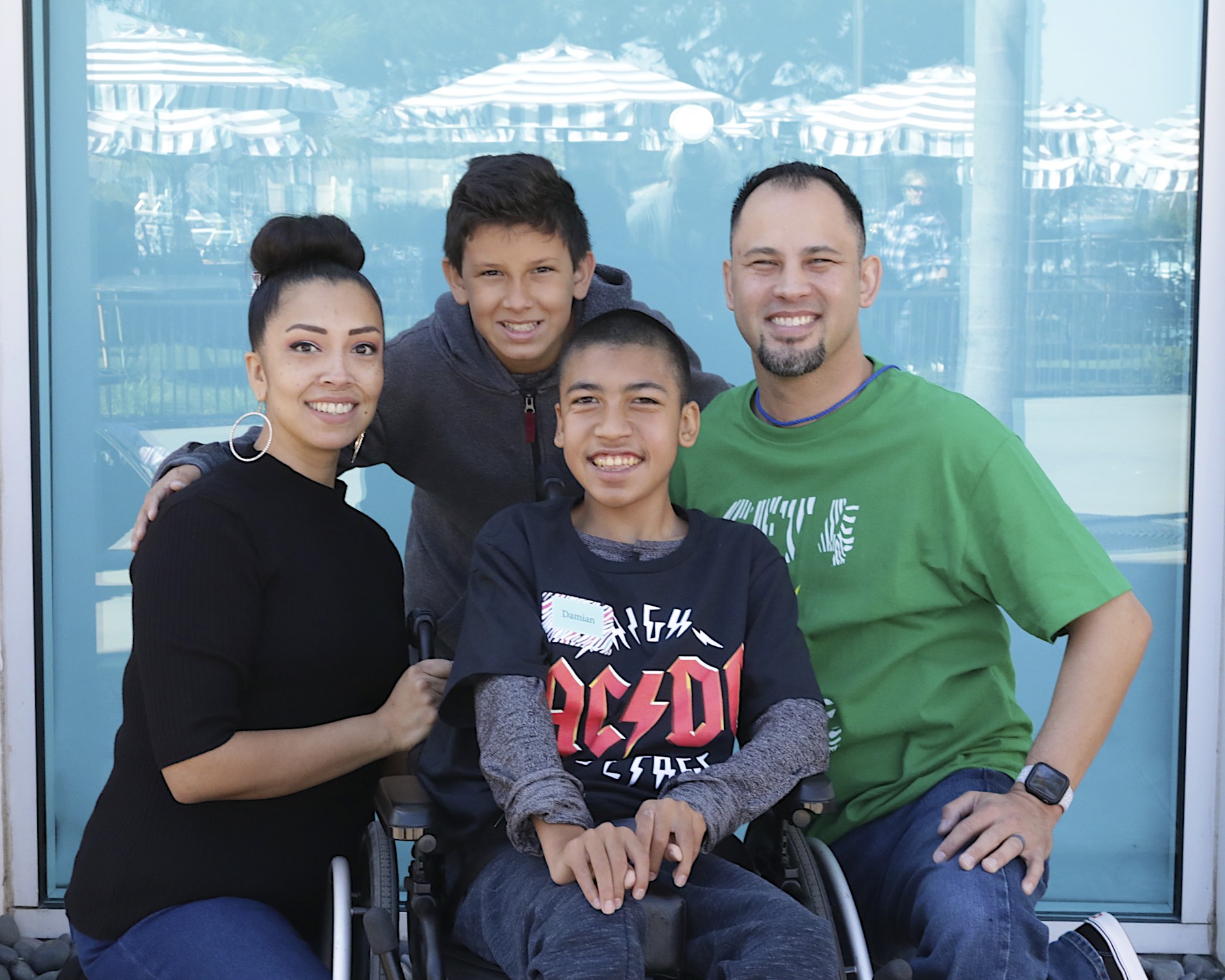
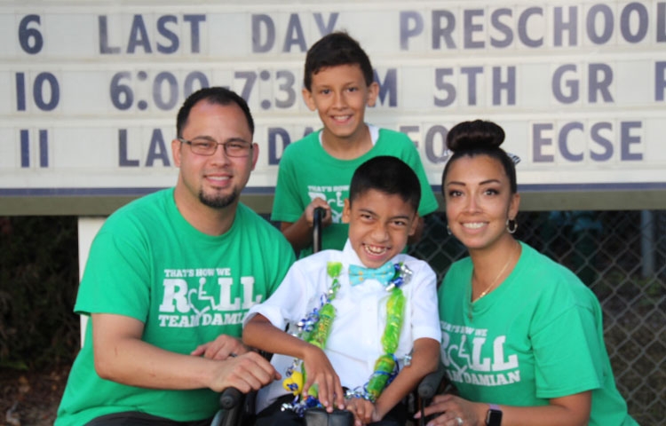
Thirteen-year-old Damian Omler is the only person in the world with his rare genetic mutation, which presents him and his parents (Donnie and Gracie) and 11-year-old brother, DJ, with major challenges every day. Damian’s condition—a congenital disorder of glycosylation, or CDG—causes him to have seizures, and requires him to have help with routine tasks such as using the restroom and dressing. And, he must use a wheelchair for mobility.
Despite these obstacles, Damian lives a rich, fulfilling life. But protecting his health during the COVID-19 pandemic threw a major wrench into the Omlers’ routine.
“In the early days of the pandemic, we didn’t know what kind of effect COVID would have on Damian, so we had to take a lot of precautions, including not seeing a lot of family and friends, which was very isolating,” says Donnie.
“Damian is also very sociable—we call him the hot potato because he just goes from person to person, so the pandemic was hard for him in that way as well,” adds Gracie. “We were so glad when we were finally able to get our family vaccinated so we could be more a part of the community.”
Staying at home had its ups and downs for the Omlers
Although most of us can relate to the isolation of the pandemic, there are unique challenges that come with being a family living with a rare disease during this time.
“Appointments were so much more difficult for Damian over Zoom,” says Gracie. “I had to help him through his physical therapy, and I was nervous that I might be doing it wrong or even hurting him.”
Despite these complications to Damian’s care, there were some unexpected silver linings to spending more time at home.
“Damian does choir and dance for his electives at school,” says Gracie. “I love that with remote learning I was able to interact with him and the class and learn the dances with him.”
“She definitely got a lot of accolades from the teachers for being one of the parents who participates,” adds Donnie, jovially.
Returning to Sanford Burnham Prebys’ Rare Disease Day
The Omlers are longtime friends of Sanford Burnham Prebys. They first visited the Institute in 2012, when Damian was 5. Before then, they’d been struggling to find a diagnosis for their son, who’d been missing developmental milestones since he was born.
With the help of Institute professor Hudson Freeze, PhD, who has dedicated his career to CDG research, doctors were finally able to diagnose Damian’s specific case in 2015.
“After the diagnosis, we sat and smiled for a long time,” says Donnie. “Just knowing was such a relief.”
Since 2016, the Omlers have also been regular participants in the Institute’s Rare Disease Symposiums, which help patients, researchers and clinicians from around the world connect in order to support one another and learn about the latest advances in rare disease research.
The most recent Rare Disease Day the Omlers attended was in 2020, just before the pandemic took hold. And although the event didn’t take place last year, this year it’s back stronger than ever. And the Omlers can’t wait to be back too.
“Meetings like this bring us hope and help us raise awareness for CDG,” says Donnie. “That gives us a sense of purpose each and every time we go. And we won’t stop, even 20 years from now.”
The 2022 Rare Disease Day Symposium & CDG/NGLY1 Family Conference will take place February 25–27 at the Dana Hotel on Mission Bay in San Diego. Scientific sessions will be held on the 25th and 26th, and the Family Conference will take place on the 27th.
And if you see a young man acting like a social “hot potato” on the 27th, that’s Damian. He’ll probably say hi to you.
Institute News
One at a time: How a Sanford Burnham Prebys professor changes patient lives
AuthorMiles Martin
Date
February 22, 2022
Having worked for decades to improve the lives of children with rare diseases, Hudson Freeze is still on the case.
Hudson Freeze, PhD is not your average researcher. His work focuses on congenital disorders of glycosylation, or CDG, a severe group of diseases that affect fewer than 2,000 children worldwide. Those conditions occur when sugar molecules on many of our proteins are absent or incomplete. That can lead to serious, often fatal, malfunctions in various organ systems throughout the body.
Although Freeze is not a clinician, he is deeply involved in identifying these rare CDG mutations, and providing families with answers to what is often a challenging diagnosis. Because CDG is a group of incurable diseases, families of children with CDG reach out to Freeze almost weekly, seeking help.
“If someone asks for help, I say, ‘Let me try,’” says Freeze. “Any glimmer of hope is a path worth pursuing, anything to make life easier for children with CDG.”
Freeze has been working on CDG for more than 25 years and has worked with more than 300 patients, and he has kept in touch with many of them over the years.
“Not a day goes by when I don’t think of them and their struggles—but mostly their smiles,” says Freeze. “It’s the reason we won’t give up on trying to understand them and maybe even finding treatments.”
Treating disease with sugar
Although CDG presents as permanent and irreversible mutations, Freeze’s research has been instrumental in discovering an approach to alleviate severe symptoms of CDG—such as seizures—in certain patients. The answer: sugar. Thanks to Freeze and others, there are about 30 patients worldwide who are now taking mannose, a simple sugar molecule, to help alleviate their CDG symptoms.
Today, the strategy of treating diseases with simple sugar molecules is being explored in other glycosylation disorders, as well as less-rare diseases such as multiple sclerosis, cancer and diabetes.
Rare Disease Day at Sanford Burnham Prebys
Freeze’s impact on the lives of families living with CDG extends well beyond the walls of his lab. Since 2010, he has organized an annual Rare Disease Day Symposium each February, where scientists, doctors and families gather from around the world to discuss the latest research and meet other families coping with rare diseases. Last year, the pandemic forced the Institute to press pause on the event, but this year, Rare Disease Day is back in San Diego and stronger than ever.
“It’s a chance for the global CDG community to come together, support one another and continue to put our heads together to find treatments,” says Freeze. “It’s always my favorite weekend of the year, and I’m thrilled that we’re able to do it again safely.”
The 2022 Rare Disease Day Symposium & CDG/NGLY1 Family Conference will take place February 25–27 at the Dana on Mission Bay Resort in San Diego. Scientific sessions will be held on the 25th and 26th, and the Family Conference will take place on the 27th.
Implicit bias in the workplace: An interview with Lydia Villa-Komaroff
AuthorMiles Martin
Date
January 26, 2022
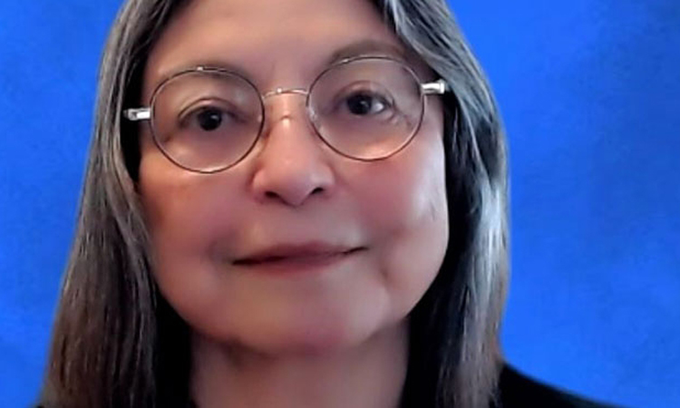
When Lydia Villa-Komaroff, PhD, graduated from MIT in 1975, she was one of only three Mexican American women in the United States to ever receive a PhD in a natural science field. Since then, she has had a diverse career, ranging from research benchwork to academic administration, as well as many years working as a biotechnology executive. As a co-founding member of the Society for the Advancement of Chicanos/Hispanics and Native Americans in Science (SACNAS), she has also been a longtime champion of diversity in STEM.
Today, she owns and operates a one-woman consulting firm, where she continues to work with biotech companies and research organizations to help them acknowledge and confront unconscious biases in the workplace.
Ahead of the implicit bias seminar she is delivering to Sanford Burnham Prebys employees on February 7, we sat down with Villa-Komaroff to discuss implicit bias and how it manifests in the sciences, as well as what organizations can do to address it.
How does implicit bias work? Villa-Komaroff: We make decisions based on the color or gender of a person we see in less time than it takes to blink. Human beings have a way of thinking about others that is influenced by unconscious assumptions. That way of thinking dominates our decision-making, particularly if we are not aware of it. Our society and the structural racism within it are the consequences of those attitudes.
How does implicit bias manifest in scientific research? Villa-Komaroff: The most obvious problem is underrepresentation of minority groups. The classic experiment is taking two identical CVs and giving one a man’s name and one a woman’s and sending them in for the same position to see how they’re received. Men are deemed to be more competent, they’re deemed to be more hirable, they’re more likely to get mentoring and they’re offered higher starting salaries. People really believe they’re making decisions based on merit, but in many cases they aren’t.
Why is it important to address implicit bias in scientific research? Villa-Komaroff: Most scientists have heard the term implicit bias, but scientists don’t fundamentally believe that they make any decisions based on biases. But they do, because this is something that’s shared by every member of the human race. It’s not unique to white men. My agenda is to convince scientists that this thing called implicit bias is real and it impacts them and their decisions.
What can individuals and institutions do to recognize and prevent implicit bias? Villa-Komaroff: The first thing is awareness. You have to be aware that this is something you need to watch out for. The second thing is that we need to put in place processes and checks and balances that help people when they’re making decisions. Those are the important things in terms of hiring faculty and recruiting students. There’s also a need for systems to support students and help them understand how structural racism affects them. People need to realize that recruiting and mentoring people of color doesn’t just benefit them, it benefits everybody.
A strange research ecosystem: Discussing Lyme disease with Victoria Blaho
AuthorMiles Martin
Date
December 22, 2021
As an infectious disease immunologist studying Lyme disease, Victoria Blaho is one of a rare breed.
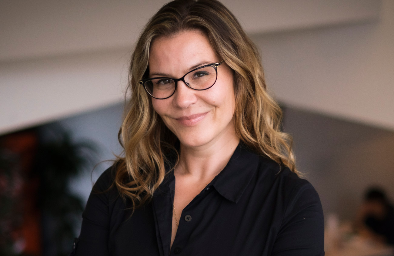
Sanford Burnham Prebys assistant professor Victoria Blaho, PhD, investigates the biochemical signals of the immune system and how they impact our bodies’ abilities to fight pathogenic infections, a branch of immunology that has become much less popular since the advent of antibiotics in the early 20th century.
Blaho’s disease of choice is Lyme disease, an unusual tick-borne bacterial infection that affects some 476,000 people in America each year, a number that is on the rise.
We caught up with Blaho to talk about why Lyme disease research is important, the progress being made and the work that remains in studying this strange and burdensome disease.
Why is Lyme disease research important? Blaho: Lyme research is a very small field for a disease that is becoming bigger and bigger every year. Case counts are increasing for Lyme disease all over the world, and people get very sick from it. Some people are infected, take antibiotics and that’s the end of it. But others have chronic symptoms like arthritis or carditis that can last for years and become completely debilitating.
What makes Lyme difficult to study? Blaho: One reason is that Lyme is an unusual infection from a microbiological standpoint. In the early days of Lyme research, there were studies showing that the bacteria that caused the disease, Borrelia burgdorferi, could change its physical form from a corkscrew shape to dormant blobs—and the blobs could be causing extended disease. This is a problem because scientists haven’t agreed on the true cause of chronic Lyme disease.
To make matters worse, a lot of the medical field still believes Lyme is easily curable with antibiotics, and if people are still having problems, then it’s psychosomatic. This makes it harder to get support for research into the longer-term inflammatory effects of Lyme. These politics make Lyme disease research a strange ecosystem of patients, physicians, researchers and funding agencies, and this is a barrier to learning more about the disease and helping people find relief.
How does your work enter the picture? Blaho: I’ve been working on Lyme disease for over 15 years, since I was PhD student. It started because Celebrex was hugely popular at the time to treat arthritis, but nobody had ever studied it in the arthritis that emerges in Lyme disease. Celebrex inhibits an enzyme of the immune system that triggers inflammation, so we figured that Celebrex might work just as well in Lyme arthritis as in other types. But research on mice didn’t bear this out.
Inflammation doesn’t just peter out when an infections clears. The immune system has to clean up the mess. We discovered in mice that Celebrex inhibits the resolution of inflammation after Lyme disease has resolved, so the arthritis never went away.
Since then, my career has focused on exploring the signaling molecules that regulate inflammation and its resolution. These molecules affect all parts of the immune system and provide us with a whole host of different potential therapeutic targets for inflammatory diseases like chronic Lyme.
What are the next steps for your Lyme research and for the field at large? Blaho: My own immediate next step is to take the work I’ve been doing here at Sanford Burnham Prebys and connect it directly back to my original work with Lyme. My team here is currently working on a signaling molecule called S1P, and while we haven’t studied it in Lyme yet, we think there are connections between it and the immune mediators we first found through those Lyme studies.
Our next steps are to look for the protein that carries S1P in mice with Lyme disease. This protein is associated with disease susceptibility in other inflammatory illnesses like diabetes and cardiovascular disease, and we think it has a role to play in Lyme as well. We’re also planning to partner with the Bay Area Lyme Foundation to see if we can find changes in this protein in their collection of human samples.
More broadly, I think this field is hungry for innovation because there have been a small number of scientists focusing on it. If older ideas about Lyme being simple to treat were the complete picture, we’d already be able to better diagnose and treat patients. But we’re just not there yet.
Lyme may be a lot cleverer than we originally thought, but if we’re able to embrace new technologies and ideas and continue to push forward with new work, we’ll be able to find innovative approaches to fight Lyme and, ultimately, to help people suffering from this horrible disease.
Institute News
San Diego Nathan Shock Center announces pilot grant awardees
AuthorMiles Martin
Date
December 21, 2021
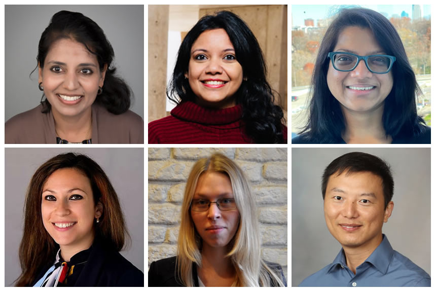
The San Diego Nathan Shock Center (SD-NSC) of Excellence in the Basic Biology of Aging, a consortium between Sanford Burnham Prebys, the Salk Institute for Biological Studies and the University of California San Diego, has announced its second-year class of pilot grant awardees. Recipients from six different institutions will receive up to $15,000 to pursue research that advances our understanding of how humans age, with the ultimate goal of extending health span, the number of years of healthy, disease-free life.
Aging is the biggest risk factor for most human diseases. Individuals age at different rates, and even specific cells and tissues within a person age differently. This depends on intrinsic properties, including genetics and where cells are in the body, and extrinsic factors, like exposure to environmental toxins and pathogens. Understanding this “heterogeneity” and how it contributes to overall human aging, risk for disease or therapeutic responses is the theme of the SD-NSC and the focus of pilot grant awards
“We are excited to support researchers who are working on these innovative, basic biology of aging research projects,” says Salk Institute Professor Gerald Shadel, who directs the SD-NSC. “The findings from this collective group of projects will deepen our understanding of the heterogeneity of aging, which is key to finding interventions to improve human health span.”
The six pilot grant awardees are:
Leena Bharath, assistant professor at Merrimack College, “Human T cell inflammation in aging”
Shefali Krishna, staff scientist at the Salk Institute for Biological Studies, “Characterization and function of mitochondrial age mosaicism and heterogeneity”
Gargi Mahapatra, postdoctoral fellow at Wake Forest School of Medicine, “Identifying mediators of bioenergetic decline in peripheral cells of older adults across a spectrum of cognitive abilities”
Chiara Nicoletti, postdoctoral fellow at Sanford Burnham Prebys, “Extracellular vesicles as soluble mediators of accelerated aging within the heterogeneous population of muscle-resident cells in Duchenne muscular dystrophy”
Anastasia Shindyapina, instructor in medicine at Brigham and Women’s Hospital and Harvard Medical School, “Unraveling heterogeneous biological aging of mouse immune cells at single-cell resolution”
Xu Zhang, research associate at the Mayo Clinic, “The dynamics and heterogeneity of cell fates during cellular senescence.”
Grant recipients will receive subsidized access to the SD-NSC Research Resource Cores (shared research facilities), necessary reagents/supplies, and access to training workshops offered by the center and its core research facilities. They will also be paired with an established aging-research investigator, who will provide career mentoring and guidance to ensure project success.
Research reported in this announcement was supported by the National Institute On Aging of the National Institutes of Health under award number P30AG068635. The content is solely the responsibility of the authors and does not necessarily represent the official views of the National Institutes of Health.
This piece was originally published by the Salk Institute for Biological Studies.
Institute News
Sanford Burnham Prebys and Roche fight back against antibiotic resistance
AuthorMiles Martin
Date
December 8, 2021
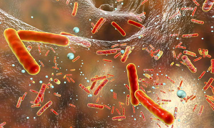
Researchers from Sanford Burnham Prebys have teamed up with prominent drug developer Roche Pharma to learn how bacteria develop antibiotic resistance.
Their new results, published in the journal mBio, are one piece of a long-standing collaboration between the two organizations, the goal of which is to mitigate the growing threat of antibiotic resistance by developing more “irresistible” drugs and by helping improve antibiotic prescribing practices.
“The emergence of antibiotic resistance is inevitable for any single drug, new or old. It’s only a question of time,” says senior author Andrei Osterman, PhD, a professor at Sanford Burnham Prebys. “But the precise time is different for every drug and every microbe, so studying when and how resistance to antibiotics evolves gives us powerful information for improving antibiotic treatment.”
Antibiotic resistance develops rapidly
When a patient is treated with antibiotics, most individual bacteria die, but a few cells will survive, usually as a lucky consequence of a random genetic mutation. These survivors go on to multiply into a whole new population of antibiotic-resistant bacteria.
“The development of antibiotic resistance is a strictly Darwinian process, very similar to evolution in larger organisms,” says Osterman. “The difference is that in bacteria, it happens much more rapidly, which makes antibiotic resistance one of the most pressing challenges facing medicine today.”
Although the speed at which evolution occurs in bacteria makes antibiotic resistance a threat, the researchers were also able to take advantage of this speed to study its development. The team cultured three species of bacteria in a morbidostat, a device that allows bacteria to grow continuously over multiple generations while being dosed with antibiotics. Although theirs was not the first morbidostat device, the team designed a new, more effective version of the system for their experiments.
“It’s like an evolution machine, letting us watch the development of antibiotic resistance in real time and in an environment that more accurately models what happens to bacteria in a clinical setting than other approaches,” says Osterman. “This gives us a clearer and more comprehensive view of resistance than we’ve ever had before.”
Different bacteria develop resistance differently
By observing the bacteria’s evolution in the morbidostat and sequencing their genomes as they evolved, the researchers found that all three species had a similar pattern of resistance development. However, they also found subtle differences in the ways certain genes were expressed, particularly those that help bacteria remove toxins, a critical process in developing resistance.
“It’s like three remakes of the same movie by three different directors, and their comparison gives us a wealth of information to guide the development and use of antibiotics,” says Osterman.
Understanding resistance is critical to reducing its harm
Working with Roche, the team has completed similar studies on several other classes of antibiotic drugs, which is helping Roche identify promising candidates for antibiotics that are less prone to resistance.
And because antibiotic resistance is often not assessed in drug candidates until years into the process, using resistance to screen for drug candidates this way could save the biomedical industry millions of dollars and help patients benefit from effective drugs sooner.
“A completely ‘irresistible’ drug is a holy grail, something we can never truly achieve,” says Osterman. “But some drugs are less resistible than others, and our methods allow us to figure out which is which in a systematic way.”
In addition to helping develop new drugs, the researchers claim that their findings are easily translatable to the clinic, where doctors can use detailed knowledge of resistance to select optimal drug combinations with less likelihood of failure due to resistance.
“We are moving away from trial-and-error approaches in medicine and moving toward being able to predict exactly what drugs will work best for each patient,” says Osterman. “It is going to take time, effort and money to make this happen, but it will all be worth it if we’re able to alleviate the threat of antibiotic resistance and help save lives, which I’m confident can be done.”
Institute News
From child neurology to stem cells: An interview with Evan Snyder
AuthorMiles Martin
Date
November 18, 2021
What do Evan Snyder and Sigmund Freud have in common? Both radically changed how we see the human brain.
In a first for San Diego, Sanford Burnham Prebys Professor Evan Y. Snyder, MD, PhD has been featured in the second edition of Child Neurology: Its Origins, Founders, Growth and Evolution, a collection of biographies detailing the lives of innovators in child neurology throughout history.
To celebrate this honor, we caught up with Snyder to discuss how his work in child neurology led him to make foundational discoveries in the fields of stem cell biology and regenerative medicine, as well as where these fields are headed in the future.
Plasticity, both at the “macro” sociological level and at the “micro” clinical level, always fascinated me. And that fascination with resilience and my curiosity about its source led to my early discoveries surrounding stem cells in the brain.
How does it feel to be included in this book? Snyder: Ever since I was a student, I thumbed through the first edition and was inspired by all these old images of people from the early days of the field. To see my chapter in there along with the likes of Sigmund Freud is mind-blowing. It means a lot to be included.
How did your work in child neurology lead you to work on stem cells?
Snyder: I was always interested in resilience. As a kid, I worked at a camp for children with various challenges: poverty, behavioral issues, disrupted home lives. These children were being raised in terribly deprived environments with challenging family situations, but they adjusted and bore up without complaint. When we took them out of that environment, even for a short time, they flourished. I never forgot that lesson.
Later, doing my pediatric and neurology residencies and neonatology fellowship at Boston Children’s Hospital, I would care for babies in the newborn intensive care unit with seemingly devastating brain injuries. As a neurologist, I would often follow up with those kids months or even years later.
Despite the horrible injuries they had sustained as newborns, by the time they were children, some had recovered to the point where it was hard to notice a deficit—they were achieving their developmental milestones, were seizure-free off medications, and playing and interacting like normal kids.
It astounded me that some children could have that resilience while I knew that adults with those same injuries would likely be incapacitated. Plasticity, both at the “macro” sociological level and at the “micro” clinical level, always fascinated me. And that fascination with resilience and my curiosity about its source led to my early discoveries surrounding stem cells in the brain.
There was no notion that there could be such regenerative cells in a solid organ, particularly one that was thought to be as static as the brain.
How did your early work shape the stem cell field?
Snyder: Before I, and a few others, began examining the molecular biology of cell types that composed the brain, there really was no research area called the “stem cell field”—at least not for solid organs like the brain. What was known clinically about stem cells in the early ’80s was confined to the making of blood cells for bone-marrow transplants.
The goal of hematologists back then was to identify the youngest cell in the bone marrow that could give rise to other types of blood cells. That cell, called the hematopoietic stem cell, was difficult to isolate and identify, but it was always assumed to exist because a healthy person’s blood is always turning over.
There was no notion that there could be such regenerative cells in a solid organ, particularly one that was thought to be as static as the brain.
But from the very beginning, I had difficulty making containers of cells with a single specific identity, even if I started with what seemed to be a single cell and its identical sibling cells. The young cells I isolated always seemed to be changing their identities, even though they started out looking the same.
Colleagues observing my initial results assumed it was just an odd tissue culture artifact, but then I thought back to those kids with brain injuries for whom I’d cared, and I started thinking that maybe what I was observing wasn’t an artifact at all; maybe these cell types were one of the repositories of plasticity in the brain I had puzzled over—a “stem cell” of sorts.
To prove that notion, I quietly transplanted those stem cells into a mouse brain and waited two years to examine the brain. The cells I’d transplanted were not only still there but they had integrated into the fabric of the brain and taken on different identities depending on which part of the brain they took up residence.
This observation—which I made alone, in the middle of the night under a microscope—gave me chills that I still recall today.
I shuddered with excitement at the implication of what I was seeing. The central nervous system was always thought to be a part of the body that could not regenerate at all, yet here I had isolated cells from one mouse brain and used them to populate another brain with multiple cell types in multiple locations. I could make pots of those donor cells to be transplanted whenever and wherever I liked, like a bone-marrow transplant in the brain.
The potential seemed enormous, and my curiosity to see where that potential might lead in terms of understanding and healing the human brain became the focus of my lab—first at Harvard and then at Sanford Burnham Prebys.
Finding how we could address unmet medical needs became somewhat like looking for a lock for which we already had a key.
How did regenerative medicine enter the picture?
Snyder: Other people started finding stem cells, not only in the brain but everywhere in the body. In the mid-1990s, the focus then became figuring out how to exploit them. Those efforts helped give rise to the new field of regenerative medicine, where the emphasis is on repairing and regrowing tissues rather than just treating the symptoms of a disease.
We found that we could put these neural stem cells almost anywhere in the nervous system, particularly if that region was diseased or injured—and hit the “reset button” for that region. I was more interested in studying the overall biology of this new cell type rather than focusing on any one illness. So, while learning about this cell, we found many different ways that its biology could be exploited therapeutically.
Finding how we could address unmet medical needs became somewhat like looking for a lock for which we already had a key. Looking for those locks—the therapeutic obstacles that stem cell biology might help circumvent—is where my clinical experience proved handy.
One example was brain tumors. I’m not an oncologist, but I’d learned from my studies that stem cells will naturally go to regions where pathology exists in the brain, even over long distances from their point of implantation. So, by putting a therapeutic gene into a neural stem cell, I could use that cell like a “heat-seeking missile” to deliver that gene and its product to where it might be needed.
Brain tumors are often so difficult to cure because we can’t safely access all the brain areas where the tumor has infiltrated. The neural stem cell seemed a perfect way to deliver a tumor-killing gene to those disseminated tumor cells simply by harnessing their natural powers. That strategy has now moved into clinical trials, and the use of stem cells to deliver therapeutic genes is being used for other conditions as well.
What came next in the history of stem cells? Snyder: If we fast-forward a few decades from the beginnings of regenerative medicine, we now know a lot more about the different “flavors” of stem cells, how they work, and how they can be therapeutic.
One of the new flavors of stem cells is induced pluripotent stem cells, or iPSCs. One can take a mature skin or blood cell and push it back to a state where it loses all of its organ identity, and then force it to become a completely different mature cell type.
In other words, plasticity can go in both directions: an immature cell type can take on multiple identities going forward in development, and a mature cell type can be pushed back in developmental time to the point where it can now make different choices.
Importantly, that induced stem cell retains many of the characteristics of the person from which it originally came, including diseases and genetic defects that individual may have had. This realization gave rise to the idea of modeling diseases in a dish.
This approach of using iPSCs works best for diseases where we know the specific cell type, pathway, gene, or protein that causes the disease, but we wondered if iPSCs could help for diseases where we have no clue which gene, protein, cell type, or pathway is causing a particular disease.
Psychiatric disorders are the poster children for such complex and mysterious diseases. To date, there’s no psychiatric disease that we can attribute to a specific defect in a particular gene or protein. Nevertheless, I felt confident that we could use iPSCs to discover the underlying cause of challenging diseases where we have no prior knowledge of what has gone wrong.
I decided to zero in on bipolar disorder because, as a physician, I knew that this condition did have an effective treatment—lithium—which had been accidentally discovered many years ago without knowing why it seemed to help some patients.
I reasoned that If we knew lithium’s target, we could work backward, recognizing that whatever lithium was changing was the underlying cause of bipolar disorder. That strategy worked. We used the iPSCs to map the lithium-response pathway, and we learned that errors in the regulation of that pathway were the likely cause of bipolar disorder. With that in mind, we could discover drugs better than lithium.
In this branch of regenerative medicine, it’s not the stem cell that goes into the patient, but rather the drug discovered by the stem cell that goes into the patient, but this would not be possible if we could not use stem cells to give us those patient-specific disease models.
Organs and their diseases never involve just one type of cell. If we want to improve the way we model diseases, we need to re-create and target that complexity.
Where is the field going from here? Snyder: Our models for diseases are only going to get better. We’re moving away from looking at small numbers of homogenous cells in a flat dish toward creating more complex three-dimensional mini-organs from a patient’s stem cells, directing those cells to become the many different interacting cell types that make an organ.
Organs and their diseases never involve just one type of cell. If we want to improve the way we model diseases, we need to re-create and target that complexity.
As far as using stem cells directly for therapy, we’re becoming much more sophisticated in deriving the cell type we need and directing it to the specific regions in need of repair. Furthermore, as our three-dimensional representations of organs in a dish become more sophisticated, these mini-organs may themselves become the material we transplant.
I’m also certain that there are applications for stem cells we haven’t conceived of yet.
Every generation of scientists needs to be reminded that one is constantly going to be surprised; one’s initial hypotheses, based on old understandings, are likely going to be wrong. One must be prepared to revise those hypotheses when the data—even if counter to expectations—leads one in an unconventional or inconvenient direction.
Although we’ve made much progress in the stem cell field over the past 30 years, there’s so much we don’t know. And, even when we think we know something, we probably don’t. None of my work would have been possible if I’d kept my blinders on and not been able to see the connections between different areas in which I was working—including allowing my work in the clinic and as a social worker to inform my scientific vision. That mindset is critical to the future of science.

Institute News
Boosting immunotherapy in aggressive brain cancer
AuthorMiles Martin
Date
November 3, 2021
Researchers from Sanford Burnham Prebys have collaborated the University of Pittsburgh Cancer Institute to reveal a new approach to enhance the effects of immunotherapy in glioblastoma, one of the most aggressive and treatment-resistant forms of brain cancer.
The study, published recently in Cancer Discovery, describes a novel method to ‘turn off’ cancer stem cells—the malignant cells that self-renew and sustain tumors—enabling the body’s own defense system to take charge and destroy tumors.
“Tumors are more than just masses of cells—each one is a complex system that relies on a vast network of chemical signals, proteins and different cell types to grow,” says senior author Charles Spruck, PhD, an assistant professor at Sanford Burnham Prebys. “This is part of why cancer is so difficult to treat, but it also presents us with opportunities to develop treatment strategies that target the machinery powering tumor cells rather than trying to destroy them outright.”
Glioblastoma is an extremely aggressive form of cancer that affects the brain and the spinal cord. Occurring more often in older adults and forming about half of all malignant brain tumors, glioblastoma causes worsening headaches, seizures and nausea. And unfortunately for the thousands of people who receive this diagnosis each year, glioblastoma is most often fatal.
“We haven’t been able to cure glioblastoma with existing treatment methods because it’s just too aggressive,” says Spruck. “Most therapies are palliative, more about reducing suffering than destroying the cancer. This is something we hope our work will change.”
Immune checkpoint inhibitors—which help prevent cancer cells from hiding from the immune system—can be effective for certain forms of cancer in the brain, but their results in glioblastoma have been disappointing. The researchers sought a way to improve the effects of these medications.
“Modern cancer treatment rarely relies on just one strategy at a time,” says Spruck. “Sometimes you have to mix and match, using treatments to complement one another.”
The researchers used genomic sequencing to investigate glioblastoma stem cells. These cells are the source of the rapid and consistent regeneration of glioblastoma tumors that make them so difficult to treat.
The team successfully identified a protein complex called YY1-CDK9 as essential to the cells’ ability to express genes and produce proteins. By modifying the activity of this protein complex in the lab, the team was able to improve the effectiveness of immune checkpoint inhibitors in these cells.
“Knocking out this transcription machinery makes it much more difficult for the cells to multiply” says Spruck. “They start to respond to chemical signals from the immune system that they would otherwise evade, giving immunotherapy a chance to take effect.”
While the approach will need to be tested in clinical settings, the researchers are optimistic that it may provide a way to improve treatment outcomes for people with glioblastoma.
“What our results tell us is that these cells are targetable by drugs we already have, so for patients, improving their treatment may just be a matter of adding another medication,” adds Spruck. “For a cancer as treatment-resistant as glioblastoma, this is a great step forward.”
Institute News
How misplaced DNA contributes to chronic illness
AuthorMiles Martin
Date
October 28, 2021
Though DNA is essential for life, it can also wreak havoc on our bodies as we age
DNA is one of the essential building blocks of life, giving our cells instructions for virtually everything they do, but researchers at Sanford Burnham Prebys are investigating what happens to our cells when DNA ends up in places where it shouldn’t normally be, particularly as we age.
The answer – as described in their recent review in the journal Cell—is disease-causing inflammation. And the researchers hope that targeting this rogue DNA will lead to new therapeutic strategies for a range of age-related illnesses, including cancer, diabetes, rheumatoid arthritis, cardiovascular disease and neurodegenerative disorders.
“Age is the primary risk factor for all of these diseases, but they share another risk factor – chronic inflammation,” says first author Karl Miller, PhD, a postdoctoral researcher in the lab of Peter Adams, PhD, Sanford Burnham Prebys. “We’re trying to understand the underlying processes behind this inflammation so we can potentially treat all these age-related diseases together”
Typically, cells have DNA safely sequestered in their nucleus and in the mitochondria, where the DNA can do its job without interfering with the rest of the cells’ activities. When cells detect DNA in other areas, they unleash a series of biochemical responses designed to protect the cell from invaders. This response is a component of the innate immune system, our body’s first line of defense against infection.
Scientists have known about this system for decades, but until recently it was mostly thought to respond to foreign DNA, such as during a bacterial or viral infection. However, over the last decade, researchers have discovered that pieces of our own DNA, called endogenous cytoplasmic DNA, can escape from the nucleus or mitochondria and trigger this inflammatory response in our own cells, even in the absence of infection. The resulting ‘sterile’ inflammation can accumulate over time, contributing to a range of age-related diseases in all systems of the body.
But this inflammation is not without its upsides. Cytoplasmic DNA is actually an important short-term protective strategy against cancer formation. The inflammation can alert the immune system at the first sign of cancer, preventing its formation. But over the long term, the sterile inflammation caused by cytoplasmic DNA is also thought to contribute to cancer risk. In fact, we’ve only been able to observe the damage associated with sterile inflammation because people are now living long enough to experience it.
“Systems like this exist because they’re beneficial in youth, but as we age, they break down,” says Miller. “100 years ago, a lot more people died from infectious diseases early in life. Over time, we’ve become better and better at treating these acute infections, and we’re living much longer. It’s in this later period in life that we see chronic diseases emerging that used to be much less common.”
Miller’s review describes four different types of cytoplasmic DNA fragments, classified according to when and how they appear. Some arise from the nucleus during mistakes in cell division. Others emerge because of errors in DNA repair or replication. Some even escape from mitochondria—energy-producing parts of the cell that have their own separate DNA. Others still are of unknown origin.
“They all look similar under a microscope, and they all can cause similar effects. That’s one of the major problems in this field. The benefit of studying how the different types emerge is that it gives us more points to target for therapeutics,” says Miller.
In the Adams Lab, Miller and his colleagues look specifically at cytoplasmic chromatin fragments, one of the four types of cytoplasmic DNA. These fragments appear in the cell when the membrane surrounding the nucleus is weakened by senescence, a cellular stress response. Senescence is also associated with aging.
“We’ve shown how this pathway works in mice, and now we’re actually moving forward with therapeutic applications for humans by doing drug screening to find compounds that can target it,” adds Miller.
And while there is still a lot of work left for the researchers, their progress is encouraging. Adams, senior author on the Cell review, was recently awarded a $13 million grant by the NIH to study the effects of aging, including the role of cytoplasmic DNA, on the progression of liver cancer.
“We like to call what we’re doing here ‘increasing the healthspan’, as opposed to the lifespan,” says Miller. “We’re hoping to maximize the healthy period of people’s lives.”