jshort, Author at Sanford Burnham Prebys - Page 2 of 13
Institute News

COVID-19: Using “mini lungs” to understand why some people fare worse than others

AuthorMonica May
Date

April 24, 2020

Evan Snyder tells us how he is using lung organoids to find effective treatments for COVID-19. 

As the novel coronavirus races around the globe, mystifying patterns are emerging. The virus seems to hit some people hard—including men, people over the age of 65 and individuals with pre-existing medical conditions. For others, particularly children, the symptoms may be mild or even nonexistent.

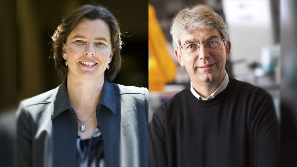
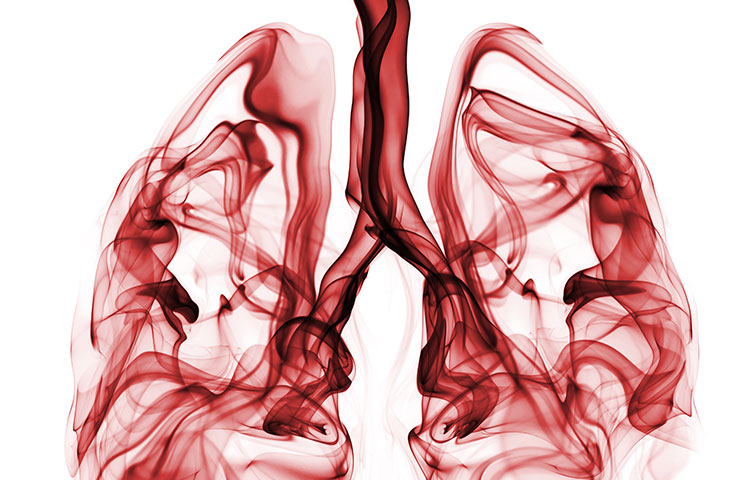
To understand why these disparities occur, stem cell expert Evan Snyder, MD, PhD, director of Sanford Burnham Prebys’ Center for Stem Cells and Regenerative Medicine, is turning to lung organoids—3D structures that replicate human lungs. 

We caught up with Snyder to learn more about these “mini lungs” in a dish—and how the outbreak has impacted his lab.

Why did you create these organoids?
A neonatologist who works in my lab, Sandra Leibel, MD, originally developed these organoids to see if we could help babies who have trouble breathing. Premature babies, and some full-term babies born with genetic conditions, can’t make surfactant—the material that allows the lungs to be flexible and breathe. The severe respiratory distress that is thought to kill some people with severe COVID-19 is actually the adult version of what these babies experience. 

I’m happy to report that, using this model, Sandra was able to find a therapy that might help these infants with the previously untreatable genetic condition. I’m hoping we can learn from this success to help people with severe COVID-19 and “adult respiratory distress syndrome.” 

How are these lung organoids made? 
We start with human induced pluripotent cells (hiPSC) created from skin cells and turn these into lung cells using the same chemical signals our body uses. Then the exciting part comes: We transform these cells from flat, two-dimensional layers into three-dimensional spheres that grow the way a lung would—aka “mini lungs in a dish.” Sandra, along with technician Alicia Winquist and Sanford Burnham Prebys graduate student Rachael McVicar, figured out the “secret sauce” the cells need to complete this process. 

What makes these lung organoids so special?
Lungs are very complicated organs. They have many cell types and structures that all interact with each other: air sac cells that make surfactant, airway cells, immune cells, blood vessel cells; and cells with cilia, tail-like organelles that whip around and clear out debris. Our organoids contain all these different parts, so they can be used to model human disease more closely than single layers of cells. They’re also easier to scrutinize, manipulate and test treatments in animal models. These organoids let us ask and answer all sorts of interesting questions.

What do you hope to learn about COVID-19 using this model? 
We want to learn the basics about the virus that causes COVID-19, called SARS-CoV-2. For example, how does the virus infect human lung cells? What does the virus do after it enters the lung cell? How does the virus move from one lung cell to another? The answers to these questions will allow us to find effective treatments for COVID-19. 

In addition, we can use this model to determine why some people fare worse than others. We can compare organoids created from men and women; younger or older people; people exposed to various environmental toxins from smoking or vaping; people with diabetes, heart or kidney disease; and even people of different racial backgrounds or individuals who have genetic variations that affect their ability to fight infection. 

If we figure out why the virus affects some people differently, we can potentially create tailored treatments. For example, we can ask whether any disparities we observe can be compensated for simply by increasing a dose of a drug or by adding another drug. With our model, we may be able to find these answers relatively quickly so that as many people as possible can benefit from any breakthroughs.

Will you partner with additional Sanford Burnham Prebys scientists on this research? 
Yes, we are in a fortunate position to collaborate with Sumit Chanda, PhD, who just completed a heroic (and I do mean heroic) effort to screen 12,000 known drugs for COVID-19. He pinpointed 30 promising drugs, and now we will help him study exactly how these drugs are working in a system that is clinically relevant. For example, in what stage of the virus’s life are they disrupting? Which part of the cell’s function is being rescued? Using lung organoids, we can rank the effectiveness of each drug—and help Sumit winnow the list to the most promising drugs for close-to-immediate use.

The best approach to treat COVID-19 will most likely be with a drug cocktail, similar to how we treat other RNA viruses. For this reason, it’s important to know how each drug is working, because we want to attack the virus at multiple points in its life cycle and block toxic downstream effects. Our model will help us map this out and advance the most promising drug combinations. 

How has the outbreak affected your lab work? 
I have time-sensitive, patient-relevant work ongoing, so I am still coming into the lab. In addition to our COVID-19 work, we are preparing to launch a clinical trial using stem cells to help newborns who are at risk for cerebral palsy

However, things are very different now. When I do go in, there are only one or two people instead of dozens, and we work in shifts. We limit our time to a few hours and only come in a few days a week. What I really miss is the meeting of the minds. Some of the most inspirational science comes from sitting around in a group and sharing ideas. Or having someone walk over and look through your microscope at the primary data. We do our best with Zoom. But the in-person human interactions are the part of science I really miss. 
 

Institute News

Rett Syndrome Foundation funds a potential cure

AuthorSusan Gammon
Date

April 23, 2020

The research may lead to a major step toward a cure.

Rett syndrome is a neurodevelopmental disorder that affects almost all aspects of a child’s life, from walking to eating to intellectual capability. There is no cure for the disease, which occurs mostly in girls, and treatments are aimed at slowing the loss of abilities and alleviating the debilitating symptoms. 

Jing Crystal Zhao, PhD, associate professor at Sanford Burnham Prebys, has received new funding from the Rett Syndrome Foundation to find ways to reverse the changes in a gene that causes Rett syndrome. The research may lead to a major step toward a cure.

“I’m very grateful to the Rett Syndrome Foundation, and excited to begin this project,” says Zhao. “While Rett syndrome may not be well known among the general public, our research may lead to treatments to improve the lives of patients around the world.”

More than 90% of Rett Syndrome cases are caused by genetic changes in a gene called MECP2. Every female carries two copies of the MECP2 gene. Rett syndrome patients carry both a normal and a mutant copy of MECP2. Unfortunately, in some cells, the normal copy of MEPC2 becomes inactive due to a biological process called X-chromosome inactivation—a process that occurs in females—and this leads to Rett syndrome. 

“Recent studies suggest that reversing X-chromosome inactivation could reactivate the normal copy of the MECP2 gene,” says Zhao. “We have identified an DNA element that plays a key role in X-chromosome inactivation. We are now going to test if we can block this element and restore the silent MECP2 gene, which could be life changing. 

“Our aim is to help individuals regain the skills and abilities stolen by Rett syndrome,” adds Zhao. “This award takes us closer to that goal.” 
 

Institute News

Scientists discover an early sign of type 2 diabetes: Misfolded proinsulin

AuthorMonica May
Date

March 19, 2020

The findings could lead to tests or treatments that help prevent type 2 diabetes.

Misfolded proinsulin—a protein the body normally processes into insulin—is an early sign of type 2 diabetes, according to a study by scientists at Sanford Burnham Prebys and the University of Michigan Medical School. The discovery, published in eLife, could lead to tests or treatments that help prevent people from developing type 2 diabetes.

“Understanding the molecular events that occur as prediabetes progresses to diabetes opens new avenues for us to detect or interrupt these processes,” says Randal Kaufman, PhD, director and professor in the Degenerative Diseases Program at Sanford Burnham Prebys and co-corresponding author of the study. “With this information, we can start to find interventions that might spare millions of people from a serious, lifelong condition.”

More than one in three Americans, or approximately 88 million people, have prediabetes—which is characterized by elevated blood sugar. If left untreated, within four years nearly 40% of people with prediabetes develop type 2 diabetes, which occurs when the body doesn’t use insulin properly. In 2017, the cost of treating diabetes exceeded $327 billion, according to the American Diabetes Association. Due to increasing obesity rates, the number of people with the condition—particularly children—is on the rise.

Identifying the molecular events that occur during progression from prediabetes to full-blown diabetes remains one of the most perplexing problems in diabetes research. In the study, the scientists set out to answer this question by tracking proinsulin folding in the beta cells of humans and mice that are healthy, prediabetic and diabetic.

These studies revealed that instead of undergoing its normal folding process, proinsulin proteins were abnormally linked to each other. Levels of the abnormal proinsulin accumulated as prediabetes progressed to type 2 diabetes. Obese mice in the earliest stages of diabetes had the highest levels of abnormal proinsulin in their beta cells.

“Proinsulin misfolding is the earliest known event that may contribute to the progression from prediabetes to diabetes,” says Kaufman. “Together, these studies show that abnormally linked proinsulin holds promise as a potential measure of how close someone may be to developing type 2 diabetes.”

Now, the researchers are set to uncover more details about this process, such as the proteins that interact with the misfolded proinsulin.

“Understanding the fundamental molecular events that lead to type 2 diabetes is critical as the number of people with prediabetes continues to rise,” says Kaufman. “If we don’t find preventive measures, we will soon have a diabetes epidemic.”


The study’s first author is Anoop Arunagiri, PhD; and the study’s senior author is Peter Arvan, both of the University of Michigan Medical School.

Additional authors include Leena Haataja and Fawnnie Pamenan of the University of Michigan Medical School; Ming Liu of the University of Michigan Medical School and Tianjin Medical University in China; Anita Pottekat and Pamela Itkin-Ansari of Sanford Burnham Prebys; Soohyun Kim of Konkuk University in South Korea; Lori M. Zeltser of Columbia University; Adrienne W. Paton and James C. Paton of the University of Adelaide in Australia; and Billy Tsai of the University of Michigan.

The study’s DOI is 10.7554/eLife.44532.

This work was supported by the National Institutes of Health (R01DK111174, R24DK110973 and R01DK48280) and the Juvenile Diabetes Research Foundation International (2-SRA-2018-539-A-B).

Institute News

First supercentenarian-derived stem cells created

AuthorMonica May
Date

March 19, 2020

Advance primes scientists to unlock the secrets of healthy aging.

People who live more than 110 years, called supercentenarians, are remarkable not only because of their age, but also because of their incredible health. This elite group appears resistant to diseases such as Alzheimer’s, heart disease and cancer that still affect even centenarians. However, we don’t know why some people become supercentenarians and others do not.

Now, for the first time, scientists have reprogrammed cells from a 114-year-old woman into induced pluripotent stem cells (iPSCs). The advance, completed by scientists at Sanford Burnham Prebys and AgeX Therapeutics, a biotechnology company, enables researchers to embark on studies that uncover why supercentenarians live such long and healthy lives. The study was published in Biochemical and Biophysical Research Communications.

“We set out to answer a big question: Can you reprogram cells this old?” says Evan Snyder, MD, PhD, professor and director of the Center for Stem Cells and Regenerative Medicine at Sanford Burnham Prebys, and study author. “Now we have shown it can be done, and we have a valuable tool for finding the genes and other factors that slow down the aging process.”

In the study, the scientists reprogrammed blood cells from three different people—the aforementioned 114-year-old woman, a healthy 43-year-old individual and an 8-year-old child with progeria, a condition that causes rapid aging—into iPSCs. These cells were then transformed into mesenchymal stem cells, a cell type that helps maintain and repair the body’s structural tissues—including bone, cartilage and fat.

The researchers found that supercentenarian cells transformed as easily as the cells from the healthy and progeria samples. As expected, telomeres—protective DNA caps that shrink as we age—were also reset. Remarkably, even the telomeres of the supercentenarian iPSCs were reset to youthful levels, akin to going from age 114 to age zero. However, telomere resetting in supercentenarian iPSCs occurred less frequently compared to other samples—indicating extreme aging may have some lasting effects that need to be overcome for more efficient resetting of cellular aging.

Now that the scientists have overcome a key technological hurdle, studies can begin that determine the “secret sauce” of supercentenarians. For example, comparing muscle cells derived from the healthy iPSCs, supercentenarian iPSCs and progeria iPSCs would reveal genes or molecular processes that are unique to supercentenarians. Drugs could then be developed that either thwart these unique processes or emulate the patterns seen in the supercentenarian cells.

“Why do supercentenarians age so slowly?” says Snyder. “We are now set to answer that question in a way no one has been able to before.”


The senior author of the paper is Dana Larocca, PhD, vice president of Discovery Research at AgeX Therapeutics, a biotechnology company focused on developing therapeutics for human aging and regeneration; and the first author is Jieun Lee, PhD, a scientist at AgeX.

Additional authors include Paola A. Bignone, PhD, of AgeX; L.S. Coles of Gerontology Research Group; and Yang Liu of Sanford Burnham Prebys and LabEaze. The work began at Sanford Burnham Prebys when Larocca, Bignone and Liu were members of the Snyder lab.

The study’s DOI is 10.1016/j.bbrc.2020.02.092.

Institute News

How to help children survive—and thrive—after a brain cancer diagnosis

AuthorMonica May
Date

January 13, 2020

Lynne Selinka knew in her heart that something was seriously wrong with her 10-year-old son, Travis. For months he had experienced dizziness, vomiting and headaches, despite his doctor’s best efforts to find a cause. A visit to Rady Children’s Hospital-San Diego revealed a heartbreaking diagnosis: Travis had a malignant brain tumor. He was operated on the next day and then endured two months of radiation treatment followed by six rounds of chemotherapy.

“That year, Travis asked Santa, ‘Can I please be done with chemo before Christmas?’” Lynne said. “It was by far the hardest year of our life.”

Brain tumors are the most common cause of cancer-related death in children—recently surpassing leukemia. To help the public learn about the latest efforts to develop better treatments for pediatric brain cancer, our Institute teamed up with the Fleet Science Center to host a panel discussion on Sunday, December 8. Travis and his parents, Lynne and Tony, shared their story alongside the clinician who treated Travis, John Crawford, MD, director of Pediatric Neuro-Oncology at Rady Children’s Hospital-San Diego; and a scientist working on personalized treatments for pediatric brain cancer, Robert Wechsler-Reya, PhD, of Sanford Burnham Prebys and Rady Children’s Institute for Genomic Medicine. 

As the speakers explained, while aggressive therapies have improved outcomes for children with brain tumors (today Travis is a junior in high school), one in four children with a malignant brain tumor does not survive. Children who do survive have an increased risk of severe long-term side effects from undergoing aggressive treatment at such a young age, including developing additional cancers or experiencing intellectual disability. Six years after he was declared cancer-free, Travis was diagnosed with chronic myeloid leukemia, a type of blood cancer caused by his previous chemotherapy. So far, his new treatment is working.

Wechsler-Reya hopes his work to develop personalized therapies based upon an individual’s tumor could help spare children from this painful experience. By analyzing patient tumor samples—obtained from Rady Children’s Hospital—his team works to understand the cancer at a molecular level, studying the tumor’s DNA mutations, changes in gene expression, responses to drugs, and much more. Armed with this information, the scientists then work to find therapies that are customized to a child’s specific tumor—and may be more effective and less toxic.

“For pediatric brain cancer, success doesn’t just mean better treatments. It also means developing treatments with fewer long-term side effects,” says Wechsler-Reya. “If successful, this work might help more children not only survive brain cancer, but also live a long, healthy life after treatment.

Travis and his family welcome this work with open arms.  

“We try to look for a silver lining in every day. Travis has become an amazing public speaker and now shares his story with other children fighting brain cancer. But each part of our journey has been so hard—from receiving the diagnosis, seeing Travis go through a painful surgery and then chemo, not knowing if the treatments would work, and then being diagnosed with another cancer almost six years later,” said Lynne. “We are so grateful for the efforts of researchers who are working toward a world where a child doesn’t have to go through what Travis did—or at least is spared from some of the hardest parts of the journey.”

This event was the last of our five-part “Cornering Cancer” series at the Fleet Science Center. Read about our past discussions focusing on lung, blood, breast and pancreatic cancers.

Institute News

Veterans still giving back in new roles at Sanford Burnham Prebys

AuthorMonica May
Date

November 8, 2019

More than 19 million Americans are veterans—the service members who have sacrificed for our nation’s safety and for the common good. Each year nearly a quarter of a million veterans transition to civilian life, where they often continue to give back in new roles. 

In honor of Veterans Day, we caught up with three veterans who now work in the information technology (IT) department at Sanford Burnham Prebys. They told us about their journey from the front lines to our research institute—and the many ways their service informs their work today.

Matt Sciaroni, lead help desk specialist

Tell us more about your military service.
As a Marine I worked on signals intelligence. While deployed to Afghanistan in 2012, I supported intelligence and counter-intelligence activities, such as cryptography and satellite communication.

What skills from this experience inform your work today? 
The Marine Corps teaches you how to develop and execute plans. Here, we always have concurrent projects that are all time sensitive. Just like in the Marines, we all do our part to get the job done. I also learned small unit-leadership skills, which I use every day—I lead a small team of about six IT professionals.

Why Sanford Burnham Prebys? 
The important work that is being done here was a really big factor for me. I actually received a second job offer from a local company that paid more. I decided that it was more important to me to make a contribution to society, even if I made a little bit less. Here, our scientists are working to save lives. I definitely made the right choice.  

Matt Sciaroni, US Marine Corps

Any advice for veterans who are thinking about moving into IT? 
Definitely contact a hiring recruiter. Transitioning out of the military takes a lot of organization and focused effort—a recruiter makes your life so much easier. Also, keep learning. In IT, everything is always changing. Our team learns new things every day—we recently invested in an IT training tool that allows us to learn new skills at our pace.

What do you do in your free time? 
I spend time with friends, family, and my bulldog, Ben.


Jeff Lustina, help desk specialist I

Tell me more about your service.
I joined the Army about 20 years ago. I was a “cable dawg”—I installed cable wires. After working for two years in active duty, I went on to reserves. Then 9/11 happened. I was deployed to Kosovo for nine months in support of Operation Enduring Freedom to help with NATO peace-keeping missions.

What types of activities did you do in this role? 
I made sure radio communications was working. At this point GPS was just starting, so I would help track all of our teams and make sure that everyone was safe. We often met with the local community as well to spread good will.

Jeeff with locols in Kosovo

Lustina (right) often met with the local community while in Kosovo.

Lustina also enjoys DJ'ing in his free time.

Lustina also enjoys DJ’ing in his free time.

Do skills gained from this experience inform your work today? 
Yes, so many. Leading by example. Always setting standards for excellence—and then exceeding those standards. Being detail oriented. Another big one: thinking three steps ahead. Earlier I was helping someone whose computer stopped working. I’m already thinking about my next steps if our next action doesn’t work. 

I joined the U.S. Army because I wanted to help people. That’s what I also like about this role. I still get to help people.

Why Sanford Burnham Prebys? 
I am incredibly proud to work at Sanford Burnham Prebys. I knew I wanted to work in IT, and helping such an important cause—which could lead to cures for cancer and more—is so important to me. When I read about our breakthroughs in pancreatic cancer, and even the stem cell therapy for hair loss, I think, “We were part of that.” We are behind the scenes, but we help the scientists focus on their research—not on fixing a faulty computer.

What do you do in your free time? 
Eat! I’m a foodie. 

Favorite restaurant? 
Sushi Ota. Best sushi in San Diego.


Hayder Al Kawaz, help desk specialist 

What is your service story? 
I was born and raised in Baghdad, the capital of Iraq. I was there the day the U.S. Army arrived, in 2003. A crowd was gathering around the soldiers, and I was helping keep people back. The soldiers noticed that I spoke English well and offered me a job on the spot. I was finishing my bachelor’s degree in software engineering, so I agreed. I worked with the U.S. Army for 14 years, until the militias made it too dangerous to continue—they started to follow me home and threaten my family. I was often shot at. My family gained rapid clearance to move to San Diego, and I joined them in 2017. Now my family—my 6-year-old twins, mother and father—all live here. We love it. This is home now. 

Do certain skills from this experience inform your work today? 
Working with the U.S. Army, I had to be fast, accurate and think ahead—all while under great stress. One of my jobs was ensuring that TV stations such as the Associated Press were able to keep broadcasting, especially during the elections in 2014. I oversaw a team of ten people who were live-broadcasting three or four different channels. It was go, go, go. All of these skills translate well to my current role.  

Any advice for other veterans who are thinking about moving into IT? 
Make sure you keep up with new updates. Technology is constantly changing. I went to Cisco academy and also obtained four certificates in IT. Now I’m studying to get a network certification. In IT, you can’t ever sit back and think you know everything—there is always something you need to learn.

Interested in working at Sanford Burnham Prebys? Visit our Careers page to learn more about open positions.
 

Institute News

2017 Faculty Retreat highlights research advances, awards and promotions

AuthorSusan Gammon PhD
Date

May 12, 2017

The 2017 Faculty Retreat, held May 10-11, gave Institute researchers a chance to present their latest discoveries, share ideas, and socialize in beautiful Carlsbad, CA.

The two-day event, organized by Bas Baaten and Cosimo Commisso, included scientific sessions on:

  • Epigenetics and Immunity—Chair: Max D’Angelo
  • Neurological Disorders—Chair: Jerold Chun
  • Metabolism/Development and Regeneration—Chair: Alex Colas
  • Cancer—Chair: Brooke Emerling
  • SBP’s Drug Discovery Portfolio—Chair: Michael Jackson

CEO Perry Nisen chaired a session on “Partnering to Accelerate Research” that covered SBP’s intellectual property resources and guidelines, how to work with Business Development, and the role of philanthropy in supporting our Institute.

A major highlight of the retreat was the presentation of the “WOW” award to Alessandra Sacco, PhD, associate professor in the Development and Aging Research Center. The “WOW” award stands for Wonderful Original Work and is awarded based on votes by scientists attending the retreat. This was the second time in six years that Alessandra has been named the recipient. Her presentation titled, “Dynamics of muscle stem cell expansion during tissue repair and maintenance” gave important insights for pharmacological treatments for age-related muscle wasting diseases.

Congratulations were also in order for Malene Hansen, PhD, who was promoted to professor in the Development, Aging and Regeneration Program.

Finally, two special presentations were given by recent recipients of NIH R35 Outstanding Investigator Awards:

  • Ze’ev Ronai presented “Improvidus, apto, quod victum,” a glimpse of his stellar career as a leader in the field of melanoma research.
  • Francesca Marassi presented “Structual analysis of Yersinia-host interactions,” a summary of her work to understand how the organism that causes bubonic plague infect humans cells

And the retreat was outstandingly organized by Esther Minotti, research administrative specialist.

Institute News

SBP steps out for science

AuthorKristen Cusato
Date

April 26, 2017

Scientists from Sanford Burnham Prebys Medical Discovery Institute (SBP) marched for science all across the country on Saturday, April 22nd. The passion and commitment to keep research alive is depicted in photos sent in by Malene Hansen, PhD, associate professor; Jarett Larraga; Sepideh Khorasanizadeh, PhD, professor at Lake Nona; Cosimo Commisso, PhD, assistant professor; Anindya Bagchi, PhD, associate professor, and others.

Several who took part in the march shared what the day meant to them.

“Science saved my life. I was born premature and couldn’t breathe on my own, and spent time in the neonatal intensive care unit. Participating in the March for Science on Saturday was a way for me to pay it forward and support scientific research. I felt a wonderful sense of solidarity marching with my SBP colleagues and the scientific community at large. Marching gave me hope for the future in an uncertain present.” —Samaire Cohen, IT

“Our son will be starting college in September 2017 and will be a biology/ecology major. His love of science and nature started when he was very young, so it was wonderful to march for him and all of the future scientists out there. He believes strongly that we all need to stand up to those who belittle scientists and the incredible work that they do, as they play an integral role in saving lives and our planet.” —Bobbie Larraga, NCI-designated Cancer Center

“I marched with my wife Connie who shares her birthday with Earth Day every April 22. We do something every year to celebrate Earth Day and this year was extra special because of the added focus to champion critical thinking and evidence based science discoveries in establishing government policy.” —Tim Osborne, PhD, professor at Lake Nona

“Marching on Saturday was a great way to show my commitment to science. I was so happy to see there were so many of us!”  —Pilar Cejudo-Martin, PhD, postdoc

 “I feel strongly that our country’s future, and that of the planet, is dependent on the scientific advances made by the researchers at our institute and around the world. I was very proud to march as a scientist and to see so many people marching with us in support of the work that we do every day.” —Karen Ocorr, PhD, assistant professor

SBP March for Science

SBP March for Science

SBp

SBP March for Science

SBP March for Science

SBP March for Science

SBP March for Science

SBP March for Science

Institute News

Muscle heat may hold key to promoting weight loss

AuthorJessica Moore
Date

July 6, 2016

If you’ve tried to lose weight, you may have wished for a pill that would help you burn calories with little or no exercise. Because such a drug could treat obesity, which affects over one-third of Americans, many researchers are working toward this goal. Treatments that boost calorie burning could enhance the limited efficacy of current weight-loss drugs that suppress appetite.

Most scientists in this field focus on brown adipose tissue, a type of fat that’s specialized to convert calories to heat to keep you warm in the cold. The challenge with that approach is that most adults have very little brown fat—therapies would have to first convert regular white fat to brown. Instead, the laboratory of Muthu Periasamy, PhD, professor in the Center for Metabolic Origins of Disease, is investigating how to stimulate another, more plentiful tissue—muscle—to do the same thing.

Periasamy and Naresh Bal, PhD, a staff scientist in his lab, got the idea that muscle could be important for generating heat from birds—they don’t have any brown fat, but they can still keep themselves warm without constant shivering. In a paper recently published in the Journal of Biological Chemistry, Bal removed the brown fat from mice to examine whether muscle can effectively generate heat in mammals.

“Not only did these mice maintain near-normal body temperatures when living in the cold,” said Bal, “but they burned more calories than mice whose brown fat remained intact—they lost three times as much fat after nine days at cold temperatures.” The extended exposure is required to eliminate the contribution of shivering, which stops after they become adapted to the cold, within the first few days.

“These results suggest that inducing muscle to generate heat could be an even more efficient way to treat obesity than doing the same in brown fat,” said Periasamy. “This is the first step toward drugs that activate this process, called nonshivering thermogenesis (NST).”

“Our next step is to determine which factors turn on NST in muscle,” added Bal.

The lab’s work so far has provided some clues. They have previously shown that the protein sarcolipin changes the way muscle cells use ATP, causing them to generate heat instead of contract. In this research, they observed much higher levels of sarcolipin in the muscles of cold-adapted mice who lack brown fat.

“Since sarcolipin acts by binding another protein, it probably wouldn’t be easy to block,” explains Bal. “To find better drug targets we plan to look at how it affects its target protein, a calcium pump, and how that changes calcium dynamics. Ultimately, we might be able to mimic those effects with a drug.”

The paper is available online here.