cancer Archives - Page 9 of 11 - Sanford Burnham Prebys
Institute News

Hearst Foundation’s new fellowship funds innovative research to fight breast cancer

AuthorJessica Moore
Date

October 20, 2016

Mark Goldberg, PhD, is working on a potential way to turn cancer stem cells into harmless cells. He and his advisor, Charles Spruck, PhD, assistant professor in the NCI-designated Cancer Center, are optimistic that they could turn this approach into new drugs that prevent breast cancer from returning.

Goldberg is supported by the first-ever research fellowship given by the David Whitmire Hearst Jr. Foundation. The funds were awarded specifically for this groundbreaking project.

“Breast cancer can spread to other organs very early, sometimes even before it’s detected,” said Spruck. “Those micrometastases—just one or a few cells—lie dormant for years, and are insensitive to anticancer drugs. Our goal is to switch those cells to a normal cell type that can’t generate a tumor.”

In as-yet unpublished research, Spruck’s lab recently discovered a protein that’s crucial for pre-cancerous cells to begin growing aggressively and out of control. Goldberg will use animal models of breast cancer to show that genetically inactivating this protein prevents secondary tumors from forming. The next step is to search for candidate drugs that inhibit the protein.

“If we find blockers of this protein that controls progression to malignancy, they could be given to breast cancer patients, after standard treatment has eradicated their primary tumor, to eliminate any remaining cancer stem cells,” added Spruck.

Goldberg’s background in bioengineering gives him a fresh perspective on cancer research. As a PhD student at Caltech, he designed implantable glucose and ion sensors using microfluidics and nanophotonics. That experience gives him a flexible, solutions-oriented approach to designing experiments.

“During Mark’s interview—the first time I met him—we came up with a really exciting way to apply what he was working on at Caltech to cancer research,” Spruck commented. “That kind of creative thinking and insight is invaluable—it’s why I hired him for this fellowship-supported spot.

“Because this research is so early-stage, it’s hard to get funded through traditional avenues. The Hearst Foundation fellowship allows us to get the evidence that this strategy works. That data will be key to getting the support we need for the drug discovery phase.” 

Institute News

To treat breast cancer, give it a lifeline

AuthorJessica Moore
Date

October 17, 2016

In honor of Breast Cancer Awareness Month, we’re highlighting the work our scientists are doing towards the next generation of breast cancer therapies.

Providing more oxygen to a tumor might seem like exactly the wrong way to treat cancer. But Masanobu Komatsu, PhD, associate professor in the Cardiovascular Metabolism Program and the NCI-designated Cancer Center, is trying to find treatments that do exactly that. Enhancing a tumor’s blood supply, which carries oxygen to cancer cells, actually lowers the chance that the cancer will spread.

“We’re aiming to minimize one of the most challenging and devastating aspects of breast cancer—metastasis,” said Komatsu. ”Mortality rates for metastatic breast cancers are still incredibly high. Of the patients with cancer that has spread and led to tumors in other organs, almost 80% will survive less than five years.”

Cancer cells become more likely to move into other tissues as they adapt to a low-oxygen environment due to the tumor’s defective vasculature. Because these blood vessels grow abnormally fast, they form improperly—oxygen and nutrients leak out before reaching the tumor’s interior. However, the cancer cells buried within continue to divide and mutate, so some can survive the lack of oxygen. The master switch that enables cancer cells to generate energy by alternate means also triggers changes that let them enter the circulation and find new homes.

Strengthening the blood supply could also help make the cancer more vulnerable to therapeutic attack, Komatsu added. “Improving the circulation inside a tumor would help anticancer drugs—and the body’s own T cells, which also help eliminate cancer—reach all the tumor cells, and increasing oxygen levels helps sensitize them to radiation and immunotherapy.”

Animal studies suggest that normalizing tumor blood vessels confers such benefits, but existing drugs known to stabilize the vasculature have shown limited benefit. Komatsu and his lab are looking for better therapies by screening microRNAs, small pieces of genetic material that regulate gene activity.

With funding from the Florida Breast Cancer Foundation, the scientific team is testing each of hundreds of microRNAs to look for those that affect signaling pathways controlling the stability of tumor blood vessels. The microRNAs that come up positive could either be developed as drugs (to be used in combination with other cancer-killing treatments), or studied further to find new drug targets.

“This strategy is relevant not only to breast cancer, but to any solid tumor,” commented Komatsu. “The therapies we hope to find could help a huge number of patients.”

Institute News

Research points to possible target to stop cancer stem cells

AuthorJessica Moore
Date

July 28, 2016

When you think of stem cells, you probably think of healing and regeneration—cells that can replace tissue lost to disease or injury. But tumors also arise from stem cells—a specific kind called cancer stem cells. Because these cells can divide indefinitely, while other cancer cells’ proliferation is more limited, therapies that get rid of them would eventually stop a tumor from growing, and from ever coming back.

Researchers in the laboratory of Dieter Wolf, MD, professor in the NCI-Designated Cancer Center, may have found a new way to do this, though in a roundabout way. While examining the function of two proteins found at high levels in tumors, they discovered that these factors are required for a type of metabolism that’s essential for cancer stem cells to survive.

“Our findings suggest that these components, eukaryotic initiation factors (eIFs) 3d and 3e, are novel targets for eliminating cancer stem cells,” said Wolf, senior author of the study, which was published in Cell Reports. “If we could find a way to turn these factors off, we could starve cancer stem cells.”

The details

eIF3d and eIF3e are part of the complex that initiates protein synthesis, also called translation. Specifically, they and other eIFs help bring the ribosome, which builds proteins by linking amino acids one by one, to messenger RNA, the molecules that carry the code the ribosome reads, specifying which amino acid should be added next.

Wolf’s team was interested in whether overproduction of eIF3d and eIF3e promotes cancer progression. To determine their function, they compared the amounts of all proteins made in cells lacking these factors to those in normal cells. A difference that stood out was in the levels of the protein complexes required to produce ATP, the cell’s energy currency, in mitochondria—eIF3d/e-deficient cells produced far lower amounts than normal.

“Cancer stem cells, unlike most tumor cells, rely on mitochondrial metabolism for energy,” Wolf explained. “Since tumor cells have much higher levels of eIF3d/e than normal cells, inhibiting those factors would preferentially block metabolism in cancer stem cells.

“Our results are somewhat surprising because eIF3 has long been thought to control the synthesis of all proteins. Instead, our data suggest that some parts of eIF3 selectively recruit certain mRNAs.”

Next steps

“We’re now examining how eIF3d/e affects cancer metabolism overall to see if these factors might be relevant to more than just cancer stem cells,” added Wolf. “Also, as a step towards advancing this research to the clinic, we’re designing screens to identify small molecule inhibitors of eIF3d/e.”

The paper is available online here.

Institute News

Genetic drivers of immune response to cancer discovered through ‘big data’ analysis

AuthorJessica Moore
Date

July 18, 2016

Scientists at the Sanford Burnham Prebys Medical Discovery Institute (SBP) have identified over 100 new genetic regions that affect the immune response to cancer. The findings, published in Cancer Immunology Research, could inform the development of future immunotherapies—treatments that enhance the immune system’s ability to kill tumors.

“By analyzing a large public genomic database, we found 122 potential immune response drivers—genetic regions in which mutations correlate with the presence or absence of immune cells infiltrating the tumors,” said lead author Eduard Porta-Pardo, PhD, a postdoctoral fellow at SBP. “While several of these correspond to proteins with known roles in immune response, many others offer new directions for cancer immunology research, which could point to new targets for immunotherapy.”

Immunotherapy has been heralded as a turning point in cancer because it can treat even advanced cases that have spread to other organs. Several drugs in this class are now widely used and often lead to remarkable success, eradicating or dramatically shrinking tumors and preventing recurrence.

Most current immunotherapies rely on a similar strategy—releasing the brakes on the immune system. These treatments are powerful if the tumor is recognized by the immune system as a threat and allows immune cell infiltration, but some cancers remain undercover or block immune cell entry into the tumor in as yet unknown ways.

“To develop immunotherapies that are relevant to a wide range of cancers, we need to know a lot more about how the immune system interacts with tumors,” said Adam Godzik, PhD, professor and director of the Bioinformatics and Structural Biology Program and senior author of the study. “Our study provides many new leads for this endeavor.”

“We are exploring cancer mutations at fine resolution by accounting for the fact that mutations can affect the encoded protein in different ways depending on where the resulting change is located,” commented Porta-Pardo. “Our algorithm, domainXplorer, identifies correlations between a phenotype, in this case the amount of immune cells in the tumor, and mutations in individual protein domains—parts of a protein with distinct functions.

“This work emphasizes the value of open data,” Godzik added. “Because we could access genomic data from over 5,000 tumor samples from The Cancer Genome Atlas (TCGA), we could jump straight to analysis without having to set up a big collaborative network to gather and sequence so many samples.”

“Our plan for the next phase of this research is to use this algorithm to search for genetic regions correlating with the levels of specific immune cell types within the tumor, which will reveal further details of cancer immunology.”

Institute News

SBP researcher receives NIH Outstanding Investigator Award to study deadly pathogens

AuthorSusan Gammon
Date

June 7, 2016

Francesca Marassi, PhD, professor in SBP’s NCI-designated Cancer Center, has been awarded an Outstanding Investigator Award from the National Institute of General Medical Sciences (NIGMS). The $4 million grant is to study how proteins on the surface of pathogens promote virulence by mediating the first-line interactions with human host cells. The project has important implications for biology and medicine.

“Our initial focus is on a protein called Ail (attachment invasion locus) that is expressed on the outer membrane of Yersinia pestis, the causative agent of plague,” said Marassi. “The Y. pestis bacterium is highly pathogenic, spreads rapidly and causes an extremely high rate of mortality. Ail is critical for suppressing the human immune defenses and for promoting bacterial invasion”

Although it is sensitive to some antibiotics, the potential use of Y. pestis as a biological weapon has led to its classification as a Tier 1 Biothreat Agent – a designation used by the U.S. Department of Health and Human Services to identify pathogens and toxins that can be misused to threaten public health or national security.

“The emerging threat of bacterial drug resistance makes our work particularly important,” added Marassi. “We will be using a technology called NMR (nuclear magnetic resonance) to determine the three-dimensional structure of Ail and examine how it associates with its human protein partners. Visualizing these biomolecular complexes helps us understand how pathogens engage their human host, and advances our ability to design effective drugs and vaccines for bacteria and viruses,” added Marassi.

Institute News

Study reveals protein that dials immune responses up and down

AuthorJessica Moore
Date

May 25, 2016

Research led by scientists at the Sanford Burnham Prebys Medical Discovery Institute (SBP) has identified a new regulator of immune responses. The study, published recently in Immunity, sheds new light on why T cells fail to clear chronic infections and eliminate tumors. The findings open the door for a new approach to modulating T cell responses in many clinical settings, including infections, autoimmune diseases, and tumors that are unresponsive to currently available therapies. Continue reading “Study reveals protein that dials immune responses up and down”

Institute News

Super-oncogenic protein that promotes development of melanoma

AuthorJessica Moore
Date

May 19, 2016

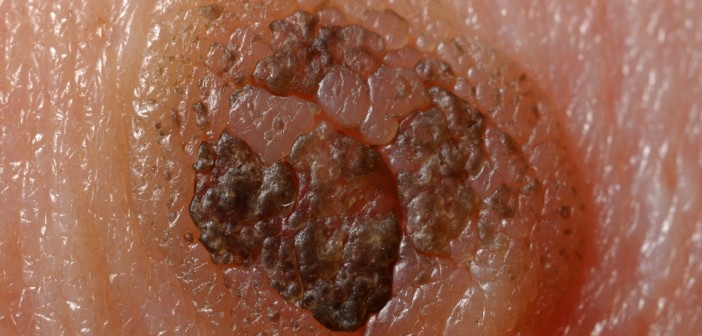
An international collaborative study led by scientists at the Sanford Burnham Prebys Medical Discovery Institute (SBP) has identified a malicious form of a protein that drives the formation of melanoma. The findings, published in Cell Reports, reveal unexpected insight into how this lethal skin cancer develops and progresses, and may help understand and develop novel therapies against these aggressive tumors.

“We found that an inactive version of a protein called activating transcription factor 2 (ATF2) elicits a tumor-promoting effect in a way not seen before,” said Ze’ev Ronai, PhD, chief scientific advisor of SBP and professor of its NCI-designated Cancer Center. “We have known for years that the active version of ATF2 promotes melanoma, but this result was a surprise because we thought ATF2 transcriptional activity was essential to activate cancer-related genes.”

Ronai’s team has been studying ATF2’s role in melanoma for two decades. Their past work led to the view that it’s dangerous when it’s in the nucleus because it controls cancer-enabling genes, but benign when it’s not.

In the current study, researchers looked at the oncogenic potential of a ‘dead’ form of ATF2 in mice with mutations in BRAF, a kinase that transmits signals promoting cell division and is often mutated in pigmented skin cells. The same mutation is found in about half of all human melanomas.

“Inactive ATF2, in mice with mutant BRAF, resulted in the formation of pigmented lesions and later, melanoma tumors,” said Ronai, senior author of the study.

“What makes this discovery relevant to human melanoma is that we identified a structurally similar form of inactive ATF2 in human melanoma samples that has the same effects on cancer cells,” added Ronai. “Inactive ATF2 could be an indicator of tumor aggressiveness in patients with BRAF mutations, and maybe other types of cancer as well.”

“Unlike models with more complex genetic changes, like the inactivation of PTEN and p16 combined with BRAF mutations that result in rapid tumorigenesis (within a few weeks), the inactive ATF2 caused BRAF mutant mice to develop melanoma much slower, more similar to the timescale seen in patients,” commented Ronai. “This improves our ability to monitor the development of melanoma and efficacy of possible interventions.”

“We’re now investigating why inactive ATF2 so potently promotes BRAF-mutant melanoma, and looking for other types of cancer where it acts the same way,” Ronai said. “Our findings may guide precision therapies for tumors with mutant ATF2.”

The paper is available online here.

Institute News

High levels of protein p62 predict liver cancer recurrence

AuthorJessica Moore
Date

May 19, 2016

CANCER METABOLISM AND SIGNALING NETWORKS PROGRAM

New research from SBP and UC San Diego shows that high levels of the protein p62 in human liver samples are strongly associated with cancer recurrence and reduced patient survival. p62 was also found to be required for liver cancer to form in mice. Continue reading “High levels of protein p62 predict liver cancer recurrence”

Institute News

Upcoming symposium: Cancer Immunology and the Tumor Microenvironment

Authorjmoore
Date

March 10, 2016

On March 17, SBP La Jolla is hosting a symposium on the interactions between the immune system and tumors, including how they can be leveraged for cancer treatment. The symposium is organized by Carl Ware, PhD and Robert Rickert, PhD, the directors of the Inflammatory and Infectious Disease Center and the Tumor Microenvironment and Cancer Immunology Program, respectively, and features presentations by leaders in the field:

Crystal Mackall, MDStanford University

Yang-Xin Fu, MD, PhDUT Southwestern

Mikala Egeblad, PhDCold Spring Harbor Laboratory

Linda Bradley, PhDSanford Burnham Prebys Medical Discovery Institute

Jose Conejo-Garcia, MD, PhDWistar Institute

Jonathan Powell, MDJohns Hopkins School of Medicine

Shannon Turley, PhDGenentech

Karen Willard-Gallo, PhDInstitut Jules Bordet – Belgium

Sandip Patel, MDUC San Diego

Adam Godzik, PhDSanford Burnham Prebys Medical Discovery Institute

The symposium will be held from 9-4:30 in Fishman Auditorium (overflow seating in the Building 12 auditorium), with a reception to follow. If you plan to attend, please register here.

Institute News

SBP’s Garth Powis talks about the search for a cure for cancer

Authorkcusato
Date

January 25, 2016

More than 1.6 million Americans will be diagnosed with cancer this year alone. President Barack Obama announced a new national effort to find a cure for cancer in his 2016 State of the Union address. It is clear that providing hope to those diagnosed with cancer and their families is a huge priority.

The search is on for a new generation of cancer drugs, and Garth Powis D.Phil., head of the NCI-designated Cancer Center at SBP appeared the KUSI news in San Diego on January 22nd to talk about recent treatment breakthroughs and what’s to come for treating this often deadly disease.

Watch the video here.