cancer Archives - Page 8 of 11 - Sanford Burnham Prebys
Institute News

How RNA splicing can trigger cancer

AuthorSusan Gammon
Date

September 21, 2017

Cancer, which is one of the leading causes of death worldwide, arises from the disruption of essential mechanisms of the normal cell life cycle, such as replication control, DNA repair and cell death. Thanks to the advances in genome sequencing techniques, biomedical researchers have been able to identify many of the genetic alterations that occur in patients that are common among and between tumor types. But until recently, only mutations in DNA were thought to cause cancer. In a new study published in the journal Cell Reports, researchers show that alterations in a process known as alternative splicing may also trigger the disease.

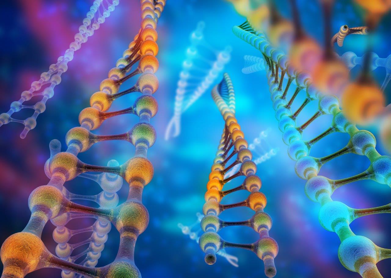
Although DNA is the instruction manual for cell growth, maturation, division, and even death, it’s proteins that actually carry out the work. The production of proteins is a highly regulated and complex mechanism: cellular machinery reads the DNA fragment that makes up a gene, transcribes it into RNA and, from the RNA, makes proteins. However, each gene can lead to several RNA molecules through alternative splicing, an essential mechanism for multiple biological processes that can be altered in disease conditions.

Using data for more than 4,000 cancer patients from The Cancer Genome Atlas (TCGA project), an international team of scientists that included Adam Godzik, PhD, professor at Sanford Burnham Prebys Medical Discovery Institute (SBP), has analyzed the changes in alternative splicing that occur in each tumor patient and studied how these changes could impact the function of genes. The results of the study show that alternative splicing changes lead to a general loss of functional protein domains, and particularly those domains related to functions that are also affected by genetic mutations in cancer patients.

From previous work, the research team learned that tumor type and stage can be predicted by observing alterations in alternative splicing. With this new study, the team discovered that changes in alternative splicing that occur in cancer impact protein functions in a way that is similar to that previously described for genetic mutations.

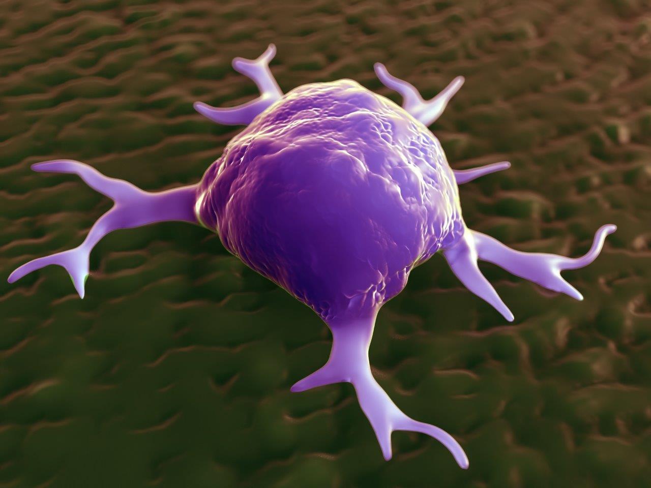
All of these alterations in protein functions would cause changes in cells morphology and function, giving them the characteristics of tumor cells, such as a high proliferative potential or the ability to avoid programmed cell death.

According to Godzik, “These changes potentially have oncogenic power in cells, which means, the ability to turn a healthy cell into a cancer cell.” A novel aspect of the study is that these changes tend to occur in genes other than those often mutated in cancer, and in patients with a low number of mutated genes.

“Changes in alternative splicing provide cancer with new ways in which it can escape fine cellular regulation. Therefore, the study of alternative splicing opens new doors in the research to cure cancer and may provide new alternatives to the treatment of this disease.

Institute News

How tumors trick immune cells

AuthorSusan Gammon
Date

September 19, 2017

The aggressiveness of a tumor is determined partly by the properties of the actual cancer cells, but also to a surprisingly large degree by the surrounding environment in which the tumor grows, including blood vessels, fat cells, fibroblasts and immune cells.

Of these elements, the roles of immune cells are arguably the most baffling. Immune cells are recruited to tumors to attack and destroy cancer cells, but their properties can change in response to specific signals they receive from the tumor. Macrophages are a good example of immune cells that play complex roles in tumor progression.

Laszlo Nagy, MD, PhD, professor and director of the Genomic Control of Metabolism Program at SBP’s Florida campus, is well-versed in many aspects of macrophage biology. Nagy comments that, “Our lab is studying the way the retinoid X receptor (RXR) controls how macrophages direct the aggressiveness of a tumor—specifically whether it spreads to other parts of the body (metastasizes). Scientists have known for some time that RXR is a regulatory protein found in the cell nucleus, but how it effects tumor metastasis has not been studied.”

Nagy’s lab recently published a study in PNAS that compared RXR macrophage knockout mice with normal mice to assess the how the protein affects the metastasis of subcutaneous tumors. Surprisingly, metastasis was increased in RXR macrophage knockout mice, even though growth of the primary tumors was not affected. Further research showed that the loss of RXR resulted in increased macrophage production of several factors that promote tumor cell colonization of sites in the lungs.

“This is a really tricky concept to grasp,” says Nagy. “The idea being developed by several research groups is that macrophages promote tumor metastasis by producing factors that modify sites in distant organs (known as pre-metastatic niches). This “conditioning” makes these sites more attractive as new homes for migrating tumor cells.

“Our contribution to understanding this phenomenon is discovering the involvement of RXR in suppressing macrophage production of metastasis-enhancing factors. The importance of RXR loss in our mouse model is underscored by finding that macrophage RXR activity is also low in patients with metastatic cancer. Since metastasis is the most dangerous aspect of cancer, we are toying with ideas for how to boost RXR activity in macrophages as a means of suppressing the metastatic phase of the disease.”

Institute News

Location matters, even for tumors

AuthorBill Stallcup, PhD
Date

September 7, 2017

Location, Location, Location! We often hear this in real estate, but it’s also true in biology. Cells need to be in the right place at the right time to help promote organ development and send the signals that keep our bodies working. But tumors rely on location as well, especially when it comes to angiogenesis—the process they use to recruit new blood vessels to “feed” their growth with oxygen and nutrients.

In the journal Cancers, William Stallcup, PhD, a professor in SBP’s NCI-Designated Cancer Center, describes the importance of location for vascular endothelial growth factor (VEGF), a protein that stimulates blood vessel formation.

“VEGF is a major stimulus for the formation of tumor blood vessels,” says Stallcup. “In fact, blocking VEGF has been one idea for slowing tumor growth by cutting off the tumor blood supply. This strategy has not been as successful as researchers had hoped, partly because blood vessel formation and the action of VEGF are both complex processes that we don’t fully understand.”

The vascular cells that form blood vessels are embedded in a fibrous meshwork called the vascular extracellular matrix (ECM). Some forms of VEGF bind tightly to this ECM, while other forms diffuse freely in tissues. The Stallcup lab’s studies reveal that the way VEGF and ECM interact is very important for tumor blood vessel formation.

According to Weon-Kyoo You, PhD, a former Stallcup postdoc and first author of the study, “When we looked at brain tumor growth in normal mice, we found that lots of VEGF was bound to the vascular ECM. But when we studied tumor growth in mutant mice that were deficient in ECM assembly, we saw that the VEGF mostly diffused away from tumor blood vessels and was dispersed in the tumor tissue.”

This change in VEGF location had large effects on the structure and function of tumor blood vessels. In normal mice, efficient binding of VEGF to the vascular ECM produced large diameter vessels that were not leaky—and brain tumors grew fast in these mice. In mice with deficient VEGF-ECM interactions, the blood vessels were thin and leaky—and brain tumors grew slowly due to lack of nutrients.

Stallcup concludes that, “We were very surprised at the correlation between healthy ECM, VEGF binding, and tumor growth. Apparently, having the VEGF located right there at the site where it can be readily used by vascular cells is a key factor in producing functional blood vessels.

“These studies add to our understanding of the tactics tumors use to grow, and also give us new clues about how we might be able to thwart cancer progression.”

Read the study here.

Institute News

Immune scavenger cells: Good guys or bad guys?

AuthorSusan Gammon
Date

July 9, 2017

The immune system is our main defense against foreign invaders (bacteria and viruses) and also against mutant cells that develop into cancer. Some of the first immune responders to these threats are scavenger cells called macrophages that destroy targets by internalizing and degrading them.

But macrophages can be tricked. When they are continually activated in a chronic disease like cancer, they can be “instructed” by cancer cells to perform functions that benefit the growing tumor instead of destroying it.

Recently, William Stallcup, PhD, professor at Sanford Burnham Prebys Medical Discovery Institute (SBP) published a study in Trends in Cell and Molecular Biology describing one way that cancer cells transform macrophages from tumor fighters into tumor helpers.

“We found that a protein called NG2 on macrophages is important for the ability of these scavengers to exit from blood vessels and migrate into tumors,” explains Stallcup. “When we knocked out NG2 on macrophages in mice, instead of allowing mouse brain tumors to grow faster (in the absence of the scavenging macrophages), the tumors actually grew much more slowly. This was somewhat surprising since we expect macrophages to help fight cancer.”

“It turns out that tumor cells can modify macrophage function, making them produce factors that stimulate the formation of tumor-nourishing blood vessels, says Pilar Cejudo Martin, PhD, a postdoc in Stallcup’s lab and first author of the paper. “By knocking out NG2, we blocked macrophage entry into tumors so that there were fewer macrophages for the tumors to use to their own advantage. So due to poor blood vessel development, tumor growth was slowed.”

So does this mean that blocking NG2 could be a general means of improving treatment of diseases involving chronic macrophage recruitment?

“It depends,” says Stallcup. “When we used a mouse model of multiple sclerosis (MS) to show that knockout of NG2 impaired macrophage entry into the damaged spinal cord, we found that recovery was hindered. That’s because macrophages are needed to produce factors that stimulate production of the cells responsible for damage repair.

“So while blocking macrophage NG2 might be useful for cancer therapy, it looks like this approach would be counter-productive for treating MS. Even though macrophages seem like attractive targets for therapy, they have such a large bag of tricks that we will probably have to deal with blocking their effects on a case-by-case basis,” adds Stallcup.

Read the paper here.

Institute News

SBP President’s Lecture highlights new approach to cancer immunotherapy

AuthorLindsay Ward-Kavanagh
Date

February 27, 2017

One of the most promising new approaches to treating cancer is immunotherapy—redirecting the immune system to detect and destroy tumor cells. That’s the topic of this year’s President’s Lecture by Andrew Weinberg, PhD, chief of the Laboratory of Basic Immunology at Providence Portland Medical Center, on 28 February 2017.

His research is related to the work of scientists at Sanford Burnham Prebys Medical Discovery Institute (SBP) working to harness the power of killer T cells that can recognize and eliminate cancerous cells.

The laboratory of Linda Bradley, PhD, professor at SBP, recently published a paper identifying PSGL-1, a protein that limits T cell responses to viruses, as a new target for checkpoint inhibition, an approach akin to taking the “brakes” off the immune system. They showed that T cells in mice that could not make PSGL-1 delayed tumor growth, suggesting that blocking PSGL-1’s function would help T cells fight tumors.  

Conversely, Carl Ware, PhD, professor and director of the Infectious and Inflammatory Diseases Center, is developing an immunotherapy strategy that’s the equivalent of “hitting the gas” to expand anti-tumor responses. Their project exploits the protein LIGHT, which turns on multiple pathways required for strong T cell responses. Ware’s current goal is to create an optimized mutant form of LIGHT with the strongest ability to drive anti-tumor immunity.

Weinberg uses a similar “hitting the gas” approach to immunotherapy, targeting the T cell co-stimulator OX40 with a drug now in clinical trials.

“Immunotherapy drugs currently approved to treat cancer block the negative signals that minimize T cell activity,” explains Ware. “Weinberg’s  work is different because the drug, called an OX40 agonist, boosts positive signals to T cells. His work has really led to this new approach of targeting immune molecules to enhance T cell function.”

Weinberg’s pioneering research revealed how OX40 identifies functional T cells in cancer and autoimmune disease patients. He confirmed the anti-tumor potential of OX40 in mice when antibodies that stimulated OX40 activity expanded the T cell population within tumors, and drove tumor regression. The identification of this anti-tumor potential ultimately led to the creation of the company AgonOx, which has commercially developed OX40 agonists in collaboration with AstraZeneca.

As clinical trials with OX40 agonists continue, Weinberg still recognizes the value of basic research. He says, “Understanding immunology in its most basic form can help us understand the principles and mechanisms involved with how these drugs work. If we understand how they work we can make them better, or choose combinations that will improve their efficacy.”

Institute News

Measuring heart toxicity of cancer drugs in a dish

AuthorJessica Moore
Date

February 22, 2017

A class of cancer drugs known as tyrosine kinase inhibitors (TKIs) are often damaging to the heart, sometimes to the degree that they can’t be used in patients. These toxic effects are not always predictable using current preclinical methods, so they may not be discovered until they make it to clinical trials.

New research could make it possible to tell which TKIs cause heart toxicity without putting any humans at risk. The collaborative study, involving Wesley McKeithan, a PhD student in the Sanford Burnham Prebys Medical Discovery Institute (SBP) graduate program and Mark Mercola, PhD, adjunct professor at SBP and a professor at Stanford University, used lab-grown heart muscle cells to assess the drugs’ potential to cause damaging effects.

“This new method of screening for cardiotoxicity should help pharma companies focus their efforts on TKIs that will be safe,” says Mercola, who collaborated with Joseph Wu, MD, PhD, also a professor at Stanford, on the study published in Science Translational Medicine. “That could mean better new TKIs will make it to the market, since we will be able to predict whether or not they cause heart problems early in the development process.”

TKIs with tolerable cardiac side effects, which include imatinib (Gleevec) and erlotinib (Tarceva), are widely used to treat multiple types of cancer. Because tumors often become resistant to these drugs, new compounds in this class continue to be developed to provide replacement treatments. Other TKIs can harm the heart in a variety of ways, from altering electrical patterns to causing arrhythmias, reducing pumping capacity, or even increasing risk of heart attacks.

Mercola and Wu’s team used heart muscle cells derived from induced pluripotent stem cells (iPSCs), which can be generated from adult skin or blood cells. After treating heart muscle cells with one of 21 TKIs, they assessed their survival, electrical activity, contractions (beating) and communication with adjacent cells. They used a new method for measuring heart cell contraction developed by the lab of Juan Carlos del Álamo, Ph.D., at UC San Diego to create a ‘cardiac safety index’, which correlates in vitro assay results with the drugs’ serum concentrations in humans. Importantly, the safety index values matched nicely with clinical reports on the cardiotoxicity of currently used TKIs.

The study also identified a possible way to protect heart muscle cells from impairment caused by TKIs—treating them with insulin or insulin-like growth factor. Although more research is needed, the findings suggest that it may be possible to alleviate some of the heart damage in patients receiving these chemotherapies.

Mercola adds, “By using cells derived from a broader group of individuals, this screening strategy could easily be adopted by the pharma industry to predict cardiotoxicity.”

This story is based in part on a press release from Stanford University School of Medicine.

Institute News

Research reveals the function of a pivotal protein impacting immunity and lymphoma

AuthorLindsay Ward-Kavanagh
Date

January 17, 2017

Robert Rickert, PhD, professor at SBP, and his team have recently published discoveries in The Journal of Immunology that may open a new avenue of treatment for B cell lymphomas. Their experiments showed that B cells make a protease called MALT1 so that they can mature into antibody-producing cells that eliminate infections, a process known as the germinal center reaction. MALT1 helps inactivate a subset of pro-apoptotic proteins, thus supporting the survival of B cells and the development of immune responses, an essential part of the process of detecting and eliminating germs.

However, in many B cell lymphomas, regulation of MALT1 is lost, and the protein instead continuously promotes survival of B cells. This occurs in the absence of infection, leading to an unhealthy accumulation of cells that fail to die and can instead accumulate cancerous mutations.

“Our study provides insight into why overexpression of MALT1 would promote lymphoma by extending B cell lifespan,” explains Rickert.

Importantly, Rickert’s group also showed that eliminating MALT1 from B cells in mice overrode survival signals, leading to the death of B cells. This result explains why other groups have found success in in vitro studies using drugs that prevent MALT1 activity to kill B cell lymphoma cells. Adding Rickert’s new observations with these earlier discoveries explains how preventing MALT1 activity in lymphoma cells could work as a treatment for patients whose tumor cells overproduce MALT1.

However, the lack of MALT1 in B cells can also be detrimental to health, preventing the body from developing a sufficiently strong immune response to eliminate infections. The new research could help doctors better understand why patients lacking functional MALT1 have trouble recovering from infections.

“It is now clear why patients that lack MALT1 function may be diagnosed with combined immunodeficiency (CID) and suffer from severe recurrent infections despite having normal numbers of B (and T) lymphocytes, since they are functionally impaired and thus do not produce antibody,” adds Rickert.

While these studies highlight a new understanding of how MALT1 in B cells could be exploited to treat patients with either cancer or immunodeficiency disorders, they also highlight the importance of balancing context and consequences in targeting MALT1. 

The paper is available online here.

Institute News

V Foundation grant to Ani Deshpande, PhD, supports pioneering research toward better leukemia treatments

AuthorJessica Moore
Date

December 2, 2016

Patients with a rare type of leukemia called acute promyelocytic leukemia (APL) have better outcomes than most leukemias because they can be treated with a very effective drug that converts their cancer cells back to normal. This success has convinced many cancer researchers that there’s a way to do the same for other leukemias. And with his recently awarded funding from the V Foundation, Ani Deshpande, PhD, assistant professor at Sanford Burnham Prebys Medical Discovery Institute, can now find targets for future drugs to do just that.

“We’re aiming to rehabilitate the cancer cells, in a sense, instead of destroying them,” said Deshpande. “The advantage to this approach is that, unlike conventional chemotherapy, it doesn’t harm normal cells, so it should have far fewer toxic side effects.”

Deshpande aims to make a big impact with this work—he’s first focusing on a group of acute myeloid leukemia (AML) with very poor survival outcomes. Worse, these leukemias, characterized by fusions of chromosome 11 with another partner chromosome, are especially common among children and infants.

This subgroup of AML is trickier than APL, where the product of the gene created by the chromosomal rearrangement directly blocks the cancer cells from becoming their normal type. In contrast, in the leukemias that Deshpande’s lab studies, the change in the cells’ programming is more complex. The mutation they carry alters the regulation of other genes, but which of these prevent AML cells from becoming normal blood-forming cells is largely unknown.

Fortunately, Deshpande is an expert in studying leukemic gene regulation. His lab specializes in epigenetics—analyzing the chemical tags on genes that influence their activity. The V Foundation funds will allow Deshpande’s team to apply an advanced sequencing-based approach to identify and validate potential targets for drugs that restore cancer cells’ epigenome to normal.

“This grant not only lets me expand my lab by hiring a new postdoc, but it also means I can take risks that wouldn’t be possible if I were proposing research to the NIH,” commented Deshpande. “I’m confident that we’ll get exciting results. The tools we’re using have gotten exponentially better over the last few decades, so we’re poised for a breakthrough.”

About the V Foundation

The V Foundation for Cancer Research was founded by ESPN and legendary basketball coach Jim Valvano with one goal in mind: to achieve victory over cancer. Since its start in 1993, the V Foundation has awarded over $170 million in cancer research grants nationwide.

Watch Dr. Deshpande talk about why foundation funding is important:

Institute News

Ze’ev Ronai wins Lifetime Achievement Award from the Society for Melanoma Research

AuthorJessica Moore
Date

November 10, 2016

Ze’ev Ronai, PhD, chief scientific advisor at Sanford Burnham Prebys Medical Research Institute (SBP) and professor in its NCI-designated Cancer Center, is the 2016 recipient of the Society for Melanoma Research’s Lifetime Achievement Award. The award honors “an individual who has made major and impactful contributions to melanoma research throughout their career.”

Ronai is being recognized for his significant contributions to melanoma research that are advancing understanding of this deadly form of skin cancer and could lead to new treatments. His studies on ultraviolet (UV) irradiation-induced changes that promote melanoma showed how they rewire signaling networks. A major discovery from those inquiries was that one player in that rewiring, a protein called ATF2, can switch from its usual tumor-preventive function to become a tumor promoter. Work by the Ronai lab also mapped how ATF2 contributes to melanoma development, and identified specific factors involved in melanoma response to therapy and metastatic potential.

In mapping the landscape of melanoma signaling, Ronai’s lab also uncovered the important role the enzyme PDK1 plays in melanoma development and metastasis. More recently, Ronai’s studies identified a mechanism underlying resistance of melanoma to BRAF inhibitor therapy, paving the road for a new clinical trial. Integral to Ronai’s research are translational initiatives, including the development of SBI-756, a small molecule that disrupts the complex that initiates protein synthesis and prevents melanoma resistance when combined with BRAF inhibition.

Ronai and his team also study how cancer cells thrive under harsh conditions, such as lack of oxygen or nutrients. That line of research has produced important insights into cancer heterogeneity and its capacity to drive the survival of the select few cancer cells that are resistant to therapy and able to metastasize. Ronai’s studies of proteins that control stress responses, such as Siah and RNF5, have furthered understanding of these processes and identified new targets for future therapies.

Ronai’s record of scientific accomplishments was recognized by the National Cancer Institute with an Outstanding Investigator Award, a seven-year grant that allows recipients to pursue projects of unusual potential. Ronai’s unique focus on how gene activity changes in cancer promises to continue establishing new paradigms for how cancers develop and respond to therapy.

About the Society for Melanoma Research

The Society for Melanoma Research (SMR) is an all-volunteer group of scientists dedicated to finding the mechanisms responsible for melanoma and, consequently, new therapies for this cancer. SMR contributes to advances in melanoma research by catalyzing collaborations among basic, translational, and clinical researchers, carrying new technology-based discoveries from bench to bedside and back.

About melanoma

The incidence of melanoma, the most lethal form of skin cancer, is rising at one of the fastest rates of all cancers in the U.S. Melanoma can strike people of all ages and is the most common form of cancer among young adults ages 25 to 29.

Institute News

Beating prostate cancer

Authorjmoore
Date

November 7, 2016

About one in seven men will be diagnosed with prostate cancer during his lifetime. Though it has one of the highest survival rates of any type of cancer—95% make it through the first ten years—diagnosis and treatment could still be improved. Since November is Prostate Cancer Awareness Month, we’re highlighting the work our scientists are doing to address key challenges and unresolved questions in prostate cancer.

Early, accurate detection—The current method of screening for prostate cancer is a blood test for prostate specific antigen, or PSA, which detects cancer early, but isn’t very specific—only one in four men with high PSA levels actually has cancer. In general, high levels of PSA mean the next step is a biopsy. A more specific test would avoid unnecessary biopsies, which are invasive and stressful for patients.

Ranjan Perera, PhD, associate professor in the Integrative Metabolism Program, is looking for biomarkers that would enable just such a test. His lab is making progress—they identified five long noncoding RNAs (RNAs that, instead of carrying genetic information to be translated, regulate the translation of other RNAs) found at higher-than-normal levels in the urine of prostate cancer patients. An RNA-based test is on the market, but could be improved—measuring multiple markers would be more sensitive and specific.

Better therapies—For advanced cases, current treatments are either insufficient or overly toxic. Prostate cancer is usually first treated with drugs that block the actions of androgens, the hormones that drive its growth. If the tumor recurs later, as happens for cancers that are already at a late stage before treatment, it forms from cells that are resistant to those drugs. Then, the only option is chemotherapy or radiation.

Towards therapies that cause less collateral damage, Nicholas Cosford, PhD, associate director of Translational Research in the NCI-designated Cancer Center, is designing new drugs against targets found to be important in prostate cancer. These drugs are intended to block two strategies by which cancer cells survive—avoiding cell death, and generating extra energy by recycling the cell’s own parts.

Understanding why obesity correlates with aggressiveness—Obese men are no more likely than others to get prostate cancer, but if they do, it’s more likely to become advanced quickly. To figure out why this happens, Jorge Moscat, PhD, director, and Maria Diaz-Meco, PhD, professor in the Cancer Metabolism and Signaling Networks Program, are looking at how fat adjacent to the prostate interacts with tumor cells. A detailed picture of how fat cells and tumor cells interact could reveal new ways to treat prostate cancer in overweight men.