cardiovascular disease Archives - Sanford Burnham Prebys
Institute News

Graduate student awarded American Heart Association Fellowship

AuthorSusan Gammon
Date

April 14, 2021

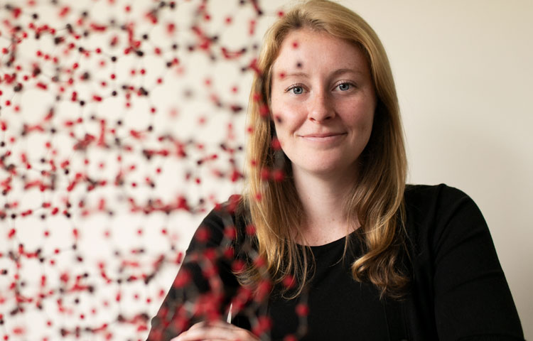
The heart is the core of life, and for PhD graduate student Katja Birker, it’s the foundation for the beginning of a career.

Birker recently received a prestigious predoctoral fellowship from the American Heart Association (AHA) to continue her research on hypoplastic left heart syndrome (HLHS), a congenital heart disease that affects between two and four of every 10,000 babies born. As of today, the only cure for HLHS is three open-heart surgeries that begin two weeks after birth.

“I’m very grateful to the AHA for supporting my research,” says Birker. “I’ve embarked on a career to study the genes that contribute to HLHS, and this award will help me continue my work that may eventually lead to targeted prevention of HLHS as well as other congenital heart diseases.”

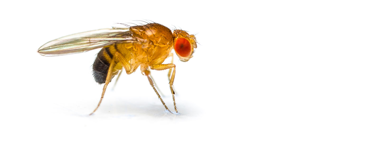
Birker is collaborating with the Mayo Clinic to identify and test whether genes found in HLHS patients—or “candidate” genes—have similar consequences in the hearts of fruit flies—a model organism for cardiovascular research. The research aims to identify novel gene functions and pathways that are likely to contribute to HLHS.

“Many believe that HLHS is a genetic disease, but the genes that are involved are not well known,” says Birker. “The fruit fly is a very good genetic system to model disease because it has many similar genes to humans and a short life span. I’m able to film videos of fly hearts to understand the impact of the candidate genes on heart function.

“My goal is to find genes that can be used in the future for the diagnosis and treatment of HLHS in babies. The research approach could also be used to screen for genes that might be involved in many other types of heart disease,” adds Birker.

The AHA supports early-career scientists with passion, commitment and focus by providing fellowships that fund their pursuit of cardiovascular research. Birker, a graduate student in the lab of Rolf Bodmer, PhD, professor and director of the Development, Aging and Regeneration Program, received her first AHA fellowship in 2018.

Institute News

American Heart Association awards postdoctoral fellowship to SBP scientist

AuthorMonica May
Date

January 23, 2019

It’s no surprise that muscles are important to our metabolism: it’s why building muscle at the gym can accelerate weight loss. 

Scientists are particularly interested in how muscle metabolism affects the heart, arguably the most important muscle in the body. With heart disease remaining the number-one killer of men and women in the U.S., the hunt is on to better understand the molecular mechanisms of the heart so we can develop better treatments. (Learn more about heart disease at our upcoming SBP Insights event.) 

Research is revealing that altered communications between skeletal and heart muscle increases the risk of heart disease. But the molecular mechanisms behind this link are currently unknown. 

Now, the American Heart Association has awarded a two-year postdoctoral fellowship to SBP’s Chiara Nicoletti, PhD, to study the genetic basis of metabolic changes in skeletal muscle that ultimately lead to heart disease. Nicoletti works in the lab of Pier Lorenzo Puri, MD, professor in the Development, Aging and Regeneration Program at SBP. 

Findings from Nicoletti’s work could uncover therapeutic targets for heart disease and/or lead to a prognostic tool that could predict heart disease risk. Both developments would be much-needed advances in the battle against heart disease. 

Interested in keeping up with SBP’s latest discoveries, upcoming events and more? Subscribe to our monthly newsletter, Discoveries.

Institute News

SBP women awarded American Heart Association Fellowships

AuthorSusan Gammon
Date

June 15, 2018

There has never been a more exciting time to embark on a career in biomedical research. Fortunately, the American Heart Association (AHA) is supporting early-career scientists with passion, commitment and focus by providing fellowships that fund their pursuit of cardiovascular research. Recently, three SBP scientists were awarded AHA grants to finance projects that align with the AHA mission of building healthier lives, free of cardiovascular disease and stroke.

Katja Birker (left)
Birker, a graduate student in the lab of Rolf Bodmer, PhD, will be studying genes that could possibly contribute to hypoplastic left heart syndrome (HLHS)—a condition that affects roughly 2–4 out of every 10,000 babies. Today, the cure for HLHS is a three-step invasive surgery that begins two weeks after the baby is born.

Birker will be collaborating with the Mayo Clinic to identify and test whether candidate HLHS genes found in patients have similar consequences in the hearts of fruit flies, which are an established model organism for cardiovascular research. She will use the flies to work toward her goal of validating novel genes that could be used in the future for diagnostic and therapeutic purposes related to cardiovascular diseases.

EePhie Tan, PhD (middle)
Tan’s research is taking a deeper dive into previous research showing that the cell recycling process called autophagy provides health benefits—including life extension—in response to reduced food intake. This project will examine the cell networks that govern autophagy, and a specialized form of autophagy called lipophagy (fat recycling). Lipophagy is a relatively new field of biomedical research, but scientists have already learned that malfunctions in lipophagy can lead to the accumulation of toxic fat deposits and contribute to heart disease.

Tan, a postdoc in the lab of Malene Hansen, PhD, will use a small worm called C. elegans as a model system to study proteins involved in the lipophagy process. Since the core machinery of lipophagy is conserved in all organisms (from humans to C. elegans), Tan’s findings may be used to find future treatments that target toxic fat deposits in heart disease.

Clara Guida, PhD (right)
Guida will study why children from obese parents have an increased risk of developing cardiovascular disease. The research may lead to the development of biomarkers that can predict heart conditions caused by parents that eat a high-fat diet (HFD), and may lead to new drugs that can prevent the negative effects of a parental HFD on the heart function of offspring.

Guida, a postdoc in Bodmer’s lab, will study the inheritance of DNA modifications called “epigenetic marks” in fruit flies fed a HFD. These epigenetic marks are thought to cause heart problems in the next generation. She will be testing potential drugs to see if they can erase the inherited abnormal gene changes and prevent the negative effects of a parental HFD. The research is especially relevant to lipotoxic cardiomyopathy—a condition associated with fat accumulation in the heart.

Institute News

Location matters, even for tumors

AuthorBill Stallcup, PhD
Date

September 7, 2017

Location, Location, Location! We often hear this in real estate, but it’s also true in biology. Cells need to be in the right place at the right time to help promote organ development and send the signals that keep our bodies working. But tumors rely on location as well, especially when it comes to angiogenesis—the process they use to recruit new blood vessels to “feed” their growth with oxygen and nutrients.

In the journal Cancers, William Stallcup, PhD, a professor in SBP’s NCI-Designated Cancer Center, describes the importance of location for vascular endothelial growth factor (VEGF), a protein that stimulates blood vessel formation.

“VEGF is a major stimulus for the formation of tumor blood vessels,” says Stallcup. “In fact, blocking VEGF has been one idea for slowing tumor growth by cutting off the tumor blood supply. This strategy has not been as successful as researchers had hoped, partly because blood vessel formation and the action of VEGF are both complex processes that we don’t fully understand.”

The vascular cells that form blood vessels are embedded in a fibrous meshwork called the vascular extracellular matrix (ECM). Some forms of VEGF bind tightly to this ECM, while other forms diffuse freely in tissues. The Stallcup lab’s studies reveal that the way VEGF and ECM interact is very important for tumor blood vessel formation.

According to Weon-Kyoo You, PhD, a former Stallcup postdoc and first author of the study, “When we looked at brain tumor growth in normal mice, we found that lots of VEGF was bound to the vascular ECM. But when we studied tumor growth in mutant mice that were deficient in ECM assembly, we saw that the VEGF mostly diffused away from tumor blood vessels and was dispersed in the tumor tissue.”

This change in VEGF location had large effects on the structure and function of tumor blood vessels. In normal mice, efficient binding of VEGF to the vascular ECM produced large diameter vessels that were not leaky—and brain tumors grew fast in these mice. In mice with deficient VEGF-ECM interactions, the blood vessels were thin and leaky—and brain tumors grew slowly due to lack of nutrients.

Stallcup concludes that, “We were very surprised at the correlation between healthy ECM, VEGF binding, and tumor growth. Apparently, having the VEGF located right there at the site where it can be readily used by vascular cells is a key factor in producing functional blood vessels.

“These studies add to our understanding of the tactics tumors use to grow, and also give us new clues about how we might be able to thwart cancer progression.”

Read the study here.

Institute News

Will flies help fix heart rhythm problems?

AuthorSusan Gammon
Date

July 12, 2017

Your heart beats about 35 million times in a single a year. That’s a whopping number of beats—each generated by electrical signals that make the heart contract. Occasionally problems with heart’s electrical system can cause irregular rhythms, or arrhythmias. Some types of arrhythmias are merely annoying; others can last long enough to affect how the heart works, or even cause sudden cardiac arrest.

Although certain arrhythmias can be successfully treated with medication, surgery and/or devices such as pacemakers, cardiac disorders and heart disease still account for more deaths than any other disease.

Finding new treatments for arrhythmias requires a deep understanding of how the heart beats, and specifically the intricate electrical system that prompts the heart to contract. It also requires a model to study. Is Drosophila melanogaster—a type of fruit fly—the answer?

Using flies to study the heart

Both the human heart and fly hearts have four chambers and both start out as linear tubes in embryos—but ours loops during development to form a more compact structure where as a the fly heart does not. Despite this structural difference there are many functional similarities between the fly and human heart.

“One of the similarities that we focus on in my lab is the way ion channels work—and don’t work—to fully understand how faulty ion channels contribute to heart arrhythmias,” says Karen Ocorr, PhD, assistant professor at SBP.

Ion channels are proteins found in cell membranes that allow specific ions such as potassium, sodium and calcium, to pass through cells. When ion currents travel into heart muscle cells, the muscle becomes depolarized, creating an electrical current that causes the heart to contract. A second set of channels are important in repolarization of the heart, which allows it to relax and refill with blood.

In her new paper published in PLOS Genetics, Ocorr describes how two repolarizing potassium ion channels called hERG and KCNQ control the rate and efficiency of fly heart contractions—similar to their role in human heart muscle. The research also shows that mutations in hERG and KCNQ lead to arrhythmias that worsen with age—as they do in humans.

“In humans, when hERG is compromised, either by drugs or inherited mutations, hearts can take longer than normal to recharge between beats, causing a potentially fatal condition called long QT syndrome. In fact, some anti-arrhythmia drugs actually cause long QT syndrome, hence the need for better, more specific therapies,” explains Ocorr.

The capacity for drugs to cause long QT syndrome has led the Food and Drug Administration (FDA) to recommend including the evaluation of new cardiac and non-cardiac drugs for this possible side effect. The FDA is the United States agency that provides licenses to market new drugs.

Interestingly, neither of the ion channels we identified in the fly heart play a major role in the adult mouse heart, ruling it out as useful model to screen for drug-related long QT effects,” says Ocorr.

“We are encouraged that Drosophila may become an easy, accurate tool to pre-clinically screen for adverse cardiac events associated new anti-arrhythmia therapies—potentially making the next drug discovery for patients happen sooner.”

Read the paper here

Institute News

Promising target for blocking buildup of fatty plaques in arteries

AuthorJessica Moore
Date

July 22, 2016

Every 34 seconds, someone in the US has a heart attack or stroke. New research from the laboratory of Erkki Ruoslahti, MD, PhD, distinguished professor in the NCI-designated Cancer Center, could lead to treatments that lower that frequency.

Heart attacks and strokes are caused by a blocked artery, which cuts off blood supply to a part of the heart or brain. These blockages occur when atherosclerotic plaques—deposits of inflamed, fat-containing cells surrounded by fibrous material inside arteries—rupture and seed blood clots. In a study published in the Journal of Controlled Release, Ruoslahti’s team shows that a specific peptide blocks expansion of these plaques at advanced stages.

“Our findings demonstrate the relevance of a new target, p32, to slowing the deposition of plaque,” said Zhi-Gang She, PhD, staff scientist in Ruoslahti’s lab and co-lead author of the paper. “We’re hopeful that drugs that act on this protein would help lower the risk for heart attacks and stroke.”

The details

The new study used a peptide called LyP-1, a ring of nine amino acids that Ruoslahti’s group has worked with for many years. LyP-1 binds to p32, a protein that’s normally located inside cells, but is found on the surface of tumor cells and active macrophages.

“Macrophages drive plaque enlargement by taking up fats and promoting inflammation, and we knew from our other investigations that LyP-1 can trigger cell death in macrophages,” explained Ruoslahti. “We thought that LyP-1 might eliminate macrophages from plaques, which would slow the advance of atherosclerosis.”

Their results confirmed this expectation—the LyP-1 peptide greatly reduced the size of plaques in mice when it was administered at advanced stages.

“Eliminating macrophages from arterial plaque is like cutting off the roots of a plant,” said She. “Not only does that get rid of a portion of the plaque, but because macrophages feed it by taking up lipids, it also keeps the plaque from getting larger.”

Clinical relevance

“The peptide itself is not a candidate drug,” added Ruoslahti. “It can only be given by injection, which isn’t practical for a chronic disease like atherosclerosis. However, we have identified small molecules that interact with p32 in a similar way to LyP-1, so they could form the basis of a drug that’s taken as a pill.”

“The key to making sure this treatment strategy is safe is confirming that it doesn’t make the plaques more likely to rupture,” commented She. “We didn’t see anything indicating that LyP-1 makes plaques less stable, but future studies should explore that issue further.”

The paper is available online here.

Institute News

Scientific breakthrough may limit damage caused by heart attacks

AuthorJessica Moore
Date

June 30, 2016

A research advance from the Sanford Burnham Prebys Medical Discovery Institute (SBP) and Stanford University could lead to new drugs that minimize the damage caused by heart attacks. The discovery, published in Nature Communications, reveals a key control point in the formation of new blood vessels in the heart, and offers a novel approach to treat heart disease patients.

“We found that a protein called RBPJ serves as the master controller of genes that regulate blood vessel growth in the adult heart,” said Mark Mercola, PhD, professor in SBP’s Development, Aging, and Regeneration Program and jointly appointed as professor of medicine at Stanford University, senior author of the study. “RBPJ acts as a brake on the formation of new blood vessels. Our findings suggest that drugs designed to block RBPJ may promote new blood supplies and improve heart attack outcomes.”

In the US, someone has a heart attack every 34 seconds. The ensuing loss of heart muscle, if it affects a large enough area, can severely reduce the heart’s pumping capacity, which causes labored breathing and makes day-to-day tasks difficult. This condition, called heart failure, arises within five years in at least one in four heart attack patients.

The reason heart muscle dies in a heart attack is that it becomes starved of oxygen—a heart attack is caused by blockage of an artery supplying the heart. If heart muscle had an alternative blood supply, more muscle would remain intact, and heart function would be preserved. Many researchers have therefore been searching for ways to promote the formation of additional blood vessels in the heart.

“Studies in animals have shown that having more blood vessels in the heart reduces the damage caused by ischemic injuries, but clinical trials of previous therapies haven’t succeeded,” said Ramon Díaz-Trelles, PhD, staff scientist at SBP and lead author of the study. “The likely reason they have failed is that these studies have evaluated single growth factors, but in fact building blood vessels requires the coordinated activity of numerous factors. Our data show that RBPJ controls the production of these factors in response to the demand for oxygen.

“We used mice that lack RBPJ to show that it plays a novel role in myocardial blood vessel formation (angiogenesis)—it acts as a master controller, repressing the genes needed to create new vessels,” added Diaz-Trelles. “What’s remarkable is that removing RBPJ in the heart muscle did not cause adverse effects—the heart remained structurally and functionally normal in mice without it, even into old age.”

“RBPJ is a promising therapeutic target. It’s druggable, and our findings suggest that blocking it could benefit patients with cardiovascular disease at risk of a heart attack. It may also be relevant to other diseases,” commented Pilar Ruiz-Lozano, PhD, associate professor of pediatrics at Stanford and adjunct professor at SBP, co-senior author. “Inhibitors of RBPJ might also be used to treat peripheral artery disease, and activators might be beneficial in cancer by inhibiting tumor angiogenesis.”

The paper is available online here.

Institute News

Doug Lewandowski, PhD, elected as a Fellow of the American Association for the Advancement of Science

AuthorJessica Moore
Date

April 28, 2016

The director of Translational Cardiovascular Research at SBP’s Lake Nona campus was recently named a Fellow of the American Association for the Advancement of Science (AAAS). E. Douglas Lewandowski, PhD, was one of 33 scientists selected to become a AAAS fellow in the Section on Medical Sciences, recognizing his “distinguished contributions to fundamental aspects of cardiac metabolism and their implications for heart disease.” Continue reading “Doug Lewandowski, PhD, elected as a Fellow of the American Association for the Advancement of Science”

Institute News

Failing hearts switch fuels to generate energy

Authorsgammon
Date

January 27, 2016

More than 5 million people in the United States suffer from heart failure, according to the American Heart Association. Less than half of those with heart failure survive five years after diagnosis. New research from scientists at SBP published in the journal Circulation may lead to a new approach to help treat heart failure early in the disease. Continue reading “Failing hearts switch fuels to generate energy”

Institute News

Novel model for cardiomyopathy paves the way for new therapies

Authorsgammon
Date

May 29, 2015

A new fruit fly model that captures key metabolic defects associated with cardiomyopathy could translate into more-effective treatments for this potentially deadly heart condition, according to a study conducted by researchers at Sanford-Burnham and the Universidad Autónoma de Madrid in Spain. The findings, published April 9 in Human Molecular Genetics, could also have broader clinical implications for human metabolic diseases affecting other organ systems such as the liver and skeletal muscle. Continue reading “Novel model for cardiomyopathy paves the way for new therapies”