Cell stress response protein implicated in cancer progression, yet it also weakens resistance to immunotherapies
Metabolic disorders such as obesity and type 2 diabetes place extra stress on the liver. Liver cells try to protect themselves from the accompanying surge in dysfunctional proteins by activating factors that help restore an appropriate protein balance.
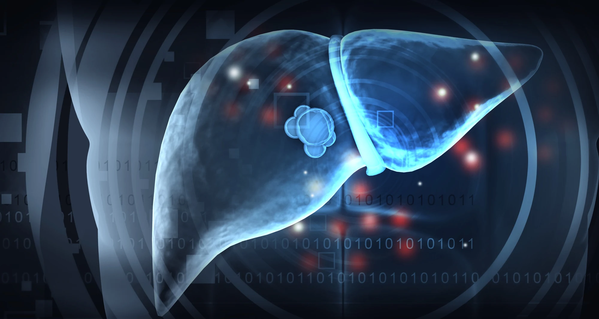
One of these factors is a protein called activating transcription factor 6 alpha (ATF6α) that was recently shown to drive the onset of liver cancer if left permanently active. In a Nature study published February 4, 2026, an international team of scientists demonstrated that activating ATF6α in mice caused liver disease that progressed to liver cancer.
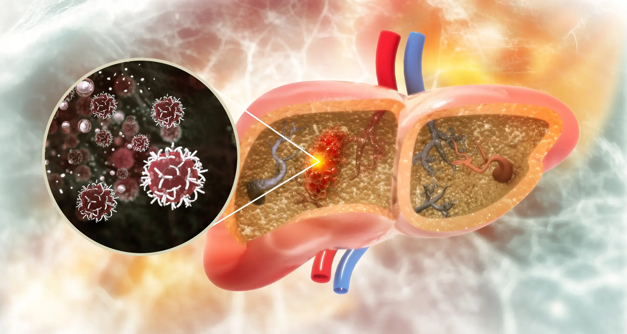
In data from human liver cancer patients, ATF6α activation was linked with more aggressive tumors, a suppressed immune system surrounding tumors and reduced patient survival.
The researchers also uncovered ways that ATF6α might be used to advance the treatment of liver cancer. Liver cells with ATF6α switched off developed fewer tumors. While high ATF6α activity levels were associated with cancer progression, they also were found to make tumors more susceptible to certain immunotherapies.
These findings suggest the need for future clinical trials to test drugs that directly target ATF6α to treat the disease. Additionally, it might prove advantageous to screen liver cancer patients for ATF6α activity to find those most likely to benefit from existing immunotherapies.

Randal Kaufman, PhD, is a professor in the Center for Metabolic and Liver Diseases at Sanford Burnham Prebys and a co-corresponding author of the study. Image credit: Sanford Burnham Prebys.
To learn more, read the German Cancer Research Center press release.
Xin Li, PhD, a postdoctoral fellow at the German Cancer Research Center (DKFZ), shares first authorship of the study with co-corresponding author Cynthia Lebeaupin, PhD, principal scientist at Pfizer and former postdoctoral researcher at Sanford Burnham Prebys Medical Discovery Institute.
The other co-corresponding authors are Dirk Haller, PhD, Technische Universität München; Randal Kaufman, PhD, Sanford Burnham Prebys; and Mathias Heikenwälder, PhD, University of Tübingen and DKFZ.