NIH funding Archives - Page 2 of 7 - Sanford Burnham Prebys
Institute News

New roles for autophagy genes in cellular waste management and aging

AuthorCommunications
Date

January 3, 2024

Autophagy genes help extrude protein aggregates from neurons in the nematode C. elegans.

Autophagy, which declines with age, may hold more mysteries than researchers previously suspected. In the January 4 issue of Nature Aging, it was noted that scientists from the Buck Institute, Sanford Burnham Prebys and Rutgers University have uncovered possible novel functions for various autophagy genes, which may control different forms of disposal including misfolded proteins—and ultimately affect aging.

“While this is very basic research, this work is a reminder that it is critical for us to understand whether we have the whole story about the different genes that have been related to aging or age-related diseases,” said Professor Malene Hansen, PhD, Buck’s chief scientific officer, who is also the study’s co-senior author. “If the mechanism we found is conserved in other organisms, we speculate that it may play a broader role in aging than has been previously appreciated and may provide a method to improve life span.”

These new observations provide another perspective to what was traditionally thought to be occurring during autophagy.

Autophagy is a cellular “housekeeping” process that promotes health by recycling or disposing of damaged DNA and RNA and other cellular components in a multi-step degradative process. It has been shown to be a key player in preventing aging and diseases of aging, including cancer, cardiovascular disease, diabetes and neurodegeneration. Notably, research has shown that autophagy genes are responsible for prolonged life span in a variety of long-lived organisms.

The classical explanation of how autophagy works is that the cellular “garbage” to be dealt with is sequestered in a membrane-surrounded vesicle, and ultimately delivered to lysosomes for degradation. However, Hansen, who has studied the role of autophagy in aging for most of her career, was intrigued by an accumulation of evidence that indicated that this was not the only process in which autophagy genes can function.

“There had been this growing notion over the last few years that genes in the early steps of autophagy were ‘moonlighting’ in processes outside of this classical lysosomal degradation,” she said. “Additionally, while it is known that multiple autophagy genes are required for increased life span, the tissue-specific roles of specific autophagy genes are not well defined.”

To comprehensively investigate the role that autophagy genes play in neurons—a key cell type for neurodegenerative diseases—the team analyzed Caenorhabditis elegans, a tiny worm that is frequently used to model the genetics of aging and which has a very well-studied nervous system. The researchers specifically inhibited autophagy genes functioning at each step of the process in the neurons of the animals, and found that neuronal inhibition of early-acting, but not late-acting, autophagy genes, extended life span.

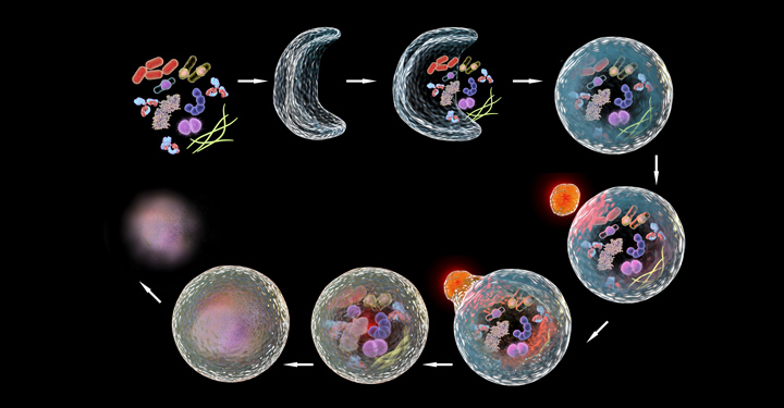
An unexpected aspect was that this life span extension was accompanied by a reduction in aggregated protein in the neurons (an increase is associated with Huntington’s disease, for example), and an increase in the formation of so-called exophers. These giant vesicles extruded from neurons were identified in 2017 by Monica Driscoll, PhD, a collaborator and professor at Rutgers University.

“Exophers are thought to be essentially another cellular garbage disposal method, a mega-bag of trash,” said Caroline Kumsta, PhD, co-senior author and assistant professor at Sanford Burnham Prebys “When there is either too much trash accumulating in neurons, or when the normal ‘in-house’ garbage disposal system is broken, the cellular waste is then being thrown out in these exophers.

“Interestingly, worms that formed exophers had reduced protein aggregation and lived significantly longer. This finding suggests a link between this process of this massive disposal event to overall health,” said Kumsta. The team found that this process was dependent on a protein called ATG-16.2.

The study identified several new functions for the autophagy protein ATG-16.2, including in exopher formation and life span determination, which led the team to speculate that this protein plays a nontraditional and unexpected role in the aging process. If this same mechanism is operating in other organisms, it may provide a method of manipulating autophagy genes to improve neuronal health and increase life span.

“But first we have to learn more—especially how ATG-16.2 is regulated and whether it is relevant in a broader sense, in other tissues and other species,” Hansen said. The Hansen and Kumsta teams are planning on following up with a number of longevity models, including nematodes, mammalian cell cultures, human blood and mice.

“Learning if there are multiple functions around autophagy genes like ATG-16.2 is going to be super important in developing potential therapies,” Kumsta said. “It is currently very basic biology, but that is where we are in terms of knowing what those genes do.”

The traditional explanation that aging and autophagy are linked because of lysosomal degradation may need to expand to include additional pathways, which would have to be targeted differently to address the diseases and the problems that are associated with that. “It will be important to know either way,” Hansen said. “The implications of such additional functions may hold a potential paradigm shift.” 
 
DOI: 10.1038/s43587-023-00548-1

Institute News

The “Eph” system may pave the way for novel cancer therapies

AuthorSusan Gammon
Date

November 27, 2023

Over the past three decades, researchers have been investigating an important cell communication system called the “Eph system,” and the evidence implicating the system in cancer is staggering.

The Eph system is comprised of multiple Eph receptors and their ligands—ephrins—and are involved in contact-dependent communication between cells. They play essential roles in regulating various cellular processes.

Modern studies have shed light on the Eph system’s role in tumor expansion, invasiveness, metastasis, cancer stem cell maintenance and therapy resistance.

This month, Elena Pasquale, PhD, published a review in Nature Reviews Cancer that summarizes the current state of research on the Eph system and its links to cancer progression and drug resistance.

“The Eph system has many critical functions during the development of tissues and organs, but it also has the capacity to either promote or suppress cancer progression and malignancy” says Pasquale. “In cancer, the activities of the Eph system can differ depending on the circumstances—for example, which Eph receptors and ligands are present in a tumor cell, the types of tumor cells in which they function, and the characteristics of these cells.”

“It’s this remarkable versatility that makes the Eph system a compelling but also challenging target for potential therapies,” says Pasquale.

“The aims of this review were to comprehensively survey the large body of data regarding various aspects related to Eph signaling in tumors and to highlight potential strategies for therapeutic targeting,” says Pasquale. “Overall, while significant progress has been made in deciphering the Eph system in cancer, there is much more to learn.

“Gaining a deeper understanding of how the Eph system functions in cancer is challenging but will be essential for the development of targeted therapies and personalized treatment approaches for patients.”

Institute News

Scientists unite to get to the heart of AFib

AuthorSusan Gammon
Date

August 15, 2023

A collaborative study led by researchers at Sanford Burnham Prebys is paving the way to identifying gene networks that cause atrial fibrillation (AFib), the most common age-related cardiac arrhythmia.

The findings, published in Disease Models & Mechanisms, validate an approach that combines multiple experimental platforms to identify genes linked to an abnormal heart rhythm.

“One of the biggest challenges to solving the AFib genetic puzzle has been the lack of experimental models that are relevant to humans,” says Alex Colas, PhD, co-senior author and assistant professor in the Development, Aging and Regeneration Program at Sanford Burnham Prebys. “By working with colleagues who focus on AFib but in different systems, we have created a robust multiplatform model that can accurately pinpoint genes associated with this condition.”

AFib is characterized by an irregular, rapid heartbeat that causes a quivering of the upper chambers of the heart, called the atria. This condition is the result of a malfunction in the heart’s electrical system that can lead to heart failure and other heart-related complications, which include stroke-inducing blood clots.

AFib impacts more than 5.1 million people in the United States, with expectations of 15.9 million by 2050. It is more common in individuals over the age of 60 but can also occur in teenagers and young adults.

“There will never be a one-size-fits-all solution to AFib, since it can be caused by many different genes—and the genes that do cause it vary from person to person,” says Karen Ocorr, PhD, also a co-senior author and assistant professor in the Development, Aging and Regeneration Program at Sanford Burnham Prebys. “A better understanding of the gene network(s) that contribute to AFib will help us design tests to predict a person’s risk, and develop individualized approaches to treat this dangerous heart condition.”

To overcome the limitations of current AFib research models, Colas, Ocorr and researchers from UC Davis and Johns Hopkins University combined forces to assemble a multi-model platform that combines:

  • A high-throughput screen using atrial-like cells (derived from human-induced pluripotent stem cells) to measure how a gene mutation alters the strength and duration of a heartbeat.
  • A Drosophila (fruit fly) model—with heart genetics and development remarkably similar to human hearts—that permits analysis of gene mutations in a functioning organ.
  • A well-established computational model that uses computers to simulate the effects of gene mutations on the electrical activity in human atrial cells.

The accuracy of the multi-model platform was confirmed when each screened 20 genes, and all three platforms identified phospholamban, a protein found in the heart muscle with known links to AFib.

“This collaboration has greatly expanded our ability to understand AFib at the genetic level,” says Colas. “Importantly, the high-throughput screening component of the model will also allow us to rapidly and effectively screen for drugs that can restore a heart to its normal rhythm.”

He adds, “Hopefully this is just the beginning. There are many more cardiac diseases to which our system can be applied.”

Institute News

José Luis Millán joins international initiative to study calcification in aging

AuthorMiles Martin
Date

July 21, 2023

Sanford Burnham Prebys professor José Luis Millán, PhD, has joined a five-year, $13 million program that will study misplaced calcification in the eyes and brains of patients suffering from age-related macular degeneration (AMD) and Alzheimer’s disease (AD).

The initiative is funded by the National Institute on Aging and will be led by Francesca Marassi, PhD, an adjunct professor at Sanford Burnham Prebys and chair of biophysics at the Medical College of Wisconsin.

AMD affects nearly 20 million adults in the U.S. and is the leading cause of central vision loss and legal blindness. AD affects more than 6 million people in the U.S., and it is the top cause of dementia across the globe. Age is a prominent risk factor for both diseases. However, how AMD and AD progress over time is not well understood, and research is needed to drive the development of effective pharmaceutical treatments.

Both diseases are associated with the progressive accumulation of mineralized deposits under the retina and in the brain. Healthy calcification processes are needed to grow and repair bones, but these same processes can cause misplaced deposits in the eye and the brain that contribute to disease. Scientists do not yet know what causes these deposits to form, and answering this question may provide clues to better understand AMD and AD, as well as aid the development of new ways to diagnose and treat these diseases.

The international research team, which also includes scientists from UC San Diego, University of Maryland School of Medicine, and Queen’s University Belfast, will explore the characteristics of misplaced calcifications in both the eye and the brain. They have devised four projects to examine calcifications at varying scales, from their atomic structure up to their accumulation in cells and animals.

Millan will direct the fourth project, which will study how cells and tissues maintain their balance of phosphorus. In human adults, approximately 90 percent of the body’s total phosphorus is crystalized in bone, and these same crystals also are part of the calcified deposits that form in AMD and AD. Dr. Millan’s team will study mice to determine how cells control phosphorus levels and how these biochemical pathways contribute to the formation of calcified deposits in the eye.


The grant, funded by the National Institute on Aging, is titled “Molecular mechanisms of calcification: roles and opportunities in diseases of aging.”

This story is adapted from a press release published by Medical College of Wisconsin.

Institute News

Heating up cold brain tumors: An emerging approach to medulloblastoma

AuthorMiles Martin
Date

July 6, 2022

Immunotherapy has revolutionized cancer treatment, but it doesn’t work on many childhood brain tumors. Researchers from Sanford Burnham Prebys are working to change that.

Brain tumors account for about a quarter of all cancer cases in children. Medulloblastoma, a particularly aggressive form of childhood brain cancer, often develops resistance to radiation and chemotherapy. Researchers from Sanford Burnham Prebys are working to solve this problem by harnessing the power of the immune system.

They describe the potential of this approach in their recently published paper in Genes & Development

“The brain’s location makes it very difficult to target medulloblastoma tumors with current therapies,” says first author Tanja Eisemann, PhD, a postdoctoral associate in the lab of Robert Wechsler-Reya, PhD “They’re also immunologically cold, which means they’re good at evading the immune system.” 

The researchers hypothesize that it may be possible to enhance the body’s immune response to medulloblastoma and help the body’s immune cells enter the brain, making treatment with immunotherapy possible.

“Immunotherapy has so much potential as a  cancer treatment, but its scope is limited right now,” says Eisemann. “We want to bring the benefits of this therapy to medulloblastoma patients and their families.”

Eisemann has been studying this approach in mice, and although the research is still at an early stage, she and her colleagues are highly optimistic about its potential.

“The brain has long been considered immune privileged, hidden from immune-system surveillance and immune responses. But we’re starting to see that this isn’t the case,” says Eisemann. “This is a rapidly evolving field, and I’m excited to be working in a lab on the forefront of that research.”

Institute News

Three Sanford Burnham Prebys faculty receive promotions

AuthorMiles Martin
Date

June 30, 2022

Sanford Burnham Prebys is proud to announce the promotion of three of our faculty from assistant to associate professor. 

The promoted faculty, all from the Institute’s NCI-designated Cancer Center, include Ani Deshpande, PhD, Brooke Emerling, PhD and Charles Spruck, PhD

Ani Deshpande, PhD

Deshpande studies developmental processes in stem cells that get hijacked by cancer, focusing specifically on acute myeloid leukemia, one of the most common types of blood cancer. Earlier last year, Deshpande published a study with researchers at the National Institutes of Health (NIH) revealing that CRISPR gene editing can sometimes favor cells with cancer mutations, encouraging a cautious approach when using CRISPR therapies for certain cancers

Deshpande joined the Institute in 2015. Prior to that, he held positions at Memorial Sloan Kettering Cancer Center and Harvard Medical School.

Brooke Emerling, PhD

Emerling studies the metabolism of cancer cells, specifically how certain signaling proteins can contribute to the uninhibited growth typical of tumors. Emerling recently received a $2.3 million grant from the NIH to continue her work over the next four years.

Emerling joined the faculty at Sanford Burnham Prebys in 2016. Prior to that, she held positions at Weill Cornell Medicine and Harvard Medical School.

Charles Spruck, PhD 

Spruck develops new, effective, nontoxic treatments for patients with advanced cancers. Specifically, his recent studies have focused on the potential to treat cancer with viral mimicry, which tricks the body into thinking it has a viral infection, stimulating immune responses that can help the body fight cancer and improve the effects of other treatments. 

Spruck joined the Institute in 2010. Prior to that, he held positions at the Sidney Kimmel Cancer Center and Scripps Research.

Institute News

Without this protein, tuberculosis is powerless

AuthorMiles Martin
Date

May 9, 2022

A new study from the lab of Francesca Marassi, PhD could help reveal new treatments for one of the world’s deadliest pathogens.

Sanford Burnham Prebys researchers have uncovered the structure of an important protein for the growth of tuberculosis bacteria. The study, published recently in Nature Communications, sheds light on an unusual metabolic system in tuberculosis, which could help yield new treatments for the disease and help make existing therapies more effective.

“Molecular discoveries like this give us valuable insight into how these bacteria survive, which is important in terms of finding cures for tuberculosis, and for other areas of health and biology,” says James Kent, a PhD candidate working in Marassi’s lab. “For example, bacteria in this family pose problems in both human health and agriculture, such as leprosy and bovine tuberculosis.”

Tuberculosis caused 1.5 million deaths in 2020 according to the World Health Organization, and this figure is expected to increase in the coming years due to the impact of the COVID-19 pandemic.

Stealing iron has its risks
The new protein, called Rv0455c, is part of a complex transportation system in Mycobacterium tuberculosis. Rv0455C helps the bacteria take up iron from the host cells they infect. This process is essential to their growth and replication.

“They produce these very small molecules called siderophores and send them out of the cell, where they bind to iron and bring it back in,” says Kent. “Rv0455C seems to be essential for secreting these molecules.”

An important step of this iron-uptake process is recycling the siderophores so they can be used again. When this process is interrupted, the leftover molecules can accumulate and poison the cell.

The study found that without Rv0455c, tuberculosis bacteria cannot secrete siderophores, which severely impairs their replication. Bacteria without Rv0455c also experienced poisoning from unrecycled siderophores. 

And while this delicate system can be interrupted by blocking previously known genes, eliminating Rv0455c does it much more efficiently.

“This seems to be the first piece of evidence that there is a single protein in this system that could be targeted by a new class of tuberculosis drugs,” adds Kent.

Structure determines function
Kent’s role in the study was to piece together the structure of the protein, which had posed a significant challenge to the researchers. Revealing the detailed structure of a protein is a critical part of understanding its function.

“The process of figuring out the structure of a protein can be time consuming and requires precise optimization of many conditions,” says Kent. “This protein is small, but it is still a three-dimensional object moving in three-dimensional space, and the way it’s shaped will affect what it does.”

Kent determined that the Rv0455c protein has an unusual “cinched” structure that could help explain its unique function in tuberculosis bacteria. The structure may also help determine whether it’s possible to target the protein with therapeutics. 

Looking ahead
The findings suggest that targeting the recycling of iron-carrying molecules may lead to the development of much-needed drugs to combat one of the world’s deadliest bacterial pathogens.

Kent is also optimistic that the findings could help augment existing treatments for tuberculosis.

“Because treatment cycles are long for tuberculosis, a common problem with is multi-drug resistance,” says Kent. “There’s a very good possibility that there will be implications for this protein in interrupting some of the processes that lead to bacterial resistance.”

Institute News

How our immune system controls gut microbes

AuthorMiles Martin
Date

April 6, 2022

And how this relationship could help fight autoimmune diseases

Sanford Burnham Prebys researchers including Carl Ware, PhD, and John Šedý, PhD, have discovered an immunological process in the gut that could help improve treatment for autoimmune and gastrointestinal diseases. The study, published March 22 in Cell Reports, found that this process regulates the activation of white blood cells in the intestines, which ultimately helps the body control the composition of the gut microbiome. 

“The immune system is like a gardener for our gut bacteria, gently monitoring and responding to their populations and keeping an eye out for unwanted pathogens” says Ware, who directs the Infectious and Inflammatory Diseases Center at Sanford Burnham Prebys. “This ultimately helps the immune system control these microbes.”

This “gardening” relies on a molecule called BTLA, one of several checkpoint proteins used by the body to control the immune system. 

“This is a signaling system we’ve known about for decades, but this is a totally new function for it that we’ve never seen before,” says Šedý, a Sanford Burnham Prebys research assistant professor, who co-led the study with Ware. “I helped discover this system two decades ago, so it’s exciting that we’re still making new discoveries about its function.”  

To explore the role of BTLA in the gut, the team zeroed in on specialized lymph nodes in the intestines called Peyer’s patches, which are full of white blood cells that help monitor and respond to pathogens and other microbes in the gut.

“Gut bacteria are in constant competition, and the populations of specific species can fluctuate,” says Ware. “In a healthy microbiome, there’s a balance, and disrupting that balance can contribute to autoimmune diseases, gastrointestinal disorders and even some brain disorders.”

The team found that BTLA is critical for maintaining this balance because it triggers white blood cells to release antibodies that control the populations of different gut bacteria.

“It’s a finely calibrated system that we’re still only just beginning to understand in detail,” adds Ware.

Immune checkpoints like BTLA are already used in immunotherapy for some cancers, and these results make the researchers confident that this system can be leveraged to treat diseases in the gut, especially those that are also autoimmune disorders, such as Crohn’s disease or ulcerative colitis. 

“The immune system is unimaginably complex, and understanding it gives us the ability to manipulate it, and that can help us treat diseases,” says Šedý. “This discovery is a step forward in that larger narrative.” 

Institute News

Randal Kaufman included in $12 million initiative to improve hemophilia treatment

AuthorMiles Martin
Date

March 8, 2022

The new project will help researchers better understanding why current gene therapy treatments aren’t working.

A multi-institute research collaboration including Sanford Burnham Prebys has just received a $12 million grant from the National Heart, Lung, and Blood Institute to improve hemophilia therapy. The award will fund three projects that could lead to safer and potentially curative treatments for the disorder. One of these projects will be led by Randal J. Kaufman, PhD, who directs the Degenerative Diseases Program at Sanford Burnham Prebys.

How viruses could be help treat hemophilia
Hemophilia is an X-linked genetic condition that prevents the blood from clotting properly. It occurs in about one out of 5,000 male births. In patients with severe forms of the disease, internal or external bleeding can be life threatening. Standard treatments for severe hemophilia involve intravenously replacing the clotting proteins that patients are unable to produce adequately on their own. However, a gene therapy approach uses viruses as a delivery mechanism to provide the body with the information it needs to start making its own clotting factors.

“Several companies have taken this forward into clinical trials, and in some of these trials, the patients initially looked like they were cured,” says principal investigator Roland W. Herzog, PhD, the Riley Children’s Foundation Professor of Immunology at Indiana University School of Medicine. “But what they all have in common is that they need to deliver a lot of the virus in order to get the desired results, and over time, clotting factor levels started to decline. So it’s clear that we need to further study the biology of this phenomenon.”

How this grant will help improve the process
In hemophilia A, which accounts for about 80% of all cases, patients do not produce enough of a clotting protein called factor VIII (FVIII). To better understand the mechanisms that are mitigating the effects of current drug candidates, Herzog is teaming up with some of the nation’s leading experts. 

Their program will focus on three major projects in gene therapy for hemophilia A:

  • Project 1 will focus on cellular toxicity and stress that can be induced by FVIII protein production. This project is led by Kaufman. 
  • Project 2 will focus on molecular virology and the development of viral vectors used in gene therapy to deliver the FVIII-encoding gene.This project is led by Indiana University School of Medicine professor of pediatrics Weidong Xiao, PhD
  • Project 3 will examine the immune system and its role in the interference of FVIII production over time. It is jointly led by Herzog and Ype P. de Jong, MD, PhD, assistant professor of medicine at Cornell University. 

Together, they hope to provide new insight that can lead to lower levels of toxicity and improved longevity of FVIII production in patients who are treated with gene therapy for hemophilia.

“This is an incredibly significant and urgent medical question, and it requires the synergy of multiple groups with different expertise to come together and solve a problem that they wouldn’t be able to solve on their own,” says Herzog. “My hope is that our studies will help the field as a whole move toward curing hemophilia A.”

The grant is titled “Toward Safer Gene Therapy for Hemophilia A” (P01HL160472). This post was adapted from a press release published by Indiana University School of Medicine.

Institute News

Boosting immunotherapy in aggressive brain cancer

AuthorMiles Martin
Date

November 3, 2021

Researchers from Sanford Burnham Prebys have collaborated the University of Pittsburgh Cancer Institute to reveal a new approach to enhance the effects of immunotherapy in glioblastoma, one of the most aggressive and treatment-resistant forms of brain cancer.

The study, published recently in Cancer Discovery, describes a novel method to ‘turn off’ cancer stem cells—the malignant cells that self-renew and sustain tumors—enabling the body’s own defense system to take charge and destroy tumors.

“Tumors are more than just masses of cells—each one is a complex system that relies on a vast network of chemical signals, proteins and different cell types to grow,” says senior author Charles Spruck, PhD, an assistant professor at Sanford Burnham Prebys. “This is part of why cancer is so difficult to treat, but it also presents us with opportunities to develop treatment strategies that target the machinery powering tumor cells rather than trying to destroy them outright.”

Glioblastoma is an extremely aggressive form of cancer that affects the brain and the spinal cord. Occurring more often in older adults and forming about half of all malignant brain tumors, glioblastoma causes worsening headaches, seizures and nausea. And unfortunately for the thousands of people who receive this diagnosis each year, glioblastoma is most often fatal.

“We haven’t been able to cure glioblastoma with existing treatment methods because it’s just too aggressive,” says Spruck. “Most therapies are palliative, more about reducing suffering than destroying the cancer. This is something we hope our work will change.”

Immune checkpoint inhibitors—which help prevent cancer cells from hiding from the immune system—can be effective for certain forms of cancer in the brain, but their results in glioblastoma have been disappointing. The researchers sought a way to improve the effects of these medications.

“Modern cancer treatment rarely relies on just one strategy at a time,” says Spruck. “Sometimes you have to mix and match, using treatments to complement one another.”

The researchers used genomic sequencing to investigate glioblastoma stem cells. These cells are the source of the rapid and consistent regeneration of glioblastoma tumors that make them so difficult to treat.

The team successfully identified a protein complex called YY1-CDK9 as essential to the cells’ ability to express genes and produce proteins. By modifying the activity of this protein complex in the lab, the team was able to improve the effectiveness of immune checkpoint inhibitors in these cells. 

“Knocking out this transcription machinery makes it much more difficult for the cells to multiply” says Spruck. “They start to respond to chemical signals from the immune system that they would otherwise evade, giving immunotherapy a chance to take effect.” 

While the approach will need to be tested in clinical settings, the researchers are optimistic that it may provide a way to improve treatment outcomes for people with glioblastoma. 

“What our results tell us is that these cells are targetable by drugs we already have, so for patients, improving their treatment may just be a matter of adding another medication,” adds Spruck. “For a cancer as treatment-resistant as glioblastoma, this is a great step forward.”