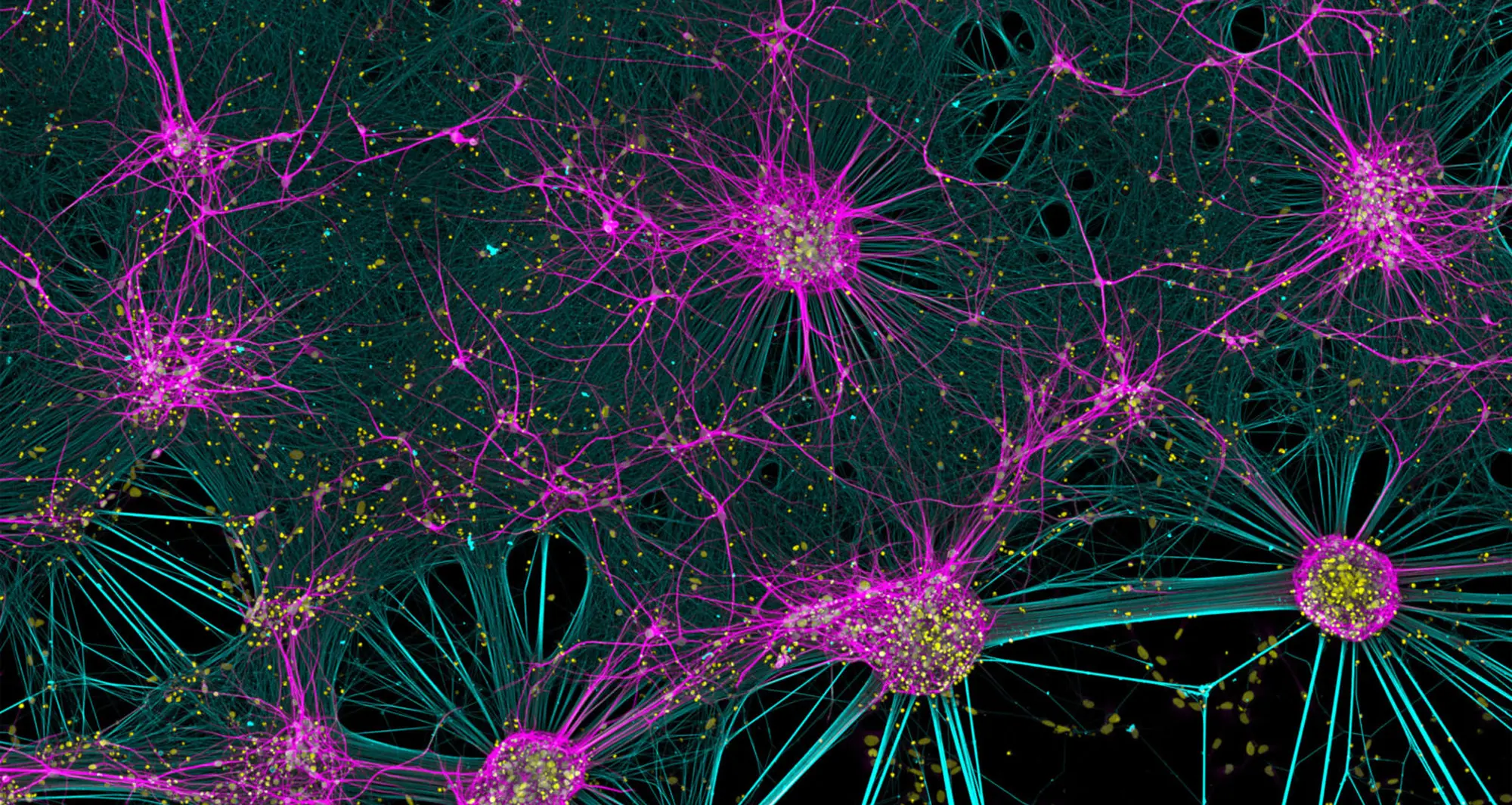
A confocal micrograph of human neurons reprogrammed from skin cells.
Image courtesy of Bruno Cisterna and Eric Vitriol, Medical College of Georgia at Augusta University.
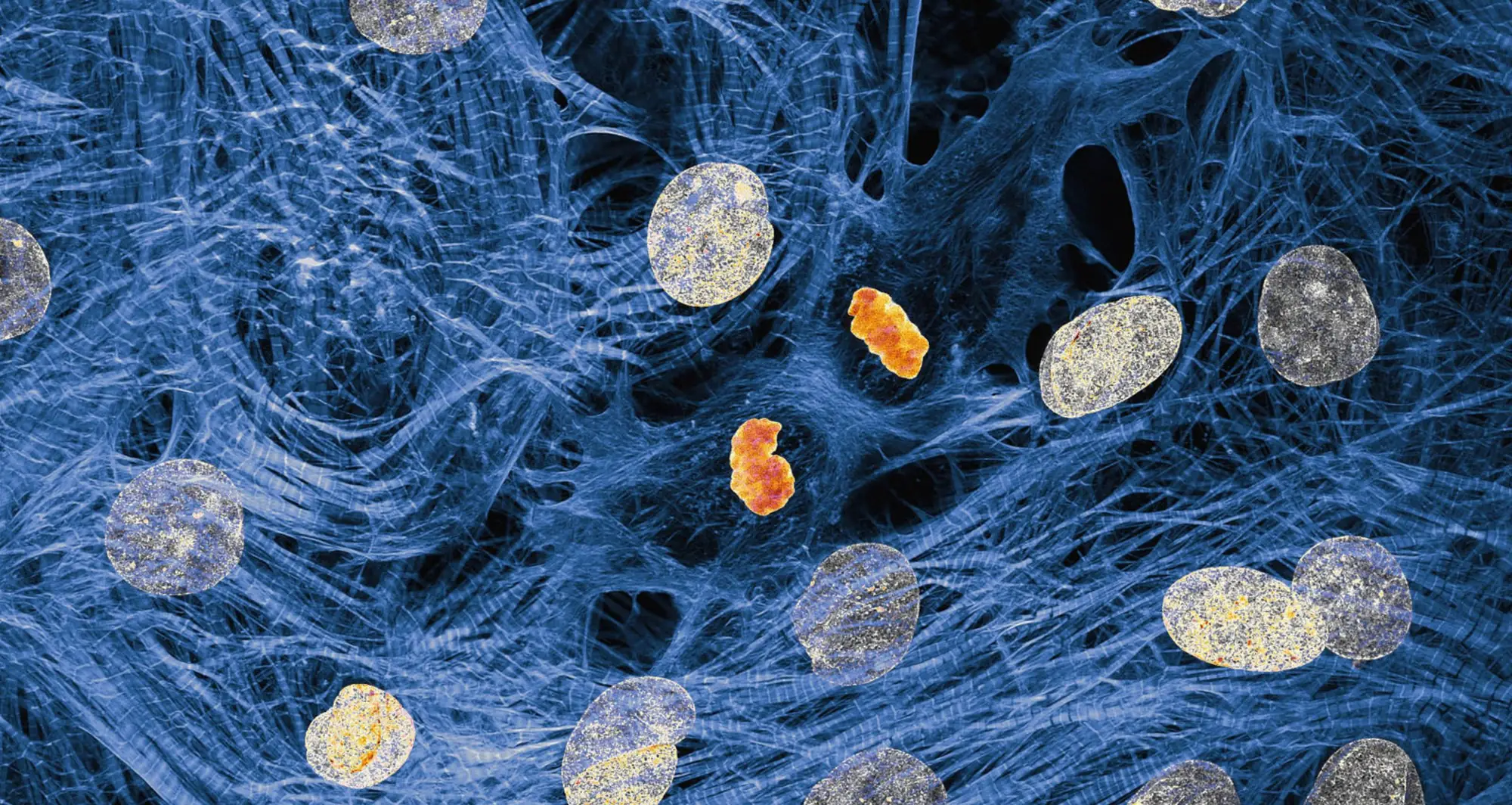
A confocal micrograph of human neurons reprogrammed from skin cells.
Image courtesy of Bruno Cisterna and Eric Vitriol, Medical College of Georgia at Augusta University.

A confocal micrograph of fluorescently marked mouse colon.
Image courtesy of Marius Mählen, Koen Oost, Prisca Liberali and Laurent Gelman, Friedrich Miescher Institute for Biomedical Research, Basel, Switzerland.
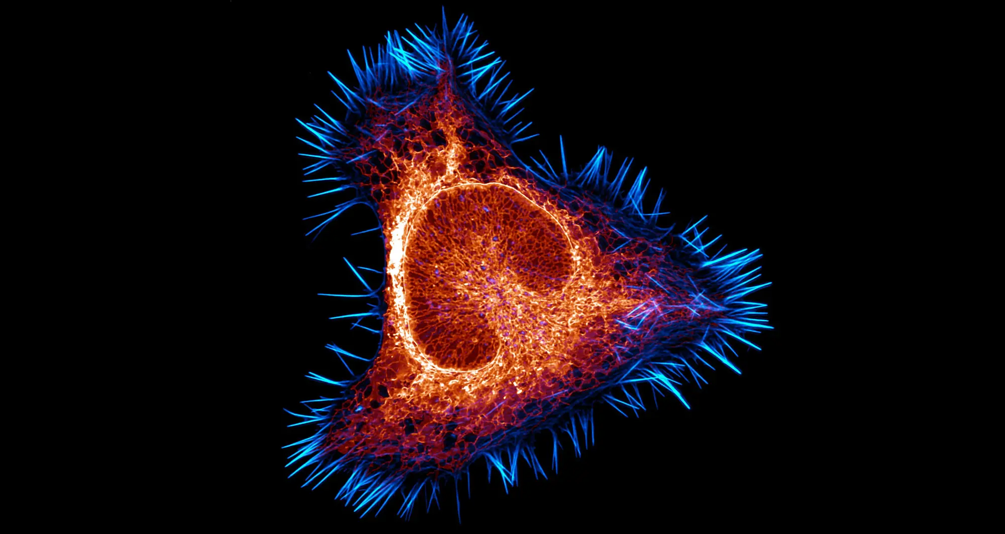
The intricate architecture of the endoplasmic reticulum, the largest organelle in most cells, is depicted in this mouse brain cancer cell. The actin cytoskeleton in cyan and the endoplasmic reticulum in red.
Image courtesy of Halli Lindamood and Eric Vitriol, Augusta University.
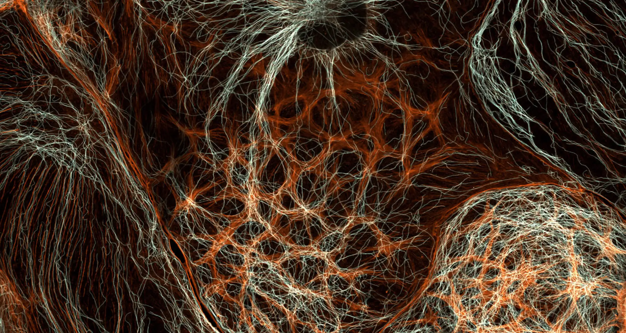
A cell’s cytoskeleton provides structural support, maintains cell shape and is involved in processes like cell movement and intracellular transport of substances. Actin and tubulin are primary components of two main types of cytoskeleton protein. In this micrograph of rat liver cells, actin is shown in orange and tubulin in white.
Image courtesy of Francisco Lázaro-Diéguez, Albert Einstein College of Medicine.
Depicted are cardiac myocytes, the beating cells of heart muscle with surrounding muscle fibers. At the center, one cell (orange) is dividing with chromosomes (DNA) being pulled into two daughter cells.
Image courtesy of James Hayes, Vanderbilt University.
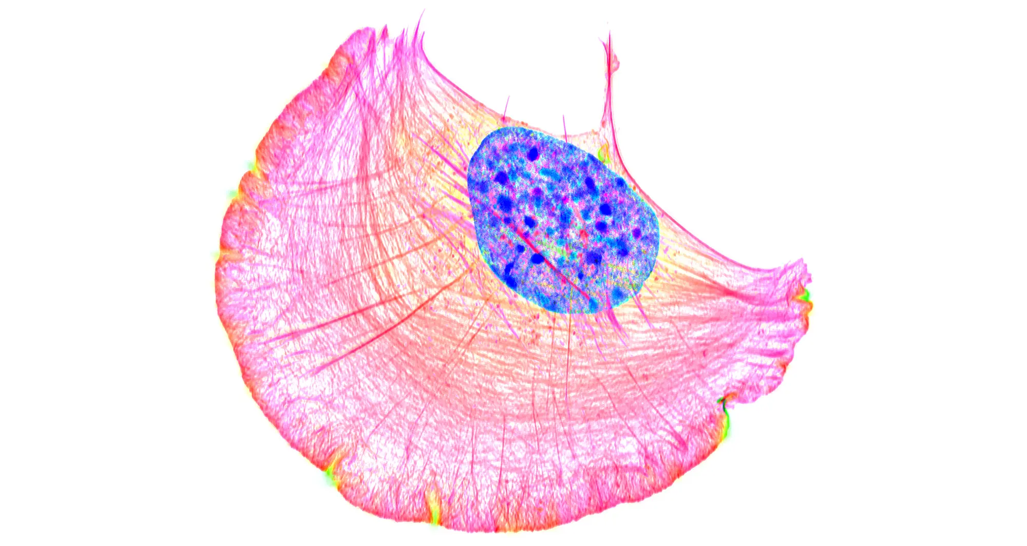
Cell crawling is a form of locomotion where a cell moves across a surface by repeatedly extending its front, anchoring it and then pulling its body forward. The movement is driven by the dynamic remodeling of the cell’s internal cytoskeleton, primarily involving the protein actin. In this structured illumination micrograph, a crawling cell is shown with DNA in blue and actin filaments in pink.
Image courtesy of Dylan T. Burnette, Vanderbilt University.
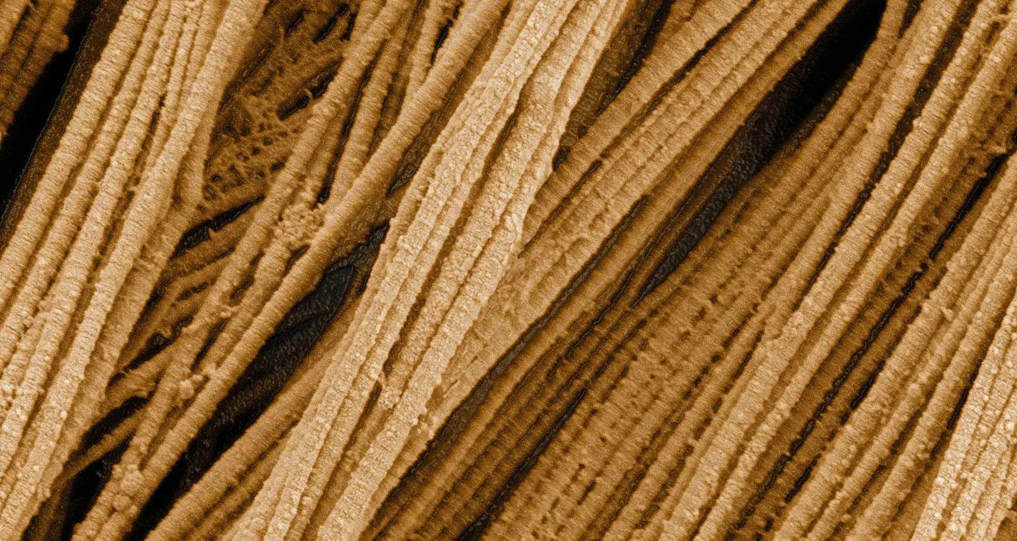
Collagen is a strong, ropelike molecule that forms stretch-resistant fibers. The most abundant protein in our bodies, collagen accounts for roughly one-quarter of our total protein mass. Among its many functions is giving strength to our tendons, ligaments and bones, and providing scaffolding for skin wounds to heal. There are about 20 different types of collagen, each adapted to the needs of specific tissues.
Image courtesy of Tom Deerinck, NCMIR, UCSD.
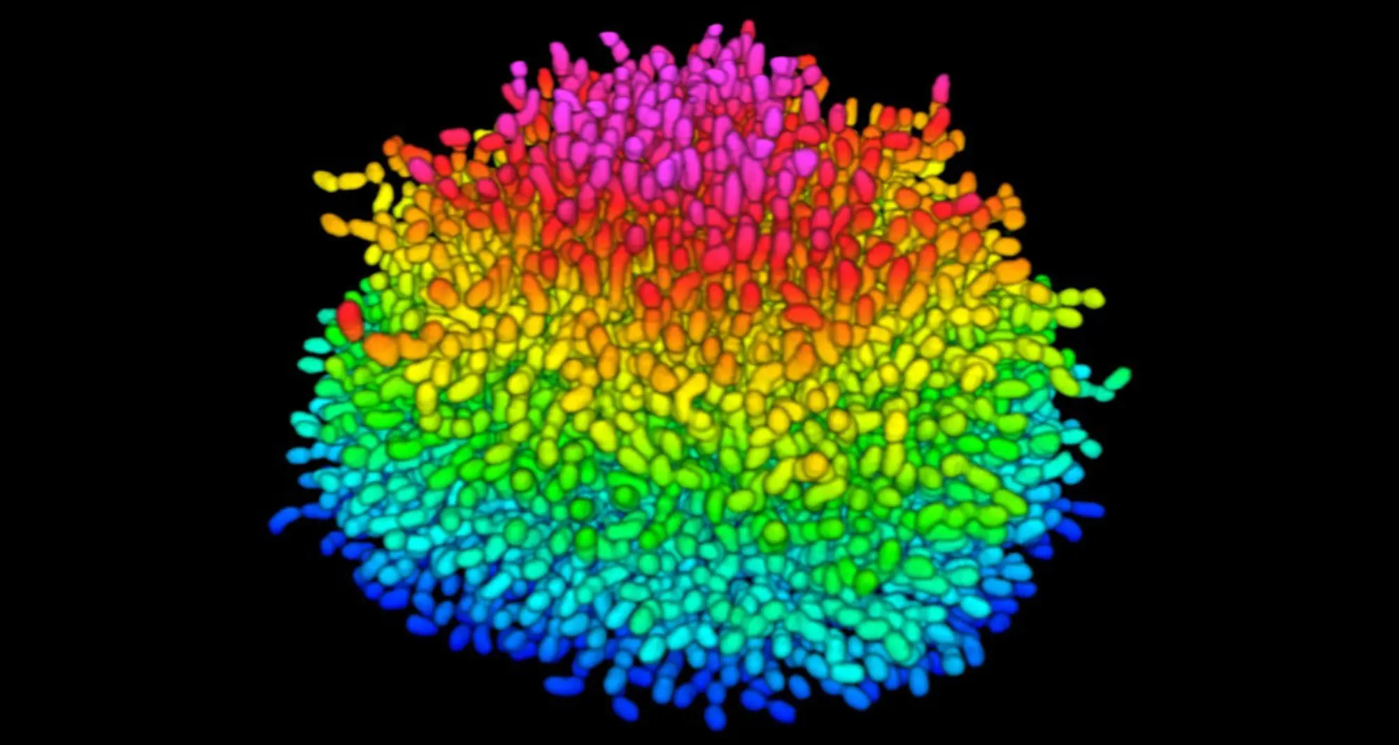
A growing Vibrio cholerae biofilm. Cholera bacteria form colonies called biofilms that enable them to resist antibiotic therapy within the body and other challenges to their growth. Each slightly curved comma shape represents an individual bacterium from assembled confocal microscopy images. Different colors show each bacterium’s position in the biofilm in relation to the surface on which the film is growing.
Image courtesy of Jing Yan and Bonnie Bassler, Princeton University.
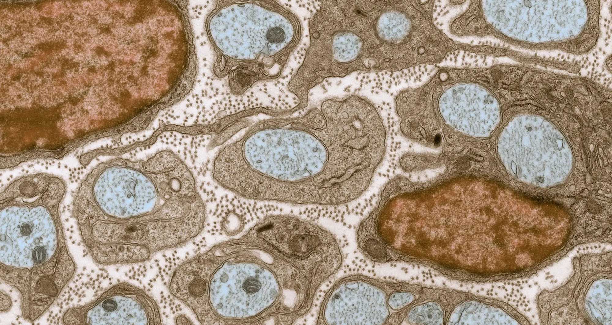
The extracellular matrix (ECM) is a network of proteins and carbohydrates that surrounds and supports cells in tissues throughout the body. In this image, the ECM (pink) is present between the axons of nerve cells. Blue-colored nerve cell axons are surrounded by brown-colored, myelin-supplying Schwann cells, which act like insulation to help speed the transmission of electric nerve impulses down the axon. The tiny brown spots within the ECM are collagen fibers.
Image courtesy of Tom Deerinck, NCMIR, UCSD.
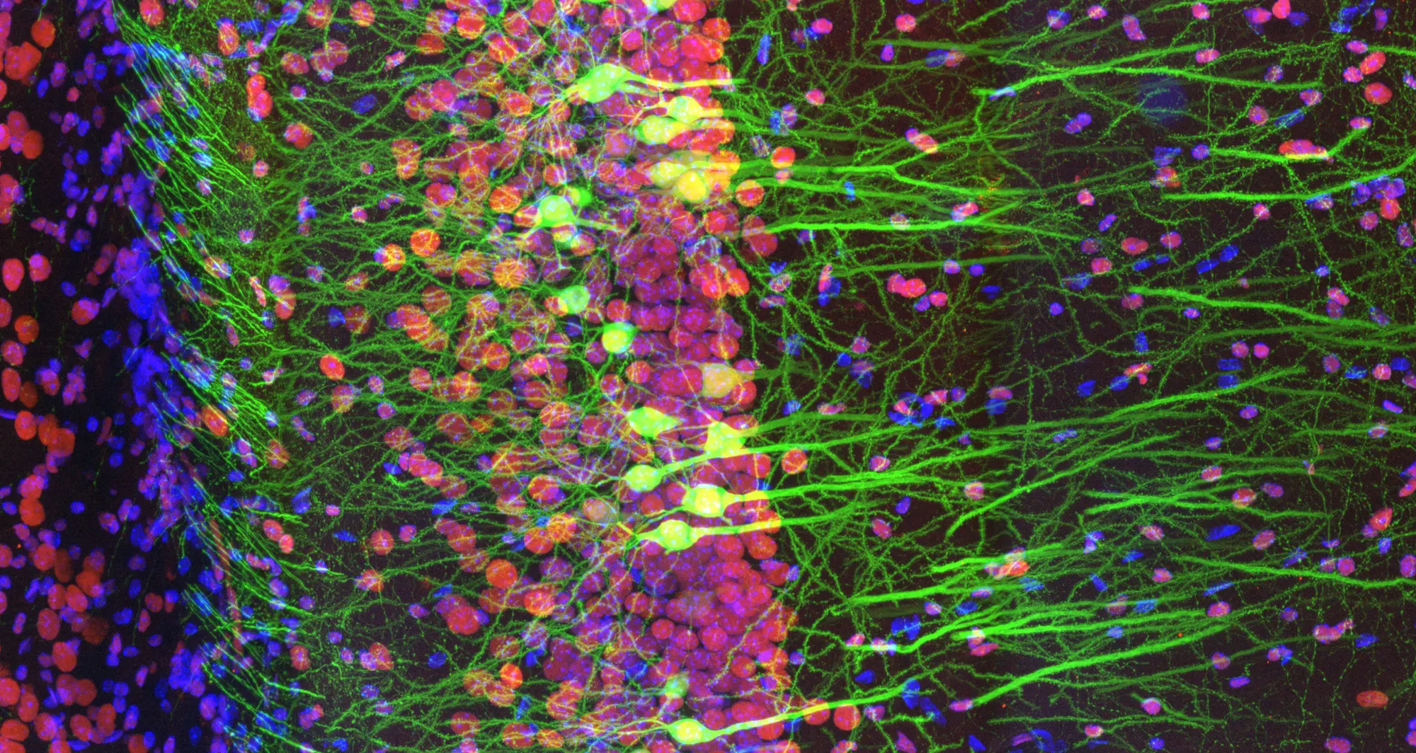
In this stained fluorescence image of a slice of mouse brain, green depicts the excitatory hippocampal neurons; in red are obesity-associated proteins and cell nuclei in blue.
Image courtesy of Ainara Pintor.