What says happy holidays and Happy New Year better than a transverse section of rachis (stem) of bracken fern, created using differential interference contrast microscopy?
Image courtesy of David Maitland and Nikon Small World.
What says happy holidays and Happy New Year better than a transverse section of rachis (stem) of bracken fern, created using differential interference contrast microscopy?
Image courtesy of David Maitland and Nikon Small World.
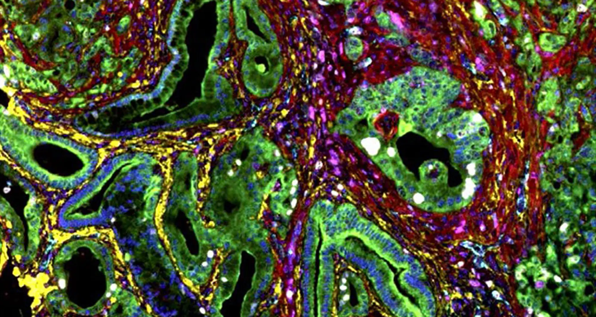
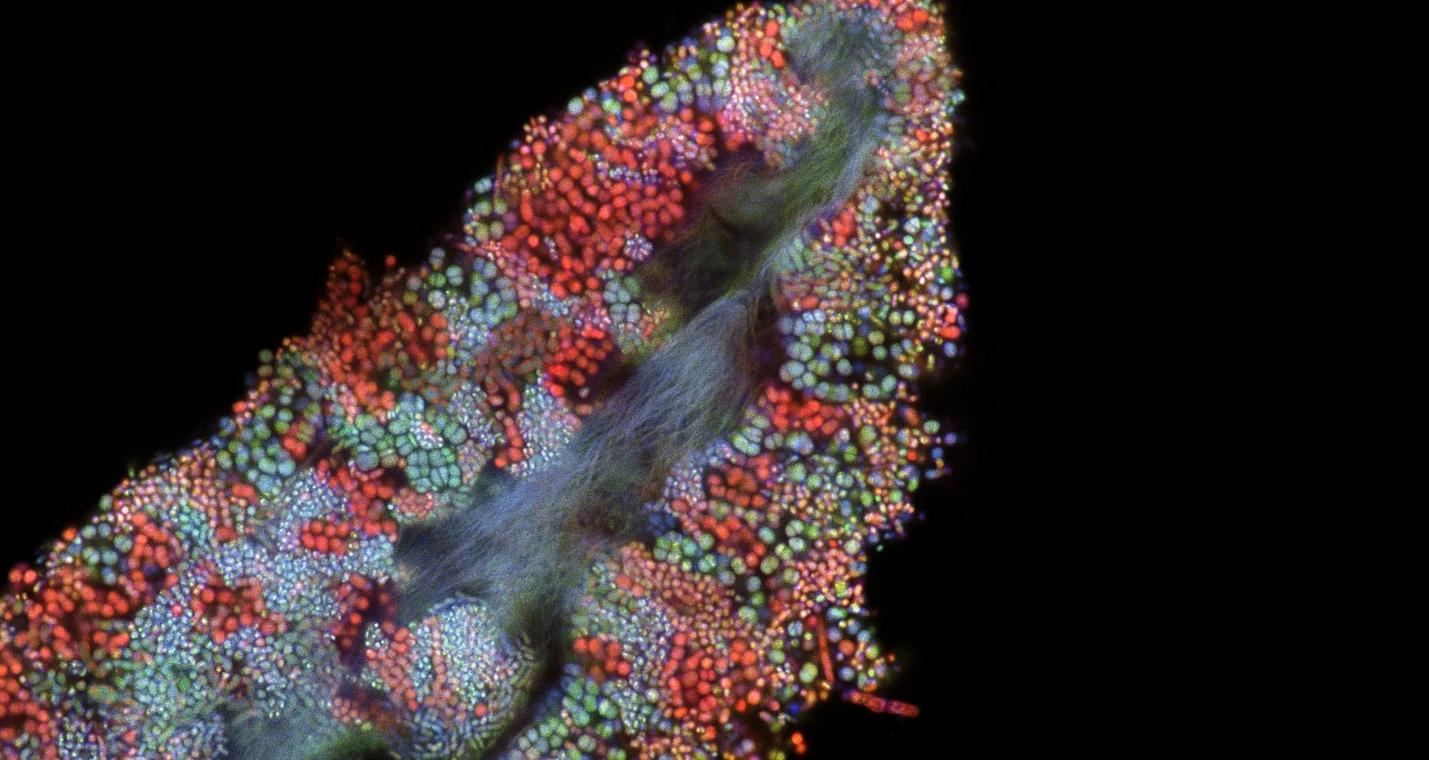
This immunohistochemistry image, stained to visualize different stroma biomarkers in a mouse liver tumor, colorfully captures the variegated heterogeneity of the tumor microenvironment.
Image courtesy of A.E. Nel, et al. National Cancer Institute.
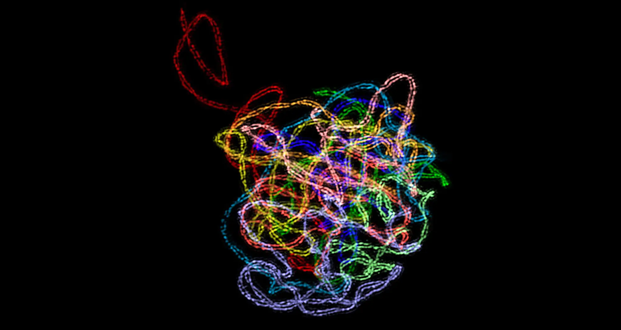
A 3D-structured illumination reveals a synaptonemal complex, a protein structure that forms between homologous chromosomes during cell division. It’s believed SCs function primarily as scaffolds to allow interacting chromatids to complete their crossover activities.
Image courtesy of Chung-Ju-Rachel Wang and Bioscapes.
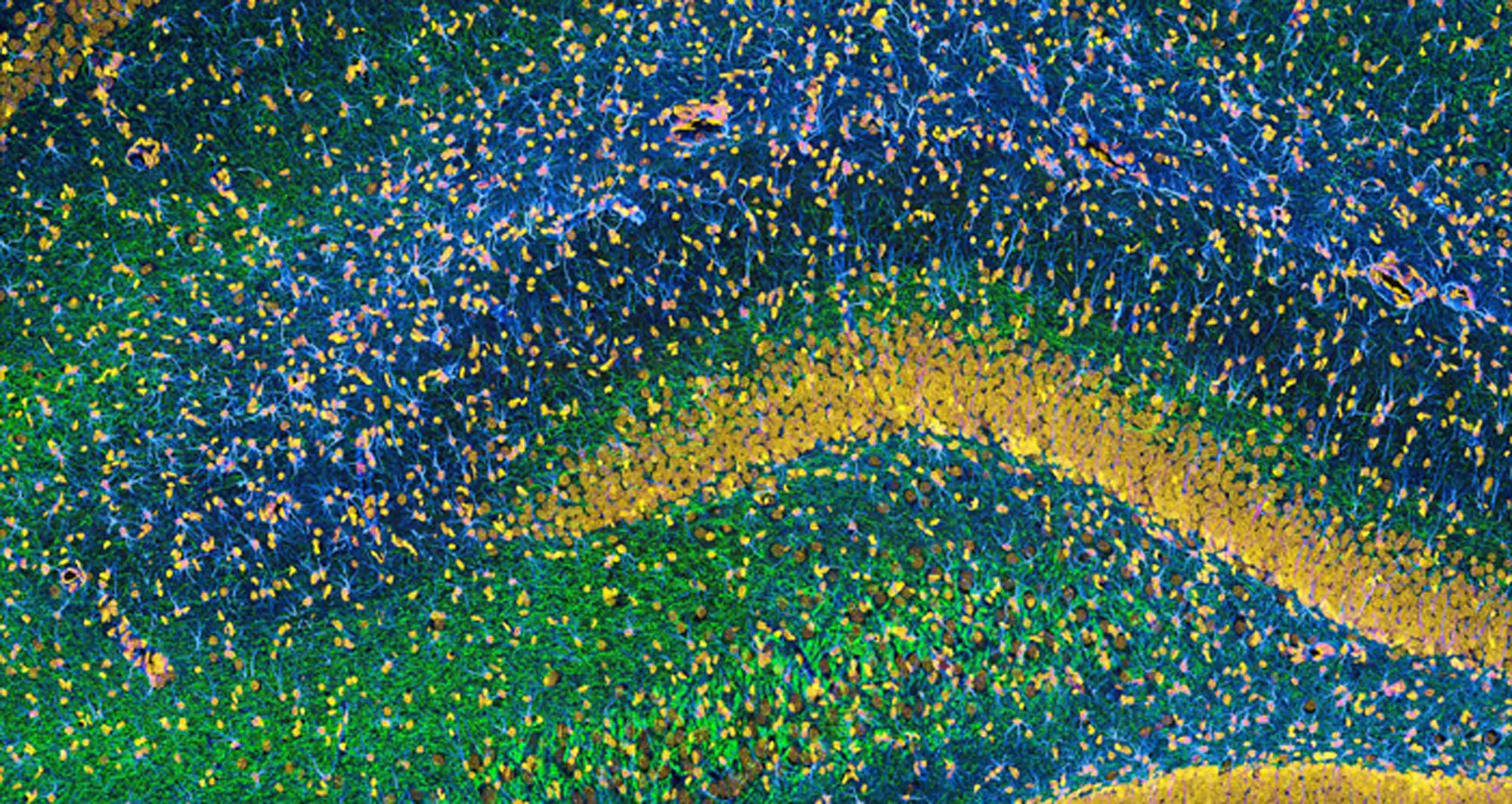
This image of the hippocampus in a rat brain was taken using an ultra-widefield high-speed multiphoton laser microscope. Tissue was stained to reveal the organization of glial cells (cyan), neurofilaments (green) and DNA (yellow).
Image courtesy of Thomas Deerinck, NCMIR and NIH.
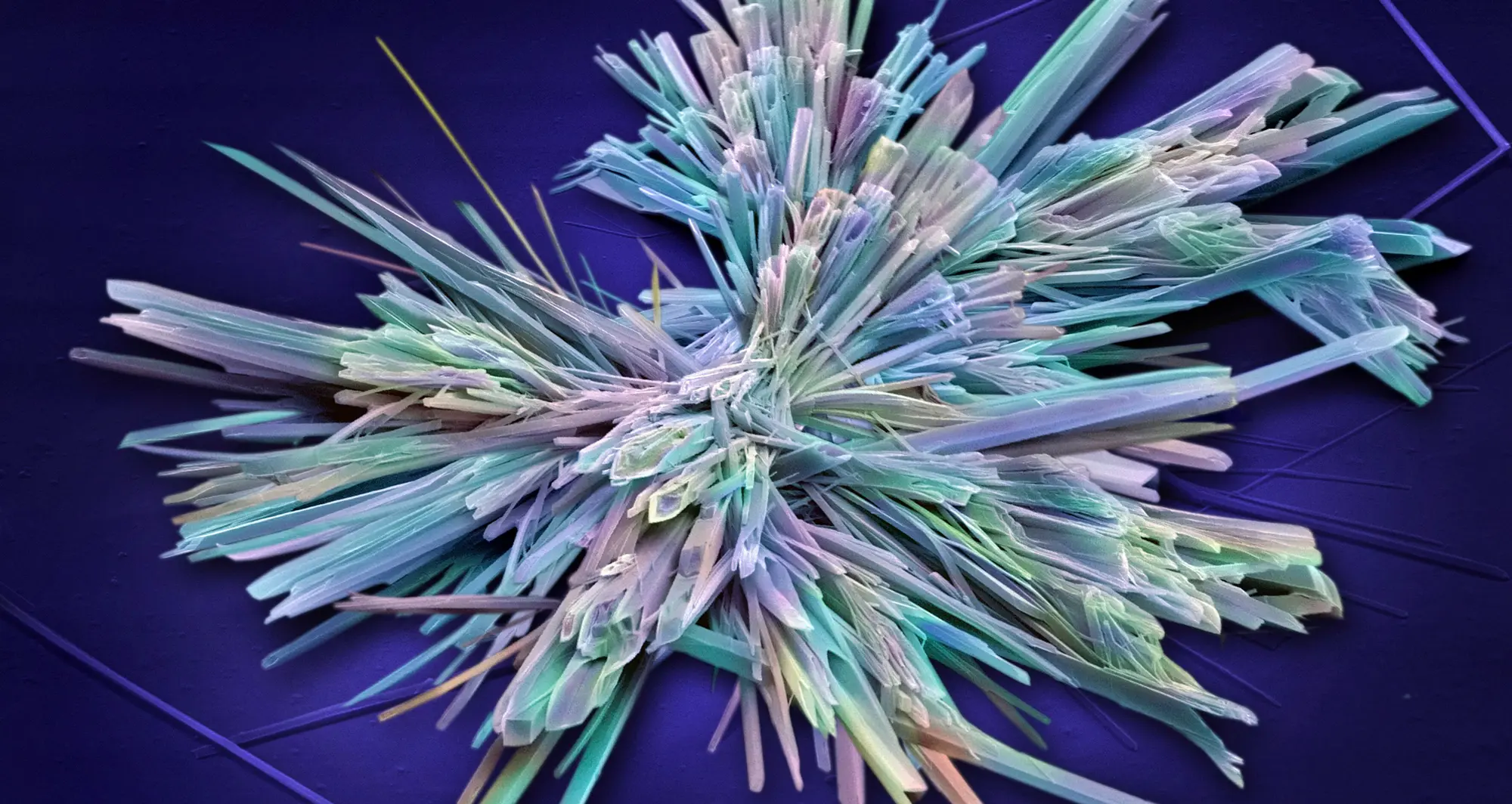
t’s as lovely as a snowflake in winter, but something entirely different: a crystal of sildenafil, the active ingredient in Viagra tablets.
Image courtesy of Annie Cavanagh, Wellcome Collection.
Confocal micrograph of bacterial biofilm on a human tongue cell. The oral cavity harbors more than 700 species of bacteria, second only to the gut.
Image courtesy of Tagide deCarvalho, University of Maryland, Baltimore County.
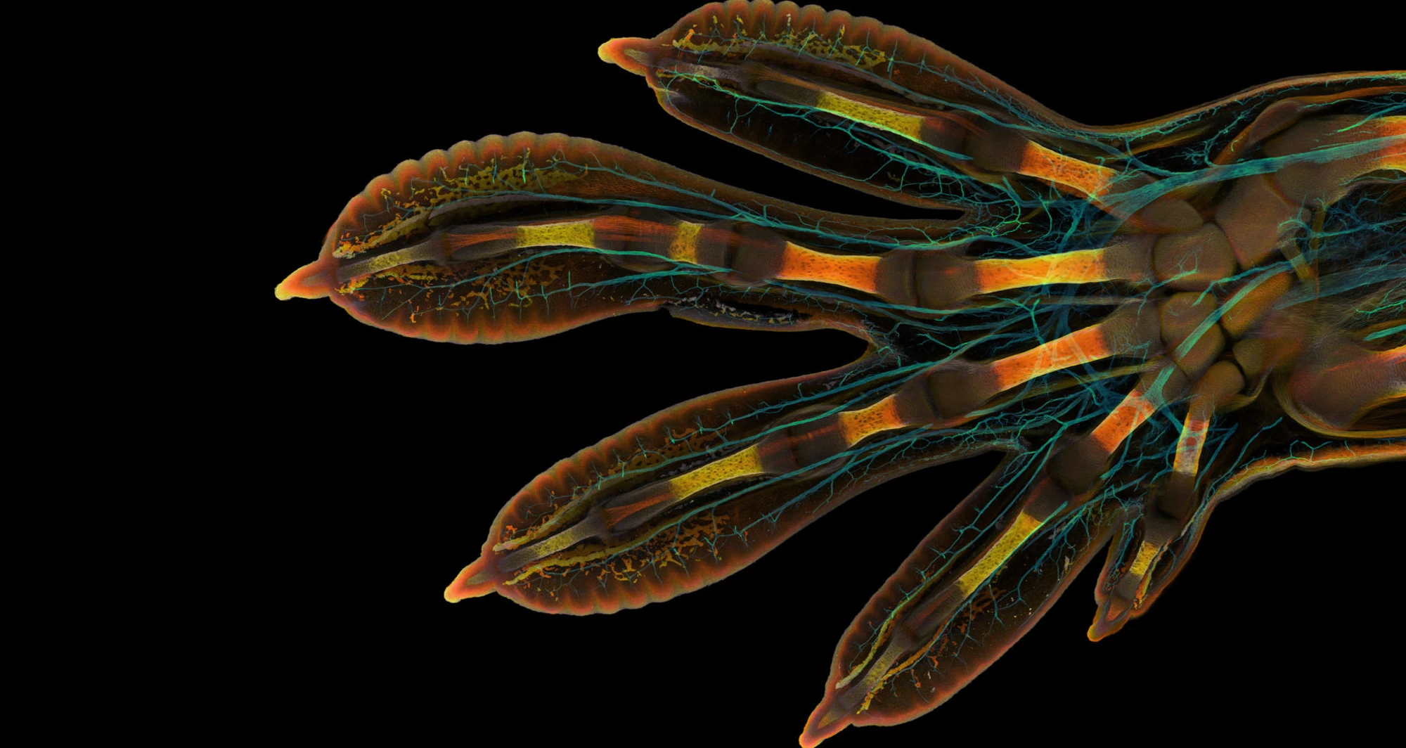
Confocal micrograph of the embryonic hand of a Madagascar giant day gecko (Phelsuma grandis), which grow to up to 12 inches and live 8-15 years.
Image courtesy of Grigori Timin and Michel Milinkovitch, University of Geneva.
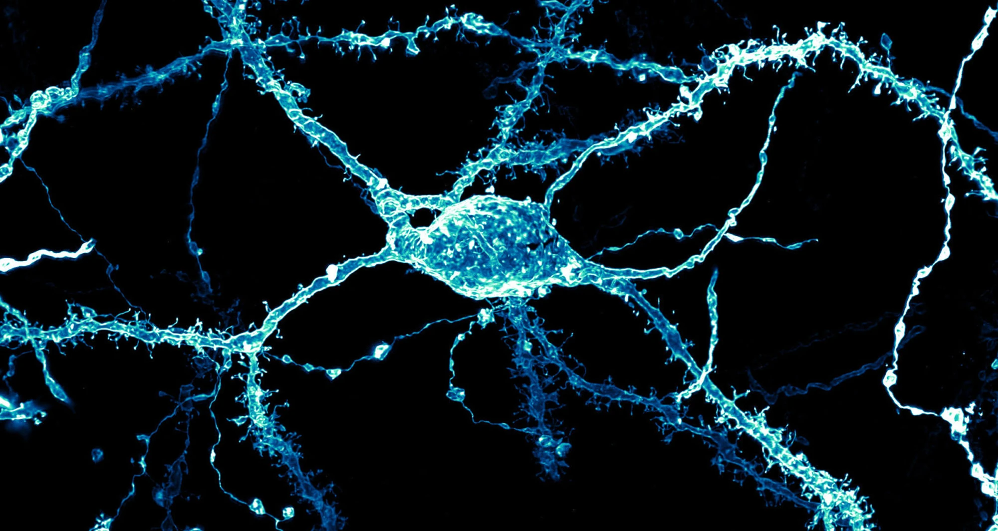
A neuron covered in dendritic spines from the striatum of an adult rat brain. The striatum is a cluster of interconnected nuclei that form a part of the basal ganglia. It is involved in decision-making functions, such as motor control, emotion, habit formation and reward.
Image courtesy of Stephanie Huang, Victoria University of Wellington School of Biological Sciences, New Zealand.
A chromosome from Drosophila melanogaster (fruit fly) salivary glands, using Brightfield microscopy. Fruit flies are model organisms, sharing 75% of the genes that cause disease in humans.
Image courtesy of Earl Nishiguchi, Kauai Community College and Nikon Small World.
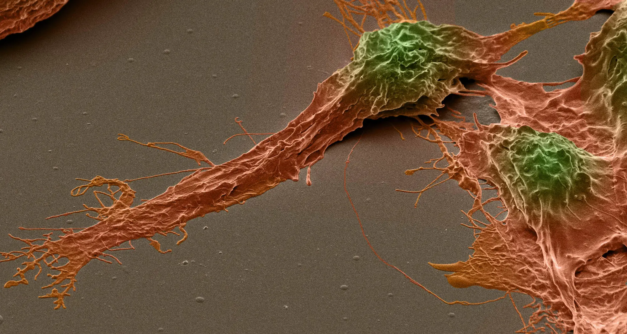
A colorized scanning electron micrograph of a human brain cancer stem cell. Cell bodies are in orange; nuclei in green. Cancer stem cells possess characteristics of normal stem cells, specifically the ability to give rise to all cell types found in a particular cancer sample. That makes them a particularly enticing target for developing cancer therapies, especially those prone to metastasis or recurrence.
Image courtesy of Izzat Suffian, Pedro Costa, Stephen Pollard, David McCarthy & Khuloud T. Al-Jamal.