A confocal micrograph depicts an embryonic mouse kidney showing the collecting duct (blue) and nephron progenitor (yellow) cells.
Image courtesy of Lori O’Brien, University of North Carolina at Chapel Hill and Nikon.
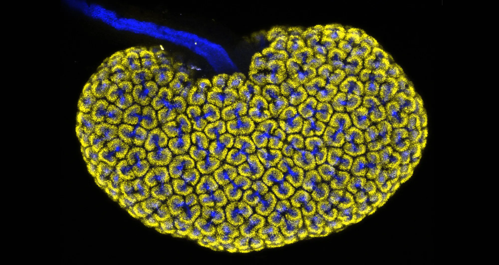
A confocal micrograph depicts an embryonic mouse kidney showing the collecting duct (blue) and nephron progenitor (yellow) cells.
Image courtesy of Lori O’Brien, University of North Carolina at Chapel Hill and Nikon.
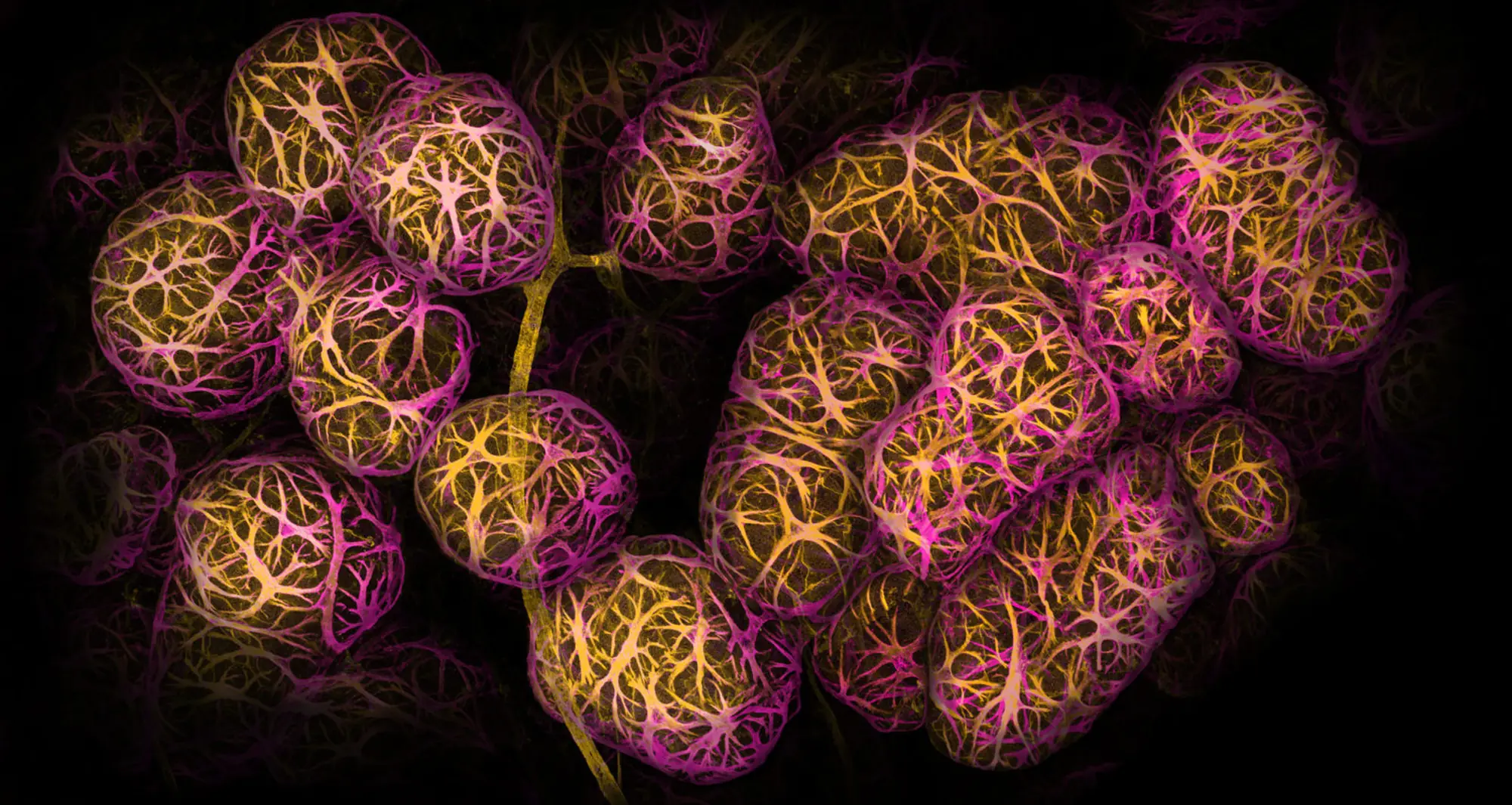
Using confocal microscopy, this image depicts breast tissue showing contractile myoepithelial cells wrapped around milk-producing alveoli.
Image courtesy of Caleb Dawson at Walter and Eliza Hall Institute of Medical Research in Australia and Nikon.
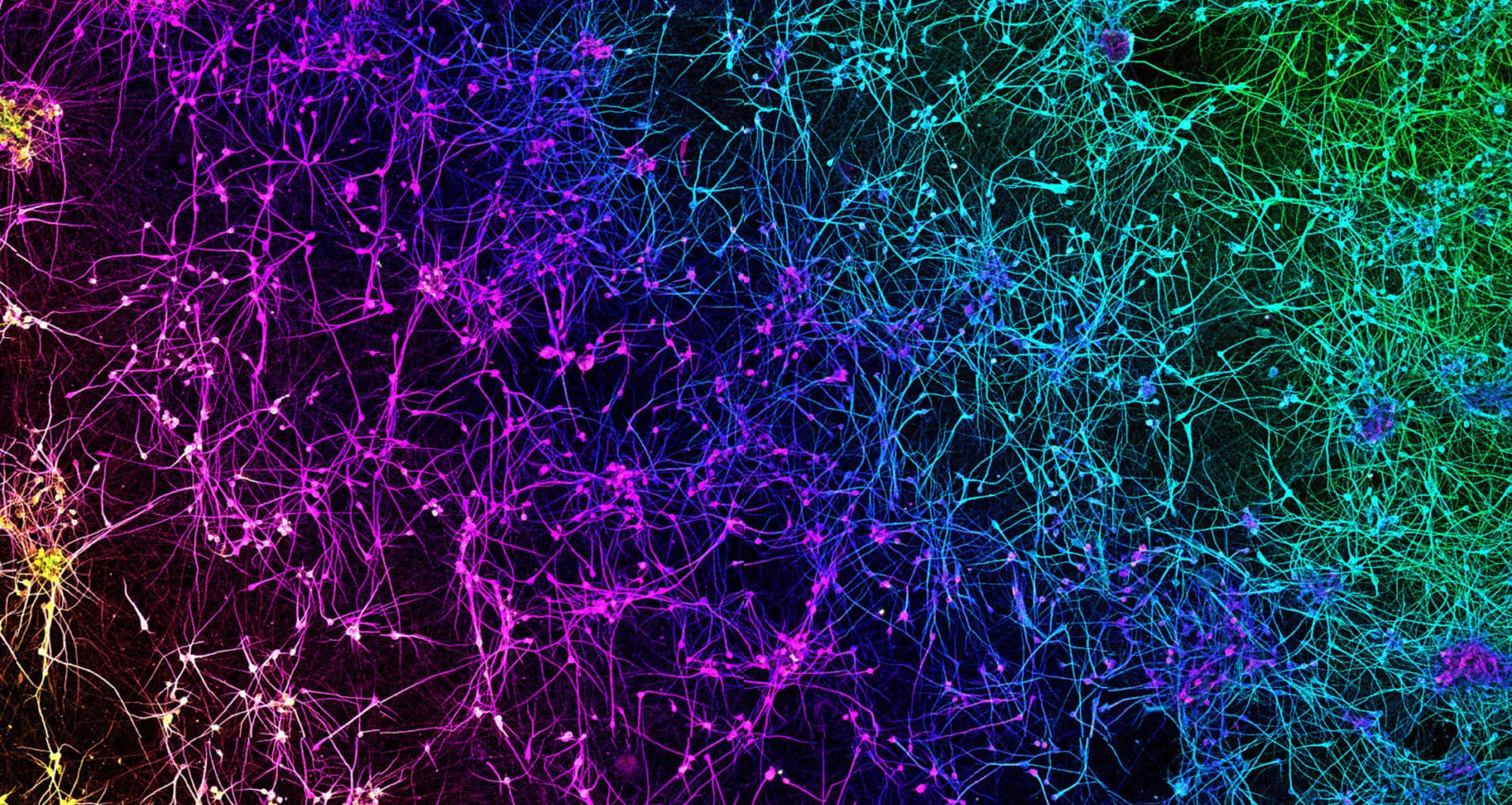
A network of dopaminergic neurons generated from human stem cells. Dopaminergic neurons regulate many brain functions, including voluntary movement, learning and reward and working memory.
Image courtesy of Nick Gatford, University of Oxford and Nikon Small World.
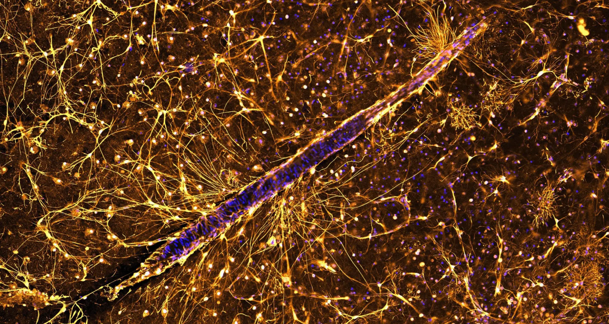
Astrocytes surrounding a blood vessel in a thin slice of human brain.
Image courtesy of Anja de Lange, University of Cape Town and Nikon Small World.
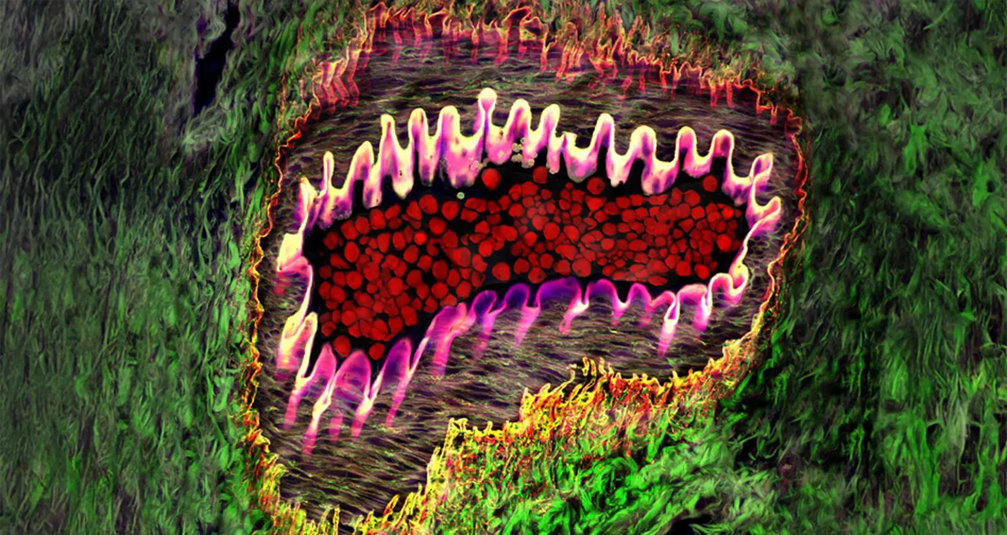
Healthy arteriole in eye, with tough, flexible elastin wall (pink), red blood cells (red) and supporting collagen fibers (web-like netting) surrounded by yellow and green areas.
Image courtesy of Donald Pottle, The Schepens Eye Research Institute, Boston and Bioscapes.
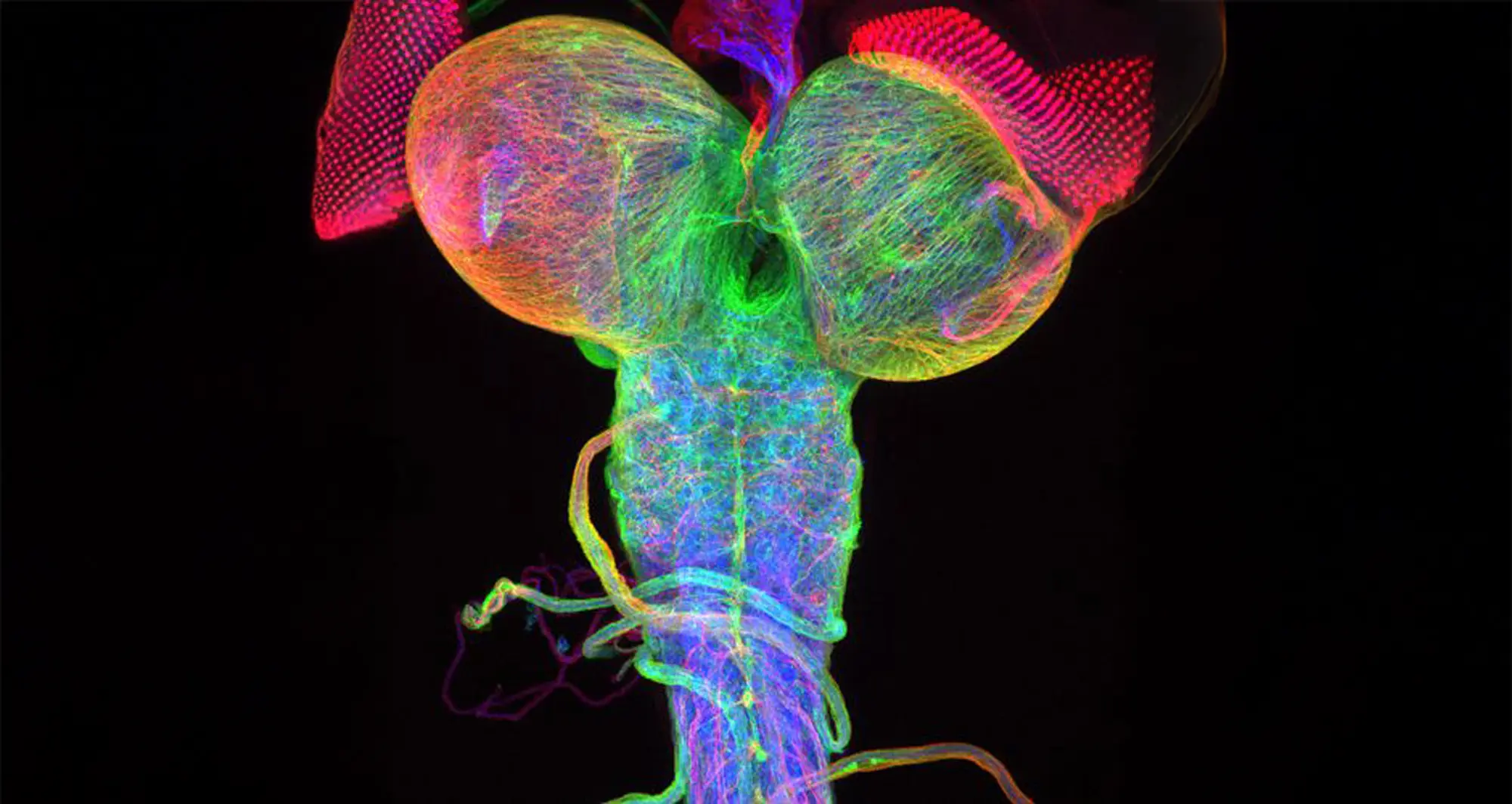
Beta-tubulin expression of a Drosophila (fruit fly) third instar larval brain, with attached eye imaginal discs.
Image courtesy of Christian Klambt and Imke Schmidt, University of Munster, Germany and Bioscapes.

What says happy holidays and Happy New Year better than a transverse section of rachis (stem) of bracken fern, created using differential interference contrast microscopy?
Image courtesy of David Maitland and Nikon Small World.
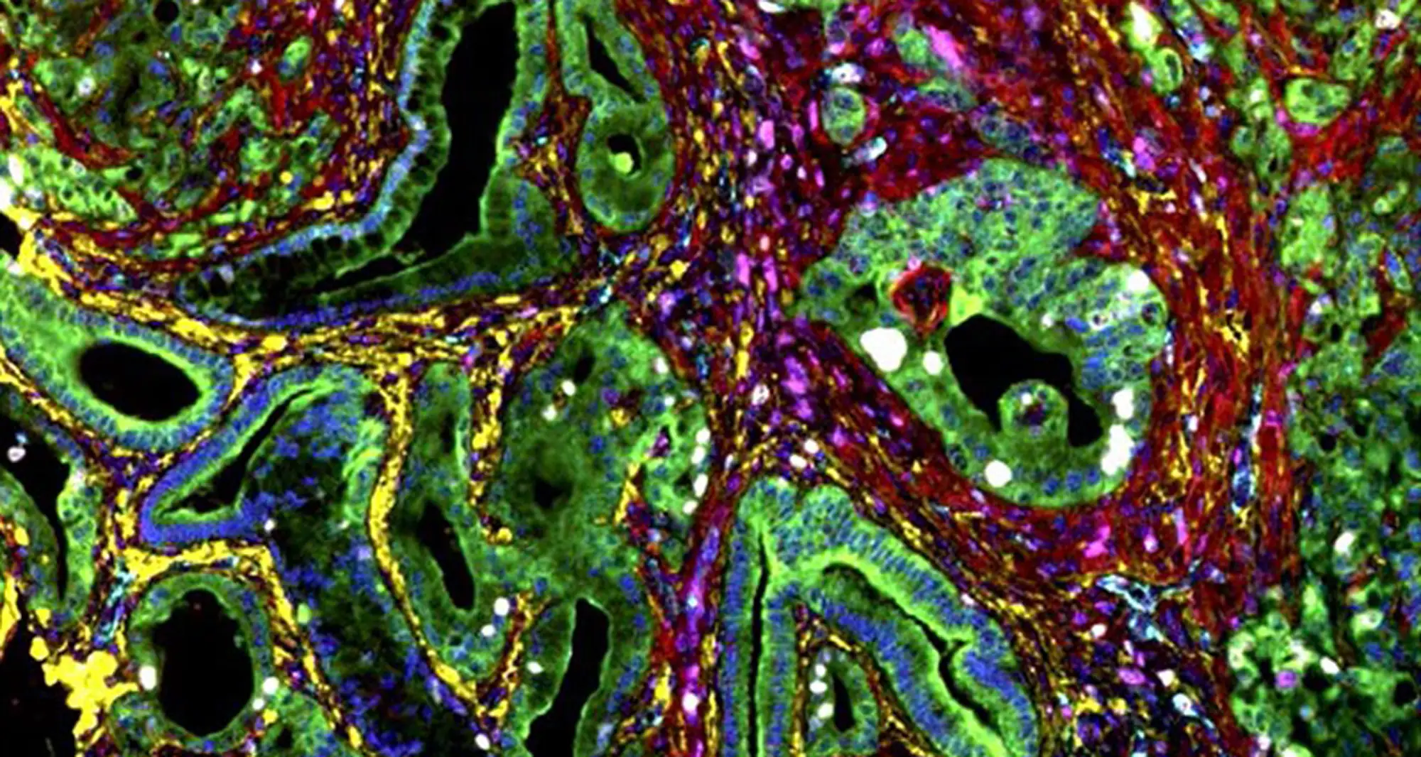
This immunohistochemistry image, stained to visualize different stroma biomarkers in a mouse liver tumor, colorfully captures the variegated heterogeneity of the tumor microenvironment.
Image courtesy of A.E. Nel, et al. National Cancer Institute.
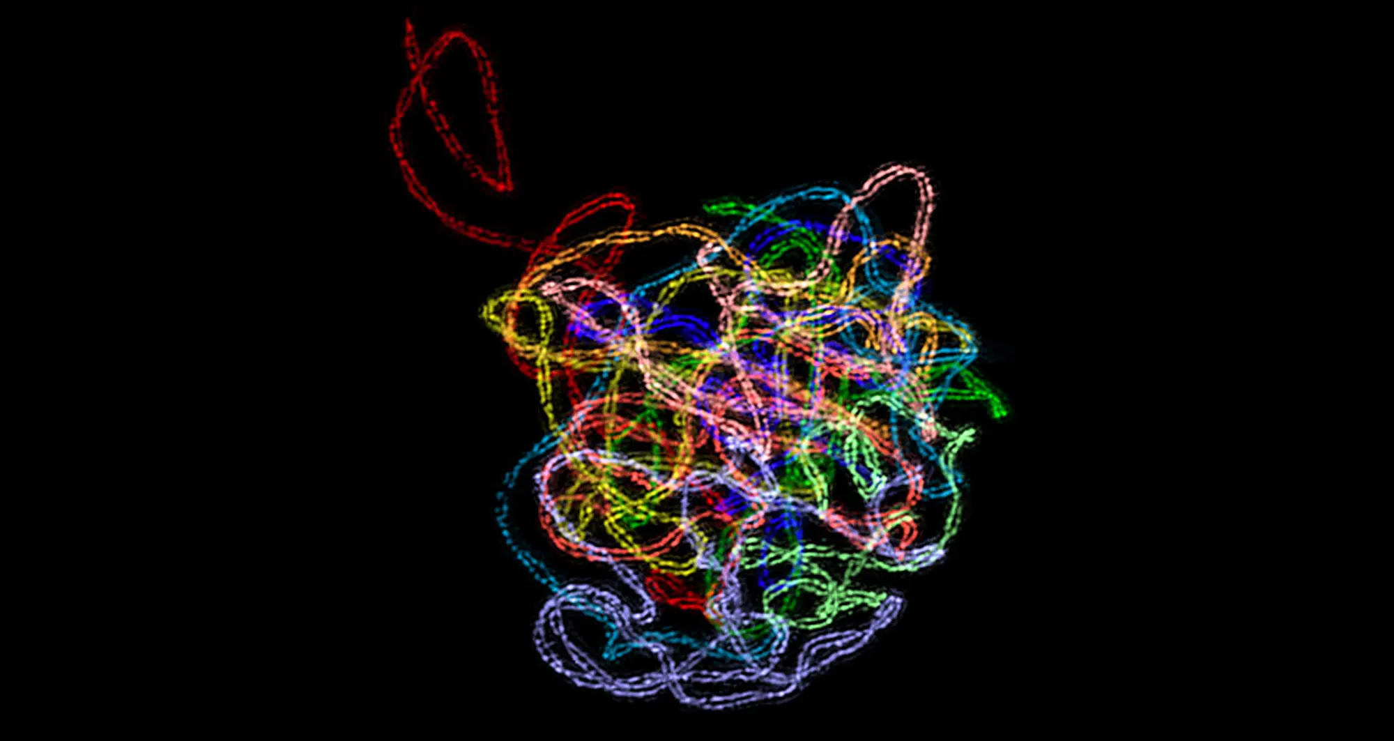
A 3D-structured illumination reveals a synaptonemal complex, a protein structure that forms between homologous chromosomes during cell division. It’s believed SCs function primarily as scaffolds to allow interacting chromatids to complete their crossover activities.
Image courtesy of Chung-Ju-Rachel Wang and Bioscapes.
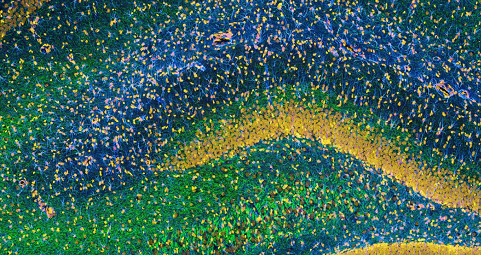
This image of the hippocampus in a rat brain was taken using an ultra-widefield high-speed multiphoton laser microscope. Tissue was stained to reveal the organization of glial cells (cyan), neurofilaments (green) and DNA (yellow).
Image courtesy of Thomas Deerinck, NCMIR and NIH.