Uncategorized Archives - Page 8 of 58 - Sanford Burnham Prebys
Institute News

Q & A with Postdoctoral Researcher Sara Ancel, PhD, from the Wang Lab

AuthorCommunications
Date

July 8, 2025

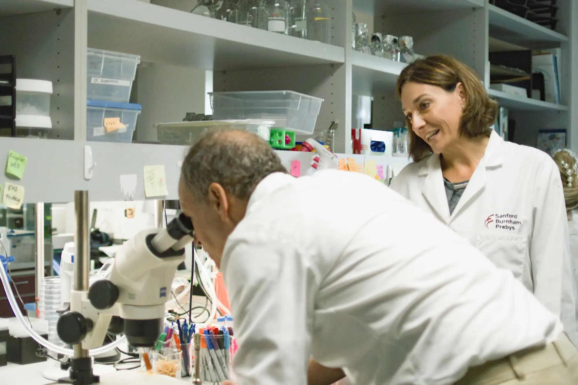
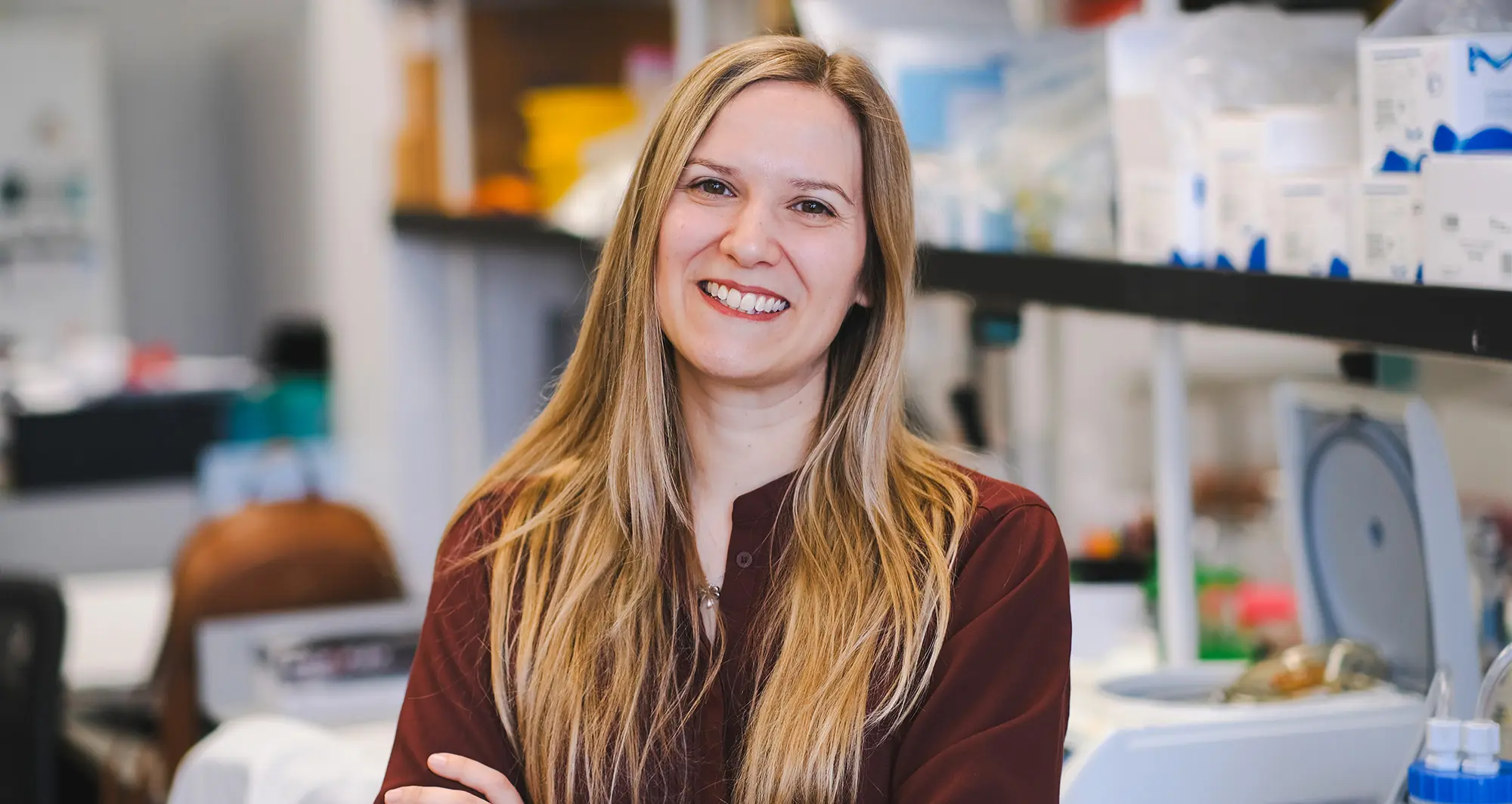
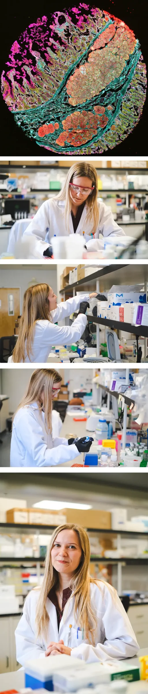
Shaping the future of science at Sanford Burnham Prebys: Sara Ancel, PhD, a postdoctoral researcher in the lab of Will Wang, PhD, who draws on a background in engineering and stem cell biology to explore tissue remodeling and disease mechanisms through cutting-edge spatial omics approaches. Originally from Switzerland, she brings together cutting-edge technology and collaborative science to push boundaries—and inspire the next generation of researchers.

How did you first become interested in science—and what brought you to Sanford Burnham Prebys? 
I didn’t grow up around science, my parents weren’t in the field, so I didn’t really get exposed to it until high school. But I’ve always been curious, especially about things I didn’t understand. That curiosity led me to study engineering, which gave me the flexibility to explore many scientific fields before focusing on one.

During my master’s studies in Switzerland, I had the opportunity to spend time at Stanford University working in Dr. Helen Blau’s lab. That’s where I met Will Wang, who would later become a principal investigator at Sanford Burnham Prebys. When I was finishing my PhD in Switzerland, he was just starting his lab here. The timing was perfect—and I became his first postdoc.
Shape

What drew you to Will Wang’s research?
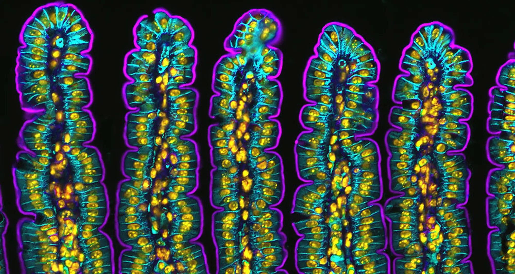
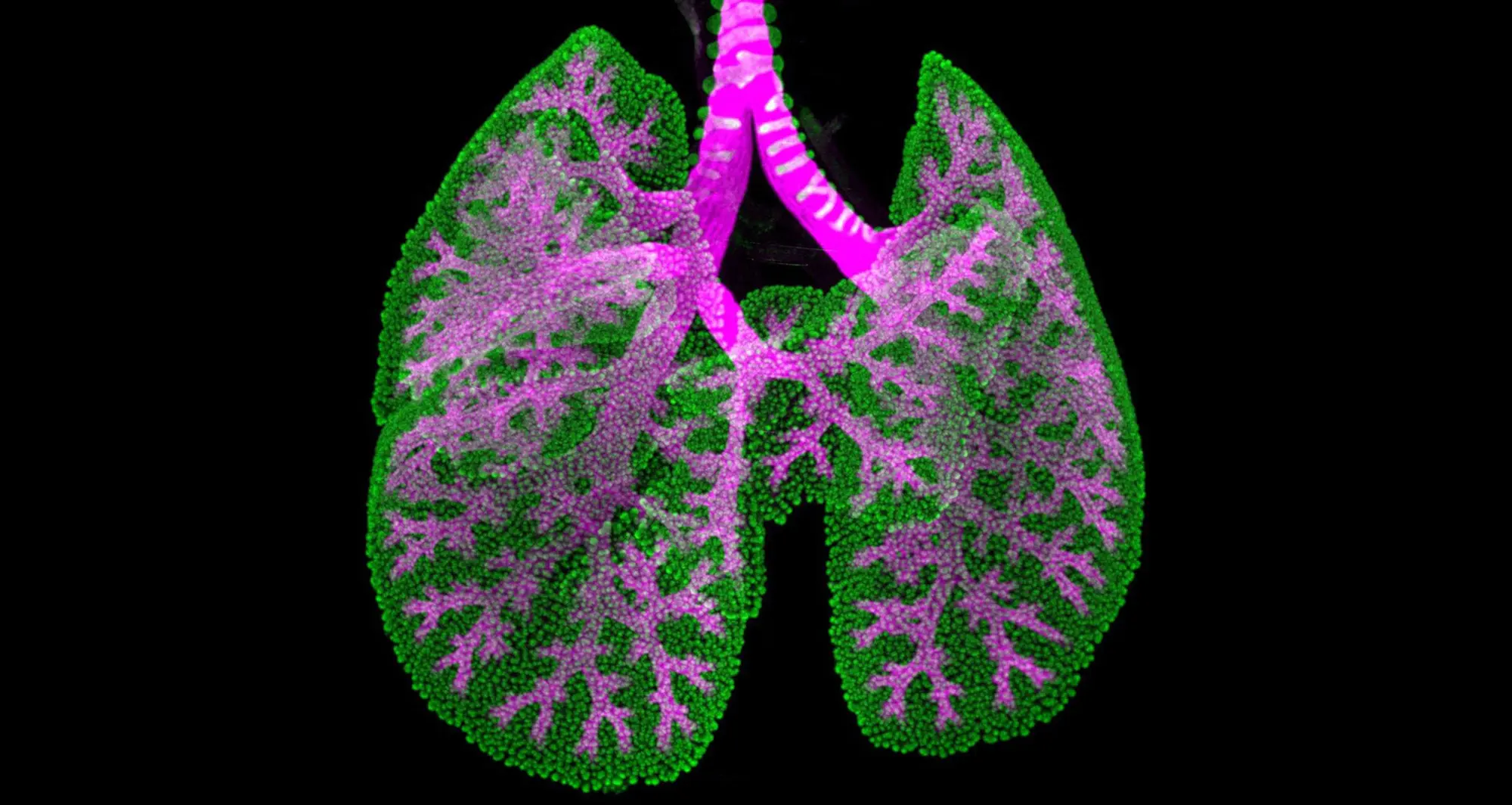
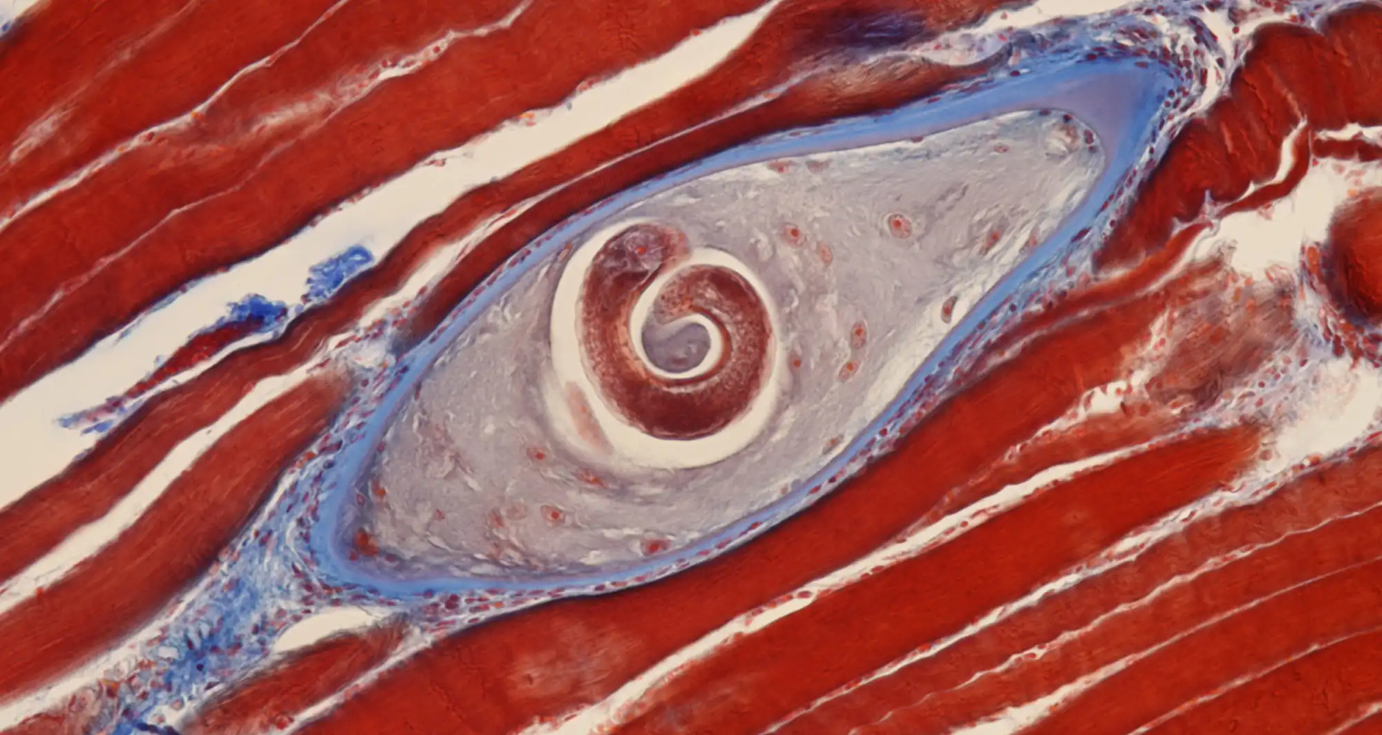
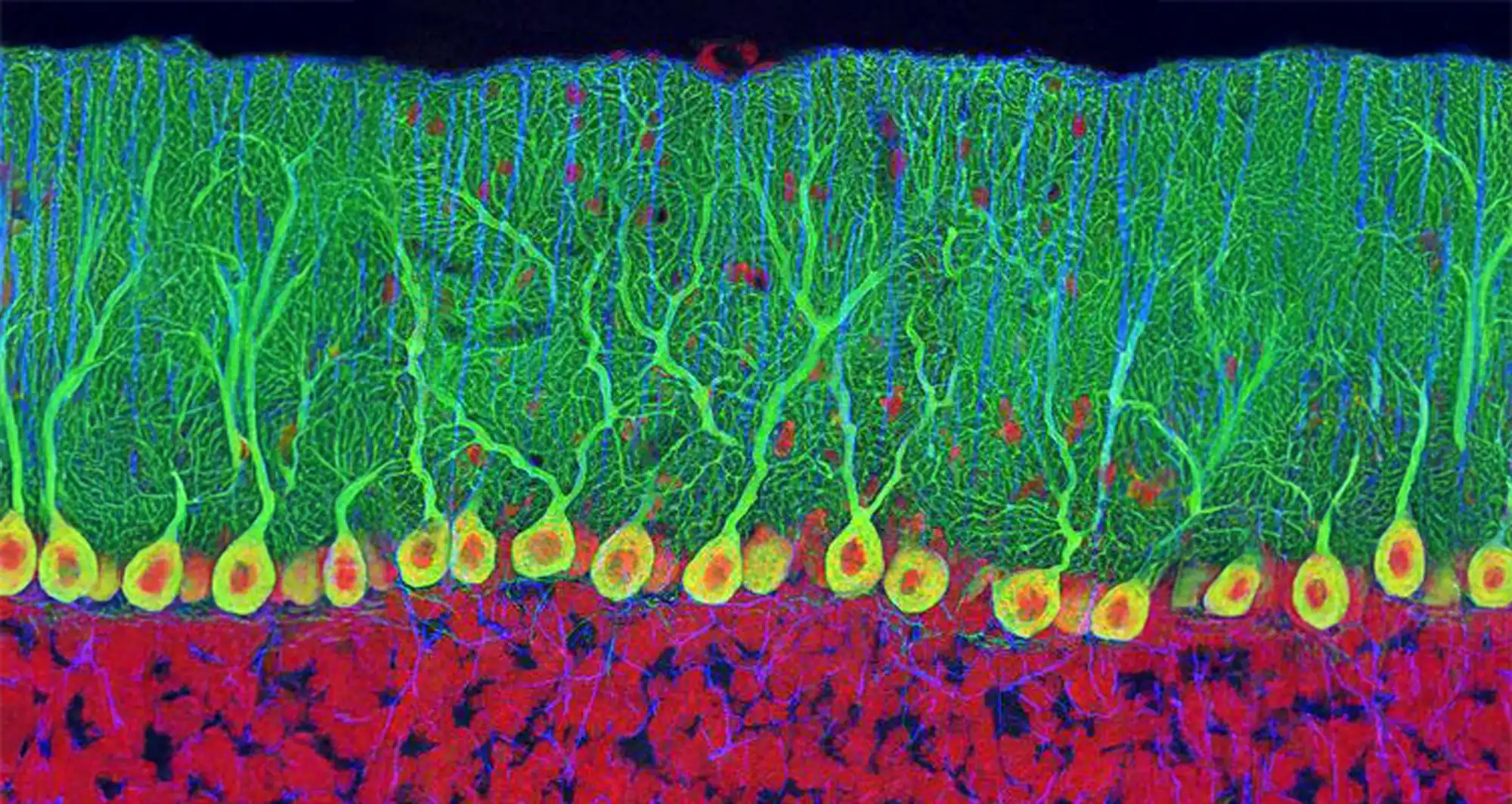
What really stood out to me was the new technology he was developing—an imaging method that lets us look at many biological markers at once. Coming from an engineering background, that kind of innovation was really exciting. I saw a chance to combine everything I’d been learning, for example, stem cell biology, muscle research, and engineering, into one meaningful project.

Plus, joining a brand-new lab was a unique opportunity. I was involved in everything from setting up experiments and training newcomers to handling operations. It was a fast-paced, all-hands-on-deck experience that taught me so much, both scientifically and personally.
Shape

What are you working on now? How would you explain it to someone outside of science?
My main project focuses on a process called glycosylation, which is how cells add sugar molecules to proteins and fats. These sugar tags might sound simple, but they play a big role in how cells function, and how things go wrong in disease.

I had no background in glycobiology when I started, but I was able to bring in new technologies and combine them with biology to explore this process in a completely new way. I’ve also been fortunate to collaborate with the Freeze Lab here at Sanford Burnham Prebys, which has been incredibly valuable.

What makes Sanford Burnham Prebys a unique place to work?
I’ve been so impressed by how collaborative this institute is. It’s a small enough community that people know each other, so reaching out for help or advice is easy. I’ve been able to train on equipment here and at nearby institutions like UC San Diego, and I’ve had the chance to connect with researchers across many fields.

One of the most exciting aspects has been working with clinicians and getting access to real patient samples. That kind of experience really deepens the impact of our research and gives me a broader view of how basic science can connect to human health.

What was one of the biggest challenges you faced when you arrived?
Moving from Switzerland to San Diego was a huge adjustment. I arrived and quickly within about a week, I was in a new culture, new lab, and new scientific environment. I was also the only person in the lab at first, which made things more intense.

But I had great support from international services and from the community of researchers here. That support helped me adapt, and it motivated me to dive in and help get the lab up and running.

What do you hope to do next in your career?
I’ve developed a wide range of skills here, not just technical, but also communication and collaboration. I’d love to build on that by moving into work that’s more closely connected to patients. Collaborating with clinicians and working with patient samples has been incredibly meaningful, and I’d like to pursue more translational or clinical science in the future.

What do you enjoy doing when you’re not in the lab?
Since moving to San Diego, I’ve gotten into climbing and bouldering, it’s something I picked up with friends from neighboring labs. I also love hiking and visiting national parks. Coming from Switzerland, I’m used to mountains, but the parks here in the U.S. are spectacular. I’ve started a list and want to see as many as I can!

What advice would you give to young scientists?
Stay curious. Don’t be intimidated by what you don’t know—see it as an opportunity to grow. Science can be frustrating when things don’t work out, but that’s part of the process. If you accept the ups and downs and keep learning, it can be incredibly rewarding.

Do you have any publications or projects in the works?
Yes! I’m finishing a methods-focused paper on the technology I’ve been developing, and we’ve filed a patent on it thanks to support from the Institute’s intellectual property team. I’m also co-authoring a review article with a researcher from Stanford on drug discovery for muscle aging. It’s been a great opportunity to step back and reflect on everything happening in the field.

Postdocs at Sanford Burnham Prebys are pushing the boundaries of science every day through curiosity, collaboration, and innovation. This series highlights their unique journeys, what inspires their work, and the impact they’re making across our labs.

Explore the Full Series

Institute News

Science in Pictures

AuthorScott LaFee
Date

July 7, 2025

A fluorescent micrograph of a section of small intestine of a mouse. The finger-like projections are villi, which line the intestinal tract and increase surface area for absorption.

Image courtesy of Amy Engevik, Medical University of South Carolina.

Institute News

Science in Pictures

AuthorScott LaFee
Date

June 30, 2025

An optical projection tomograph depicts the lung of a 16 ½ day old embryonic mouse, with airways highlighted in pink and epithelial progenitors in green.

Image courtesy of Kamryn Gerner-Mauro and Jichao Chen at University of Texas MD Anderson Cancer Center.

Institute News

Science in Pictures

AuthorScott LaFee
Date

June 23, 2025

A trichinella cyst is depicted in pork muscle. Trichinella is a parasitic worm known to cause trichinosis, an intestinal infection that, untreated, can progress to serious inflammation of the heart and lungs.

Image courtesy of Nathan P. Myhrvold, Modernist Cuisine.

Institute News

Opinion: Health database could provide key insights to improve care

AuthorDavid A. Brenner
Date

June 23, 2025

In every body and in everybody, there is an enormous, diverse and changing array of medical and health information, from longitudinal data like our weight and blood pressure over the years to the biological samples, such as blood and tissues, that your doctor or a medical professional may ask you to provide.

There is also the non-medical social and demographic information that we share, from lifestyle choices like smoking, drinking and how much we exercise to socioeconomic status and how often we actually seek or need health care.

To some degree, we provide this information to our doctors during visits to clinics and hospitals. It’s part of how they work to keep each of us as healthy as possible.

But for everyone else, not so much, which is a shame.

The amount of health information collected by physicians is vast, unprecedented and exponentially growing, fueled by now-standard electronic health records (EHR) that make it easier, simpler and faster to collect and share patient information.

That last word “share” is critical. While having a digital record of your health is useful and convenient for individual doctors and patients, it is the untapped power and potential of new insights and discoveries lying within our combined health data that promises improved biomedical research and better answers to our relentless need for new drugs, treatments and therapies.

The term “translational medicine,” whose intent is to specifically apply basic scientific discoveries to human health, was introduced in the 1990s and gained widespread use through the emergence of popular terms like “bench-to-bedside” and “precision medicine” in the early 2000s.

But there is a massive gap between the generation of clinical data with its hidden treasures and the reality of companies and institutions leveraging those insights into new drugs and treatments that actually help people.

I know because I stand in that gap, a member of the scientific infrastructure necessary to translate research into health. I’m not alone, but it can sometimes feel like a lonely crusade.

Thanks to the biomedical revolution fueled by new technologies, we now have much deeper, empirically based understanding of how life works, from molecules and cells to networks of tissues and organs. Correspondingly, we better understand the pathology of disease, albeit not perfectly. There is still much to learn.

Where we always struggle is translating basic knowledge into action, into healthier and saved lives.

Humans are 99.9% identical in their genetic makeup. It is the remaining 0.1% that makes each of us unique. These genomic differences include “variances of uncertain significance.” or VUS. They are slight differences in DNA. Unlike gene variations specifically associated with disease, such as cystic fibrosis and sickle cell anemia, it is not clear whether variances of uncertain significances are actually connected to a specific health condition.

They are riddles wrapped in a mystery inside a cell membrane. We don’t know what variances of uncertain significances do, if they do anything, but within them perhaps lie many of the answers and remedies for what ails us.

Everyday clinics and hospitals collect human cells and tissue samples for examination. If a sample reveals a known, recognizable condition or disease, it can inform the physician about next treatment steps. If it does not, it is likely stored and ignored.

Doctors lack the expertise and capacity to study variances of uncertain significances in detail. Current and future biotechnology companies might have the expertise and ability to test and market new remedies, but they need someone to first to figure out how biology translates to medicine.

Some places, like where I work, already do this. It’s part of our vision and mission. The translational journey requires taking real steps. Systematically tapping the troves of clinical samples for new knowledge is one of them.

Another approach is to introduce the variances of uncertain significance into a preclinical model of a disease and see whether it makes the phenotype (the disease characteristics) worse. If the disease gets worse in the model system, the variances of uncertain significance is probably a disease causing mutation, if the disease is unchanged, the variances of uncertain significance is probably a harmless genetic variant.

Biomedical repositories and basic researchers can be doing more, collaborating more. The benefits are both certain and significant.

View the original piece in the San Diego Union-Tribune

Institute News

LEAP scholars share research and celebrate a year of growth

AuthorCommunications
Date

June 16, 2025

Last week, scholars in the LEAP (Lab Experience As Pathway) program shared their research at a capstone presentation event, marking the culmination of a year-long journey in the lab. Designed to bridge the gap between college graduation and graduate school, the LEAP program provides recent grads with hands-on research experience, professional development, and mentorship to prepare them for advanced studies in STEM.

The program was first launched by Dr. Ani Deshpande and has since been championed by Dr. Kevin Yip, who continues to lead and shape its success. With generous support from the Prebys Foundation, the LEAP program is led by the Sanford Burnham Prebys NCI-designated Cancer Center, with significant contributions from the Office of Education, Training and International Services (OETIS), Workforce Engagement and Belonging (WEB), a dedicated team of mentors, and many other Sanford Burnham Prebys colleagues.

In his closing remarks, Dr. Deshpande reflected on the purpose and promise of the program. “This is about you,” he told the scholars. “Your energy, your growth, your potential. I’ve been so impressed by your scientific curiosity and your confidence.”

Dr. Yip added, “It’s been a privilege to work with such a talented and motivated group. Watching them grow as scientists and individuals over the past year has been truly inspiring.”

As the event wrapped up, students headed to a poster session and photo session with plenty to celebrate: a year of lab experience, new skills, and a clearer path to graduate school and beyond.

The LEAP program students include:
Emerald Adeyan (Chavez and Heynen-Genel Labs)
Developing an Innovative Assay for High-Throughput Detection of ecDNA in MYC-Amplified Medulloblastoma
Kyle Alvarez (Jackson and Sinha Labs)
Tissue Morphology Predicts Telomere Shortening in Human Tissues​
Monica Jensen (Huang Lab)
Development and Validation of TREM2 Overexpression Lines in H9 Embryonic Stem Cells
Rachel Khoury  (Wang Lab)
Motor Neuron Rewiring in Aging & Cachectic Muscle Wasting Conditions​
Kai Rauda (Osterman Lab)
Skin-Deep Discovery: A Novel Vitamin C Metabolic Pathway in Cutibacterium acnes​
Sarina Safavi (Yip Lab)
Exploring Differential Expression in Alzheimer’s Disease Resilience​
Isabel Sakowicz (Kumsta and Tharp Labs)
Glucose-Driven Fibrotic Pathways: Connecting Metabolic Stress to Ovarian Fibrosis​
Mahek Shah (Spruck Lab)
Validating F5446 as an Inducer of Viral Mimicry in Human Breast Cancer Cells​

Institute News

Science in Pictures

AuthorScott LaFee
Date

June 16, 2025

A micrograph using confocal, fluorescence and image stacking technologies depicts the optic nerve head of a rodent. Astrocytes in yellow, contractile proteins in red and vasculature in green.

Image courtesy of Hassanin Qambari and Jayden Dickson, Lions Eye Institute, Australia.

Institute News

Science in Pictures

AuthorScott LaFee
Date

June 9, 2025

A darkfield image of a mammal heart.

Image courtesy of Hillary Guzik, Rochester Institute of Technology.

Institute News

Sanford Burnham Prebys celebrates newest graduate school alumni

AuthorGreg Calhoun
Date

June 4, 2025

The Institute’s Graduate School of Biomedical Sciences held its second Commencement ceremony to celebrate four recent graduates

Family, friends and colleagues gathered at the end of May 2025 to applaud the four newest alumni of the Sanford Burnham Prebys Graduate School of Biomedical Sciences. These early-career scientists are the latest graduates to leave their mark on the institute and carry forth the graduate program’s motto, “Knowledge is the power to heal.”

On Friday, May 30, 2025, the Graduate School of Biomedical Sciences held its graduation ceremony at the institute’s La Jolla campus in the Victor E. LaFave III Memorial Auditorium.

“Each of you has made significant contributions to your field of science, created new knowledge and demonstrated the ability to perform independent research,” said Alessandra Sacco, PhD, dean of the institute’s graduate school and professor and director of the Development, Aging and Regeneration Program in the Center for Cardiovascular and Muscular Diseases.

“Today, we celebrate not only your academic journey, but also your years of dedication to get to this point, and the perseverance and intellectual curiosity that enabled you to reach this milestone.”

Following her remarks, Sacco introduced Ryan Loughran, PhD ’24, to speak on behalf of his fellow graduates.

“Graduate school is an experience unlike any other,” said Loughran. “You’re constantly drinking from a fire hose, learning new techniques, running experiments, coding data pipelines, writing and reviewing papers, preparing posters and giving talks.

‘I came to realize that it’s overwhelming by design. Somewhere in that chaos, something incredible happens. You’re forged by the fire. You begin to absorb information with an insatiable hunger. You think more critically and more creatively when confronting problems, and that is the true gift of the PhD experience.”

Loughran turned the podium over to Guy Salvesen, PhD, the inaugural dean of the graduate school, who provided the Commencement address.

“The graduates in front of you display the success of the program,” he said. “More importantly and more fundamentally, though, they have reached this milestone based on the merits of the hard-won accomplishments that they share with their mentors.”

Diane Klotz, PhD, chief learning officer at Sanford Burnham Prebys, then discussed the meaning of the hoods and symbols that are used in Commencement ceremonies. She invited the graduates forward and instructed faculty members to adorn them with the doctoral academic hoods signifying completion of a PhD program.

This year, the following graduates were recognized:

Andrei Osterman, PhD, the graduate school’s vice dean and associate dean of curriculum and a professor in the Center for Metabolic and Liver Diseases, provided the ceremony’s closing remarks.

“While today is primarily a celebration of individual intellectual achievements, it also recognizes your meaningful contributions to and interactions with the scientific community,” he said. “You have grown through peer review and collaborating with others, and these experiences will benefit you wherever your career takes you.”

More on this year’s graduates

Zong Ming Chua, PhD ’24, was born in Singapore. He developed a deep and early interest in biology after reading Darwin’s seminal work “On the Origin of Species” and various books by Richard Dawkins.

At Sanford Burnham Prebys, Zong Ming investigated the mechanisms that link cellular senescence and epigenetics. He discovered a new role of the histone variant H2A.Z R80C. He found that the histone variant influenced the transcriptomic profile of senescent cells.

Zong Ming moved to the Bay Area after graduating and is currently a computational biologist at GigaGen.

Jordan Friedlein, PhD ’24, was born and raised in Minnesota. From an early age, he expressed a desire to understand how the world worked. In high school, biology and physiology were his favorite classes.

Jordan joined the Bagchi Lab in 2019 and worked on investigating the role of circular RNA derived proteins in Myc-driven cancers. During his five years, Jordan enjoyed contributing to cutting-edge research and building lasting relationships with other grad students and institute members.

Ryan Loughran, PhD ’24, was born and raised in Greensburg, Penn. Growing up in a family with four generations of pharmacists, Ryan always imagined following in their footsteps and taking over the family-owned Loughran’s Pharmacy.

However, his path took a different turn during his undergraduate studies when he had the opportunity to spend a summer in New York City as an intern in the Emerling lab at Weill Cornell Medicine. He later moved with Emerling to help establish her new lab at Sanford Burnham Prebys as her lab manager. He later joined the Emerling lab as a graduate student.

Ryan plans to undertake a short postdoctoral position before transitioning into the pharmaceutical industry, where he aims to apply his knowledge and experience in cancer research to real-world applications.

Zhouting Zhu, PhD ’24, grew up in Changzhou city in the Jiangsu province of China. After completing a Master of Science in Surgery degree from Nanjing University in 2018, Zhouting joined the graduate program at Sanford Burnham Prebys.

She conducted her doctoral research in the lab of Tariq Rana, PhD, an educational affiliate professor in the Graduate School of Biomedical Sciences. Zhouting focused on RNA biology and cancer immunotherapy. Her work explored immune cell dynamics in tumors and spleens under various treatment conditions in mouse cancer models.

Zhouting is currently working toward MD-equivalency certification, with the long-term goal of becoming a board-certified physician-scientist.

Institute News

Science in Pictures

AuthorScott LaFee
Date

June 2, 2025

Purkinje neurons are located in the cerebellar cortex of the brain. With their flask-shaped cell bodies, many branching dendrites and a single long axon, these cells are essential for controlling motor activity.

Image courtesy of Thomas Deerinck, NCMIR, UC San Diego.