Dr. Boutros was trained in Chemistry at the University of Waterloo (BSc, 2004) and Medical Biophysics at the University of Toronto (PhD, 2008). He then began his independent research career at the newly-formed Ontario Institute for Cancer Research, where he remained until 2018. That year, he relocated to UCLA, where he served as the inaugural Director of Cancer Data Science, Interim Vice-Dean for Research, and was appointed as a full professor in the departments of Human Genetics & Urology.
Dr. Boutros is renowned for his work on integrating computational and data science into cancer research. He has worked extensively with artificial intelligence and machine learning to identify new cancer biology, new drug targets and new biomarkers. Linked to these discoveries have been major new datasets and software that serve as resources to accelerate global research in oncology, and beyond.
Dr. Boutros has published over 400 peer-reviewed research papers, and has received awards for his mentorship at the undergraduate, graduate and post-graduate levels. In 2018 he was recognized with the prestigious Dorval Prize by the Canadian Cancer Society.
Education
2016: MBA, Rotman School of Management, University of Toronto 2008: PhD, Medical Biophysics, University of Toronto 2004: BSc, Chemistry, University of Waterloo
Honors and Recognition
2023: Excellence in Postdoctoral Mentoring Award, UCLA 2023: Outstanding BIG Research Mentorship Award, UCL 2018: Bernard and Francine Dorval Prize, Canadian Cancer Society 2016: Excellence in Graduate Mentorship Award, University of Toronto
Related Disease
Head And Neck Cancer, Prostate Cancer, Sarcoma, Thyroid Cancer
Techniques and Technologies
Computational Biology, Genomics, Machine Learning, Proteomics
Out of every five Americans, two will be diagnosed with cancer during their lifetimes. Over two million people receive a new cancer diagnosis each and every year. These millions of cancers all share many common features, like changes in their DNA and an ability to rapidly grow. But each and every cancer is also different because it develops in a unique human being, with their own distinct genetics, lifestyle and behaviour.
Dr. Boutros’ research focuses on understanding the features that lead cancer to develop in different ways, using computational techniques to mine unexpected insights from very large datasets. By understanding the development of cancer, his team identifies new ways to prevent, diagnose early and effectively treat the disease.
This often involves identifying generating very large new datasets by analyzing the DNA and protein content of cancers, and then creating new algorithms and AI/ML methods to analyze these data. For example, in recent work his team created a method to analyze the “dark proteome” – proteins that have been undetectable to standard analysis methods. They are now applying that method in phase 1 and phase 2 clinical trials of exercise, studying how targeted exercise therapy impacts the progression of cancer, and seeking to understand how and why exercise can be so beneficial.
Alexander Strongin earned his PhD from Moscow State University in Russia in 1972 and his D.Sci. degree from the Institute of Microbial Genetics in Moscow in 1983. From 1982 to 1988, Dr. Strongin was head of the Laboratory of Functional Enzymology at the Institute of Genetics of Microorganisms in Moscow. He served as head of the Department of Biotechnology and Laboratory of Protein Engineering, Institute of Molecular Genetics, Russian Academy of Sciences, Moscow, from 1988 to 1990. From 1990 to 1994, he was a visiting professor of biochemistry in the Division of Dermatology at Washington University School of Medicine, St. Louis, Missouri. Dr. Strongin has worked in the La Jolla area since 1994, as senior staff scientist in the Biology Division at General Atomics, 1994-1995, and as senior staff scientist at the La Jolla Institute for Experimental Medicine, 1995-1999. Dr. Strongin joined Sanford Burnham Prebys on September 1, 1999.
Related Disease
Anthrax, Arthritis, Brain Cancer, Breast Cancer, Cancer, Hepatitis C, Multiple Sclerosis, Prostate Cancer, Type 1 Diabetes
Tumors produce unique enzymes, which by degrading the normal tissue provide an opportunity for tumor to grow in size and metastasize. These enzymes are called matrix metalloproteinases or MMPs. MMPs are a primary target for the design of anti-cancer pharmaceuticals. Our work aims to lay the foundation from which efficient therapeutics could ultimately be derived. Dr Strongin’s research is focused on the fundamental mechanisms involving MMPs in the processes of cell migration, cell proliferation and metastasis. The far-reaching goals of this pioneering research are to gain an understanding of how MMPs by cleaving the surrounding tissue and by affecting cell surface receptors govern locomotion of malignant cells. This knowledge is critical for design of efficient pharmaceuticals that may find applications in a variety of disease conditions including cancer, arthritis and stroke.
Alex Strongin’s Research Report
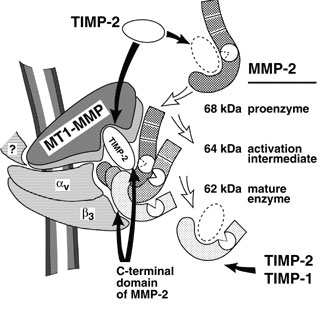
Degradation of the extracellular matrix and tissue remodeling play critical roles in tumor progression, particularly in invasion, metastasis, and neovascularization. Matrix metalloproteinases (MMPs) are essential for matrix proteolysis. Understanding the roles of MMPs in tumor growth and angiogenesis is of paramount importance to novel strategies for treatment of malignant tumors. Our primary goal is to characterize mechanisms of MMP activation involved in spatial and temporal control of focal proteolysis and to increase knowledge of cell-mediated matrix remodeling. Recently, we showed that heteromolecular assemblies involving MMPs and integrins that can simultaneously activate and dock MMPs are present on the surfaces of tumor cells. Apparently, these mechanisms of MMP activation are instrumental in clustering activated proteinases and integrins at the protrusions at the invasive front of migrating cells and at the invasive front of tumors (see Figure).
Interactions between MMPs and integrins, critical to spatial and temporal control of proteolysis, most likely are common for many cell types. To test our hypotheses and to outline novel approaches to control the activity of MMPs, we are examining mechanisms involved in activation, docking, and cross talk of MMPs and integrins in vivo and in vitro. We expect to experimentally show at the cell and protein sequence levels how the temporal and spatial regulation of matrix degradation is linked to dynamic changes of cell shape and cytoskeleton and to identify molecular mechanisms of cell motility. The immediate results of these studies will be improved diagnosis and treatment of malignant neoplasms.
Schematic depiction of the MMP-integrin molecular assembly. Inhibitory effects of tissue inhibitors of metalloproteinases (TIMPs) and the C-terminal domain of MMP-2 are shown by black arrows. The conversions of MMP-2 are shown by open arrows. The question mark stands for a cytoskeletal protein involved in a complex with membrane type-1 matrix metalloproteinase (MT1-MMP). This assembly is critical for docking, activation, and cross talk of MMPs and integrins and is intimately involved in regulating focalized matrix degradation and cell locomotion. MT1-MMP is in immediate proximity to integrin alpha vbeta 3. Both MT1-MMP and the integrin associate with the cytoskeleton. TIMP-2 links MT1-MMP (“a receptor”) and the secretory MMP-2 proenzyme. The second molecule of MT1-MMP (“an activator” that is free of TIMP-2) activates integrin alpha vbeta 3 by limited proteolysis of the beta 3 integrin subunit. The activator initiates the activation of pro-MMP-2 by cleaving the N-terminal part of the 68-kDa MMP-2 latent zymogen. The 64-kDa activation intermediate of MMP-2 efficiently associates with activated integrin alpha vbeta 3 via the C-terminal domain of the enzyme. The 64-kDa to 62-kDa autocatalytic maturation occurs if MMP-2 is complexed with the integrin. The mature MMP-2 enzyme transiently associated with the integrin at the surface of tumor cells accelerates the directional invasion of the cells. Additionally, if transiently complexed with the integrin at the surface of host stromal cells, the enzyme facilitates tumor neovascularization. TIMPs, including TIMP-2 and TIMP-1, efficiently inhibit the activity of soluble MMP-2. By providing links between integrins, MMPs, and TIMPs and, more generally, between cell shape and focal proteolysis, this model represents basic mechanisms of pro-MMP-2 activation and illustrates a coordinated interplay of inhibitors, proteinases, and integrin adhesion receptors at cell surfaces.
Arnold C. Satterthwait earned his PhD In Biochemistry with William Jencks from Brandeis University in 1973. He carried out postdoctoral research in Chemistry at Harvard University with Frank Westheimer, Imperial College with Alan Fersht and MIT with the Nobel laureate Gobind Khorana. In 1984, he joined The Scripps Research Institute in La Jolla, CA as an Assistant Professor. He moved to Sanford Burnham Prebys in 1998.
Related Disease
Anthrax, Breast Cancer, Cancer, HIV/AIDS, Prostate Cancer
The development of diagnostic reagents, drugs and vaccines is the visible outcome of a long process that spans the researcher’s laboratory and doctor’s office. The translation of disease discoveries into early detection, treatment, and prevention both tests and shapes our understanding of disease. Traditionally, drug companies have screened large collections of compounds against diseases to identify drugs. The Satterthwait lab seeks to take advantage of the explosion of new discoveries at the molecular level. We have developed synthetic methods that allow us to independently make and manipulate the critical three-dimensional regions of proteins that are being implicated in many diseases. These mini proteins are being used to assess new theories of disease at the molecular level to identify targets for various uses. We are currently using mini proteins to identify new antibodies (HIV-1), cancer drugs (prostate, breast and lung), and vaccines (anthrax).
Arnold Satterthwait’s Research Report
Peptide engineering relies on synthetic procedures to fold peptides into bioactive structures. It seeks to bridge a gap between chemistry and molecular biology by reducing the active sites of proteins to smaller molecules. Although synthetic peptides show occasional activity they are, unlike proteins, disordered and because of this often inactive. By refolding peptides into three-dimensional structures, they become active, opening up new avenues for studies on protein structure and function as well as providing leads for drugs and vaccines.
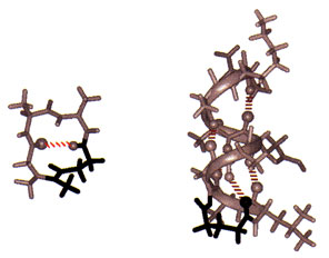
Solid-phase synthesis of a peptide constrained with a hydrazone covalent hydrogen bond mimic. The NMR structure of Sar-Ala-Ala-Gly (left) stabilized as a Type I turn (10% of protein structure) with an amidinium link (in black). The NMR structure of acetyl-Gly-Leu- Ala- Gly-Ala-Glu-Ala-Ala-Lys-Ala-amide (right) stabilized as an a-helix at its N-terminus with a hydrazone link (in black).
To fold peptides, we developed covalent hydrogen bond mimics. On average, greater than 60% of the amino acids in globular proteins engage in main-chain to main-chain hydrogen bonding (NH –> O=CRNH). In addition, protein substructures are defined by distinct hydrogen bonding patterns. Because hydrogen bonds are weak bonds, insufficient for stabilizing peptide structure, we replace them at structure defining positions in peptides with amidinium (N-C(R) = NH-CH2CH2) and hydrazone (N-N=CH-CH2CH2) covalent links. To simplify these transformations, we developed machine-assisted, multiple-peptide-synthesis procedures for inserting the hydrazone link into peptides which we link to automated multiple purifications. While these procedures, like peptide synthesis, remain labor intensive and often problematic, the critical problems have been breached.
Conformational analysis is as much a part of peptide engineering as synthesis because any claim to structure requires rigorous proof and further advances rely on understanding the relation between structure and chemistry. We have made considerable use of 2D NMR spectroscopy for structural analysis and calculations and with the help of collaborators, X-ray crystallography. These studies show unequivocally that covalent hydrogen bond mimics can stabilize peptides as b-turns, the a-helix, and even complex loops, which together make up the majority of protein substructures found on the surfaces of globular proteins.
Because protein substructure mimetics are now accessible, we have been examining the relationship between structure and activity by comparing the activities of peptides with substructure mimetics. From the several examples we have studied in detail, it is clear that remarkable gains in activity can be achieved.
Erkki Ruoslahti earned his MD and PhD from the University of Helsinki in Finland in 1967. After postdoctoral training at the California Institute of Technology, he held various academic appointments with the University of Helsinki and the University of Turku in Finland and City of Hope National Medical Center in Duarte, California. He joined Sanford Burnham Prebys in 1979 and served as its President from 1989-2002. He was a Distinguished Professor at University of California Santa Barbara in Biological Sciences 2005-2015. His honors include elected membership to the U.S. National Academy of Sciences, National Academy of Medicine, American Academy of Arts and Sciences, and the European Molecular Biology Organization, the Japan Prize, Gairdner Foundation International Award, G.H.A. Clowes Award, Robert J. and Claire Pasarow Foundation Award, and Jacobaeus International Prize. He was a Nobel Fellow at the Karolinska Institute in Stockholm in 1995, and is an Honorary Doctor of Medicine from the University of Lund, as well as a Knight and Commander of the Orders of the White Rose the the Lion of Finland. In 2022, Dr. Ruoslahti was announced as one of three winners of the Albert Lasker Basic Medical Research Award.
Education
1966: MD, University of Helsinki in Finland 1967: PhD, University of Helsinki in Finland
Awards and Honors
2022: Albert Lasker Award for Basic Medical Research Commander of the Order of the Lion of Finland Knight of the Order of the White Rose of Finland 2012: Thomson Reuters Citation Laureate 2005: Japan Prize in Cell Biology 2003: Jubilee Lecturer, Biochemical Society 1998: Jacobaeus International Prize 1997: Gairdner Foundation International Award 1995: Nobel Fellow at the Karolinska Institutet in Stockholm 1991: Honorary doctorate in medicine from Lund University, Sweden 1990: American Association for Cancer Research – G.H.A. Clowes Memorial Award
Member
National Academy of Sciences National Academy of Medicine American Academy of Arts and Sciences European Molecular Biology Organization
Related Disease
Alzheimer’s Disease, Atherosclerosis, Brain Cancer, Breast Cancer, Cancer, Prostate Cancer
The Ruoslahti laboratory studies peptides that home to specific targets in the body, such as tumors, atherosclerotic plaques and injured tissues. These peptides, which usually bind to receptors in the vessels of the target tissue, can be used to selectively deliver diagnostic probes and drugs to the target. The latest development is the discovery of homing peptides with tumor-penetrating properties. The CendR tissue penetration pathway is a new endocytosis/trans-tissue transport pathway (Pang et al., Nat Comm. 2014). The current focus is on enhancing the effects of coupled and co-injected drugs with the tumor-homing peptides, particularly in mouse models of breast cancer and glioblastoma. This laboratory also studies the receptors for the peptides and the mechanism of their tumor penetration activity.
Erkki Ruoslahti’s Research Report
Dr. Ruoslahti’s main scientific contributions are in the field of cell adhesion. He was one of the discoverers of fibronectin. His laboratory subsequently discovered the RGD cell attachment sequence in fibronectin and isolated RGD-directed cellular receptors, now known as integrins. The RGD discovery has led to the development of drugs for diseases ranging from vascular thrombosis to cancer.
Dr. Ruoslahti current studies deal with peptides that specifically target a diseased tissue, particularly its blood vessels. The peptides can be used to deliver drugs and nanoparticles to sites of disease, such as a tumor. The molecules targeted by such disease-specific peptides are of interest regarding their possible role in the disease and potential targets for drug development.
Vascular Zip Codes
The Ruoslahti laboratory screens large collections (“libraries”) of random peptides to identify those that bind to specific targets in tissues. The peptides in the library are displayed on the surface of phage (a virus that infects bacteria), and the screening is done in vivo. When the library is injected into the circulation of a mouse, phage particles that display peptides capable of binding to a selected target tissue, such as a tumor, accumulate at the target where they can be collected and their peptide identified. The process primarily probes the vasculature of the target tissue, unless the vasculature is very leaky. The method has revealed a wealth of specific features, or “vascular zip codes”, in the vessels of individual tissues and tumors. Peptides that specifically home to tumors because they recognize angiogenesis-associated or tumor-type specific markers in tumor blood vessels and can even distinguish the vessels of pre-malignant lesions from those of fully malignant tumors. Homing peptides have also revealed a zip code system of molecular changes in tumor lymphatics.
Synthetic homing peptides have been used to target drugs, biologicals, and nanoparticles into tumors. The targeting can increase the efficacy of a drug while reducing its side effects. Even a non-specifically toxic compound can be converted into a compound that selectively affects the targeted tissue. The peptides make it possible to identify the target molecules (receptors) for the peptides. The receptors of tumor-homing peptides often play a functionally important role in tumor vasculature, and because of this are candidates for drug development.
Tumor-penetrating Peptides
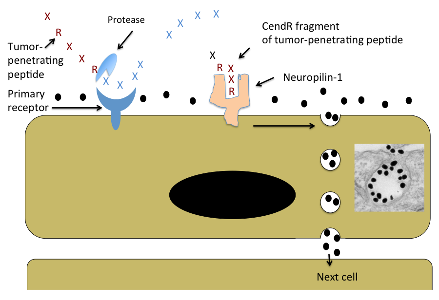
A few years ago the laboratory discovered peptides that not only home to tumor vessels, but are transported through the vascular wall and deep into tumor tissue. The key feature of these peptides is a R/KXXR/K sequence motif, named C-end Rule (CendR) motif or element. In tumor-penetrating peptides, the CendR element is cryptic. These peptides penetrate into tumor tissue in a 3-step process: (i) The peptide binds to a primary receptor on tumor endothelium. In iRGD, the RGD motif recognizes the avb3/avb5 integrins; the primary receptor for the LyP-1 family of peptides is cell surface p32/gC1qR. (ii) The peptide is then cleaved by a protease to expose the CendR element at the C-terminus of the peptide; and, (iii) the CendR element mediates binding to neuropilin-1 (NRP-1), to induce vascular and tissue penetration. The CendR transport pathway triggered through NRP-1 resembles macropinocytosis, but differs from it in being receptor-mediated. Importantly, the responsiveness of the pathway to triggering through NRP-1 is regulated by the nutrient status of cells and tissues. Its physiological function is likely to be to transport nutrients into tissues that lack them. Our ability to trigger the pathway specifically in tumors makes it useful in delivering drugs into tumors.
Targeting the Brain
The Ruoslahti laboratory has recently also applied phage screening to the identification of peptides that target brain diseases. So far, a peptide that specifically recognizes sites of brain injury, and a panel of peptides that are specific for Alzheimer’s brain have been obtained. This topic will be an expanding focus of the laboratory in the near future.
Nanomedicine
A major focus is to use homing peptides as targeting elements to deliver nanoparticles into tumors and other sites of disease. Nanoparticles are considered a promising new approach in medicine because they can be designed to perform more functions than a simple drug. The vasculature is an excellent target for nanoparticles because tumor vessels are readily available for circulating particles. In collaboration with chemistry and bioengineering laboratories, multifunctional nanoparticles for tumor targeting have been constructed. These particles can be directed into tumors in a highly selective manner as demonstrated by histology, non-invasive imaging, and tumor treatment results. The laboratory has constructed nanoparticles with the ability to amplify their own homing to tumors, and are currently working on nanoparticles coated with tumor-penetrating peptides. More recent work has dealt with nanoparticles that target brain diseases or atherosclerotic plaques. The general goal is to engineer nanoparticles with multiple functions. In addition to the specific targeting, such functions include avoidance of the reticuloendothelial system, self-amplification of the targeting, exit from vessels into tissue, ability to send signals for imaging, and controlled drug delivery.
Schematic Representation of the CendR Trans-tissue Transport Pathway
Note that CendR effect enhances the tissue penetration of molecules (depicted here as a black dots) that are co-administered with the peptide, as well as of cargo coupled to the peptide. The inset shows an electron microscopic image of a CendR endocytic vesicle that is budding from the cell surface into the cytoplasm and contains CendR peptide-coated gold nanoparticles (dark dots) See Ruoslahti, Adv. Drug Deliv. Rev. 2016.
Minoru Fukuda earned his PhD in biochemistry from the University of Tokyo in 1973 and did his postdoctoral training at the Yale University School of Medicine. Following a period with joint appointments at University of Washington and Fred Hutchinson Cancer Research Center in Seattle, he was recruited to Sanford-Burnham Medical Research Institute in 1982 as Director of the Glycobiology Program. Dr. Fukuda directs the program project grant, which consolidates the research efforts of the members of the Glycobiology Program.
Dr. Fukuda is a recipient of a Merit Award from the National Cancer Institute and the 1997 recipient of the Karl Meyer Award from the Society of Glycobiology. He served as an Executive Editor for Biochimica et Biophysica Acta, as an Associate Editor for Cancer Research and Editorial Member for Journal of Biological Chemistry. He also has edited 11 books including three books from Oxford University Press and three volumes of Methods in Enzymology and holds an Adjunct Professor appointment at the University of California, San Diego.
Education
1973: PhD, University of Tokyo, Biochemistry 1970: MS, University of Tokyo, Biochemistry 1968: BS, University of Tokyo, Biochemistry
Related Disease
Brain Cancer, Colorectal Cancer, Gastric Cancer, Helicobacter pylori, Prostate Cancer
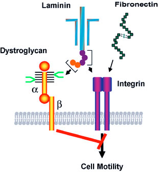
The cell surface is heavily coated with carbohydrates. The structure of those cell surface carbohydrates displays a dramatic change during development, and mature cells express cell surface carbohydrates specific to different organs and tissues. Cell surface carbohydrates thus serve as a zip code for different organs and tissues. Sialyl Lewis X represents such an oligosaccharide. After discovery of sialyl Lewis X in neutrophils by Dr. Minoru Fukuda, his laboratory demonstrated that sulfated form of sialyl Lewis X is essential for lymphocyte homing and recruitment of natural killer cells in preventing tumor metastasis to the peripheral lymph node. With colleagues from Japan, Dr. Fukuda discovered that certain carbohydrates function as antibiotics against Helicobacter pylori infection, which is a leading cause for peptic ulcer and gastric carcinoma. Most recent studies in Dr. Fukuda’s laboratory revealed that decrease of the laminin-binding glycans on α-dystroglycan in carcinoma cells leads to tumor cell migration, invasion, and metastasis. The restoration of the unique glycans by the expression of distinct β3-N-acetylglucosaminyltransferase renders these cells act like normal cells. The results indicate that certain carbohydrates on normal cells and enzymes that synthesize those glycans, such as β3-N-acetylglucosaminyltransferase, function as tumor suppressors These findings will be useful in developing carbohydrate-based therapy for the treatment of inflammation and tumor metastasis.
Minoru Fukuda’s Research Report
Cell Surface Carbohydrates as Tumor Suppressor
Many studies have focused on carbohydrates that increase in cancer cells, but only a few have looked at carbohydrates that appear in normal cells but decrease or disappear in cancer cells. A specific mucin-type O-glycans (core 3 O-glycans) is one of such glycans, and we found core 3 O-glycans suppress tumor formation and metastasis. When core 3 O-glycans were forced to express on human prostate cancer cell lines, those prostate cancer cells produced much smaller tumors and almost no metastasis. By contrast, the parent cancer cells, which did not express core 3 O-glycans, produced robust primary and metastatic tumors. We showed that the expression of core 3 O-glycans decreases a formation of α2β1-integrin complex, receptors that mediate cell adhesion, diminishing cancer cell migration.
We also revealed tumor suppressor function in the unique laminin-binding glycans on dystrophin complex, — carbohydrates located on α-dystroglycan, which is also associated with cell adhesion. We discovered that the unique glycans play a critical role in epithelial-basement membrane interaction in normal cells, and the decrease or loss of the glycans, due to downregulation of β3-N-acetylglucosaminyltransferase, leads to increased malignancy by invasive carcinoma cells. Restoration of the laminin-binding glycans by forced expression of β3-N-acetylglucosaminyltransferase, on the other hand, results in reduced cell migration, thus dramatic decrease in tumor formation and metastasis. We demonstrated that interaction of laminin with the unique glycans on α-dystroglycan counteracts the cell migration signals that are mediated by integrin binding to its ligands, thereby decreasing tumor formation and metastasis. These findings also suggest that the laminin-binding glycans can be an excellent marker for epithelial-mesenchymal transitions.
These results indicate that certain carbohydrates on normal cells and enzymes that synthesize those glycans, such as β3-N-acetylglucosaminyltransferase, function as tumor suppressors. Upregulation of those key enzymes may become a novel way to treat cancer.
Neural Cell-specific Glycans in Development and Cancer
Polysialic acid and HNK-1 glycan represent carbohydrates enriched in neural cells. Polysialic acid is mainly attached to NCAM in embryos, while the majority of NCAM in adults lack this carbohydrate. To understand the roles of these glycans in neural development, we have cloned cDNAs encoding human polysialyltransferase, PST, and HNK-1 sulfotransferase, HNK-1ST, that are responsible for the synthesis of polysialic acid and HNK-1 glycan, respectively. By using these cloned cDNAs, we demonstrated that polysialic acid facilitates the invasion of glioma, the most common form of adult brain tumor. Our studies also showed that mutant mice with deficient STX, another polysialyltransferase, exhibit reduced behavioral response to fear conditioning, apparently due to anomalies in mossy fibers of the hippocampus. Our studies also demonstrated that neural development is significantly impaired in mutant mice that entirely lack polysialic acid due to inactivation of two polysialyltransferases. We found that this defect is caused by impairment of neural cell migration.
Mucin-type O-glycans in Immune Cell Interactions
Previously, we found that an increase of core 2-branched oligosaccharides is associated with leukemia and immunodeficiency, such as in Wiskott-Aldrich syndrome and AIDS. To determine the roles of core 2-branched O-glycans in immune cell interactions, the enzyme (C2GnT-1) responsible for the core 2-branched oligosaccharide was knocked-out by gene targeting. Compared to wild-type mice, leukocytes from the gene knockout mice exhibited a reduced binding to L-, E- and P-selectin in this order. In contrast, homing of lymphocytes was moderately reduced. Lymphocyte homing is mediated by binding of L-selectin on lymphocytes to sulfated L-selectin oligosaccharide ligands, 6-sulfo sialyl Lewis X in high endothelial venules (HEV) of secondary lymphoid organs. By analyzing remaining L-selectin ligands in C2GnT-1 knockout mice, we discovered novel L-selectin ligands that are based on extended core 1 oligosaccharides. The core portion of this novel L-selectin ligand is also an epitope for MECA-79 antibody that inhibits lymphocyte homing in vivo. Moreover, crossbreeding between mutant mice with deficient L-selectin ligand sulfotransferase and another sulfotransferase led to our findings that these two enzymes in cooperation synthesize L-selectin ligands. These mutant mice lack 6-sulfate group in L-selectin ligands that results in impaired inflammatory response. More recently, mice deficient in L-selectin ligands on mucin-type O-glycans were generated. The studies on the mutant mice revealed novel functions of N-glycan-based L-selectin ligand, which supports both lymphocyte homing and inflammatory response. This finding brought a new paradigm in selectin-carbohydrate interaction.
Carbohydrate-dependent Adhesion in Tumor Metastasis
Previously we found that the amount of core 2 O-glycans is significantly increased in colon and lung carcinomas and the increase of core 2 O-glycans is highly correlated to vessel invasion and lymph node metastasis. More recently, we discovered that forced expression of core 2 O-glycans by transfecting C2GnT-1 in prostate cancer cell lines resulted in increased tumor formation.
In parallel, we discovered that forced expression of selectin ligands, sialyl Lewis X on B16 melanoma cells leads to increased lung tumor formation. We also showed that tumor formation in lymph node is suppressed by natural killer (NK) cells, which are recruited by L-selectin mediated homing of NK cells to lymph nodes.
Roles of Carbohydrates in Helicobacter pylori-mediated Inflammation and Cancer
Helicobacter pylori is a leading cause of peptic ulcer and gastric cancer. Previously it was shown by others that H. pylori adhere to gastric mucosa in a carbohydrate-dependent manner. The infection of H. pylori leads to chronic inflammation, which apparently leads to peptic ulcer and gastric cancer.
Our recent studies showed that H. pylori-induced inflammation is associated with the formation of peripheral lymph node addressin (PNAd) characterized by binding to MECA-79 antibody and L-selectin. The number of HEV-like vessels expressing PNAd increases as H. pylori-induced inflammation progresses. Moreover, PNAd disappears once H. pyloriis eradicated by antibiotic treatment. These findings indicate that H. pylori-induced inflammation is facilitated by de novo formation of PNAd thereby recruiting lymphocytes. It may be possible to attenuate or prevent the formation of peptic ulcers or gastric cancer by inhibiting L-selectin ligand synthesis, for example by inhibiting the sulfotransferases.
Binding of laminin to the specific carbohydrate (shown in bright green) on α-dystroglycan counteracts the migration signals initiated by integrin binding to extracellular matrix proteins such as laminin and fibronectin. The synthesis of this specific carbohydrate requires a unique β3-N-cetylglucosaminyltransferase, and the downregulation of the glycosyltransferase in carcinoma cells leads to increased cell migration, thereby increased tumor formation and metastasis. Thus the specific carbohydrate structure at cell surface functions as a tumor suppressor, which is controlled by the unique β3-N-acetylglucosaminyltransferase.
While over half of the world’s population is infected with H. pylori, only a fraction of those individuals progress to peptic ulcer and gastric cancer. In relation to these observations, α1,4-N-acteylglucosaminyl capping structure (α4GlcNAc) is present in deeper portions of the gastric mucosa, where H. pylori rarely colonizes. We discovered that α4GlcNAc capping structure functions as an antibiotic against H. pylori infection by inhibition of the synthesis of α-glucosyl cholesterol, a major component of the H. pyloricell wall. This unprecedented discovery should be useful in developing drugs to inhibit H. pylori colonization, through inhibition of cholesterol α-glucosyltransferase. Such drugs lead to a novel treatment for prevention and potential treatment of peptic ulcer and gastric carcinoma.
Michiko N. Fukuda earned her PhD in biochemistry at the University of Tokyo in 1980. She did postdoctoral work at Fred Hutchinson Cancer Research Center in Seattle prior to her recruitment to Sanford-Burnham Medical Research Institute in 1982.
Education
1980: PhD, University of Tokyo, Biochemistry 1970: MS, University of Tokyo, Biochemistry 1968: BS, Tokyo University of Education, Botany
Related Disease
Breast Cancer, Cancer, Congenital Disorders of Glycosylation, Endometriosis, Glycosylation-Related Disorders, Inherited Disorders, Ovarian Cancer, Prostate Cancer, Testicular Cancer
Michiko Fukuda’s Research Report
Identification of Peptide that Delivers Drugs to Tumors
Chemotherapy effectiveness is often limited by drug toxicity in healthy tissues, although methods that spare normal cells by delivering drugs specifically to tumors may help to overcome this constraint. We have identified a promising tumor-targeted drug delivery vehicle known as the IF7 peptide. Using in vitro assays, we found that the IF7 peptide bound to the protein annexin 1 (Anxa1), which is known from previous studies by others to be enriched on the surface of tumor vasculature in several tumor types. When we injected a fluorescently labeled IF7 peptide into mice with tumors, fluorescent signals appeared in the tumors within one minute of injection. By contrast the tumors showed no fluorescence when mice were pre-injected with anti-Anxa1 antibodies that inhibits IF7-Anxa1 binding, suggesting that the peptide targets tumor by homing to Anxa1.
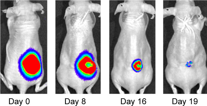
Effect of IF7-conjugated anti-cancer drug SN-38 on colon cancer mouse model. Activa cancer cells in a large tumor produce chemiluminescence which is captured by imaging. (Hatakeyama et al, 2011).
When IF7 peptide was conjugated with potent anti-cancer drug SN-38 and IF7-SN38 conjugate was injected intravenously to mice with tumors, IF7 could deliver SN-38 to tumors. We found that daily injections of an IF7-SN38 conjugate reduced a large tumor in the mouse without apparent side effects, whereas non-homing peptide-SN38 conjugate or with SN-38 alone did not reduce the tumors (Hatakeyama et al, 2011). The findings suggest that IF7 peptide may represent a clinically relevant vehicle for anti-cancer drugs.
Role of Trophinin in Human Embryo Implantation and Cancer
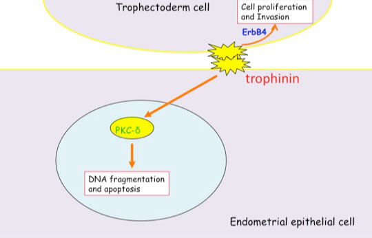
Invasion of the trophoblast into the endometrium, an essential element of embryo implantation, resembles invasion of malignant tumors. At the initial phase of implantation, the trophoblast and the uterine epithelium establish their first contact via their respective apical cell membranes. We have identified new molecules, trophinin, tastin, and bystin that mediate cell adhesion between trophoblastic cells and endometrial epithelial cells at the respective apical cell membranes. Trophinin is an intrinsic membrane protein, and tastin and bystin are cytoplasmic proteins. All of these molecules are strongly expressed in cells involved in embryo implantation in humans. However, trophinin is not expressed in human endometrial epithelia throughout the hormonal cycle, except only those cells located close to the implanting blastocyst. Trophinin expression by endometrial epithelia is induced by human chorionic gonadotrophin (hCG) derived from the implanting embryo (Sugihara et al, 2008). While embryos invade maternal cells (Sugihara et al, 2007), maternal tissue accepts embryos. We asked what happens in the maternal epithelia when trophinin-mediated adhesion takes place, and found that trophinin-mediated cell adhesion triggers an apoptotic signal in maternal epithelial cells (Tamura et al, 2011).
Distinct signal transduction of trophinin-mediated cell adhesion in trophectoderm cell and endometrial epithelial cells. In trophoblastic cells, ErbB4 (receptor tyrosine kinase) is arrested by bystin/trophinin complex. When trophinin-mediated cell adhesion takes palce, ErbB4 is released from bystin. This allows ErbB4 to be activated by phosphorylation. In endometrial epithelial cells, trophinin-mediated cell adhesion releases PKCd from trophinin. PKCd was then translocated to the nucleus, where it activates caspase 3 for apoptosis (Tamura et al., 2011).
References Cited
Hatakeyama S, Sugihara K, Shibata TK, Nakayama J, Akama TO, Tamura N, Wong SM, Bobkov AA, Takano Y, Ohyama C, Fukuda M, Fukuda MN (2011) Targeted drug delivery to tumor vasculature by a carbohydrate mimetic peptide. Proc Natl Acad Sci U S A 108: 19587-19592
Sugihara K, Kabir-Salmani M, Byrne J, Wolf DP, Lessey B, Iwashita M, Aoki D, Nakayama J, Fukuda MN (2008) Induction of trophinin in human endometrial surface epithelia by CGbeta and IL-1beta. FEBS Lett 582: 197-202
Sugihara K, Sugiyama D, Byrne J, Wolf DP, Lowitz KP, Kobayashi Y, Kabir-Salmani M, Nadano D, Aoki D, Nozawa S, Nakayama J, Mustelin T, Ruoslahti E, Yamaguchi N, Fukuda MN (2007) Trophoblast cell activation by trophinin ligation is implicated in human embryo implantation. Proc Natl Acad Sci U S A 104: 3799-3804
Tamura N, Sugihara K, Akama TO, Fukuda MN (2011) Trophinin-mediated cell adhesion induces apoptosis of human endometrial epithelial cells through PKC-delta. Cell Cycle 10 : 135-143
Kristiina Vuori earned her MD and PhD at University of Oulu, Finland. After completion of internship and residency, she received postdoctoral training at the Institute and was appointed to faculty in 1996. Dr. Vuori was selected as a PEW Scholar in the Biomedical Sciences in 1997. She has been co-Director of the Conrad Prebys Center for Chemical Genomics, housed at Sanford Burnham Prebys, since its inception in 2005. She was appointed Deputy Director of the Institute’s NCI-Designated Cancer Center in 2003, and Director of the Cancer Center in 2006. In 2008, she was appointed Executive Vice President for Scientific Affairs at Sanford Burnham Prebys. She was President of the Institute from 2010 to 2022.
Related Disease
Brain Cancer, Breast Cancer, Cancer, Leukemia/Lymphoma, Lung Cancer, Ovarian Cancer, Prostate Cancer
Dr. Vuori’s research is aimed at unraveling the cell mechanisms of the most life-threatening aspect of cancer, which is cancer metastasis. Metastasis is responsible for nearly all deaths in cancer patients, and understanding of the mechanisms that turn a cancer from a locally growing tumor into highly metastatic cancer cells will provide clues how to prevent this step in cancer progression. All cells in our body stick to one another and to the packaging material, or extracellular matrix, around them. This adhesion is essential for cell survival; if cells become detached from their microenvironment, they will die through a process known as apoptosis. This phenomenon, which is called adhesion dependency of survival, is one of the safeguards that maintain the integrity and normal function of tissues, and prevent cells from becoming cancerous. Normal cells cannot detach from their tissue and establish themselves somewhere else, because they will die on the way. Yet cancer cells somehow get around this requirement; they trespass aggressively into other tissues and metastasize to distant sites in the body without dying. Dr. Vuori’s work is aimed at identifying the molecular mechanisms that in normal cells makes them adhesion-dependent; false action of the very same mechanisms is likely to be the key step in allowing cancer cells to metastasize.
Nicholas Cosford, PhD has served on the Sanford Burnham Prebys Board of Trustees since 2023. He is the first faculty member to do so.
Cosford joined the Sanford Burnham Prebys faculty in 2008 as an associate professor. In 2013, he became a full professor. His lab investigates the interactions of small molecule compounds with therapeutically important proteins and cellular signaling pathways. With a specific focus on the discovery and optimization of compounds that might treat cancer, central nervous system diseases and infectious diseases.
Prior to joining Sanford Burnham Prebys in 2005, Cosford worked in both the biotechnology and pharmaceutical industries. At Sibia Neurosciences and at Merck Research Laboratories, he directed multidisciplinary research teams focused on small-molecule hit-to-lead optimization and was responsible for moving several lead compounds through to the clinical phase, including a nicotinic agonist (Altinicline) from research to Phase II clinical trials for treating Parkinson’s disease.
He is an author of more than 90 peer-reviewed, published scientific papers, and has been issued more than 40 issued patents, with an additional 40 patent applications pending.
Cosford has a Bachelor of Science degree in chemistry from the University of Bath in England and Doctor of Philosophy degree in organic chemistry from Emory University in Atlanta, GA.
Related Disease
Alzheimer’s Disease, Amyotrophic Lateral Sclerosis (Lou Gehrig’s Disease), Bone Mineralization Disorders, Breast Cancer, Cancer, Neurodegenerative and Neuromuscular Diseases, Neurological and Psychiatric Disorders, Ovarian Cancer, Pancreatic Cancer, Prostate Cancer
We are interested in investigating the interactions of small molecule compounds with therapeutically important proteins and cellular signaling pathways. One aspect of our research emphasizes the use of medicinal chemistry and chemical biology approaches to probe intracellular pathways that regulate cell survival and cell growth. Another area of active research is the development of synthetic chemistry methodology using microfluidic technology for the rapid synthesis of biologically active small molecules. Therapeutically, we are primarily focused on the discovery and optimization of compounds that have the potential to treat cancer, CNS diseases and infectious diseases.
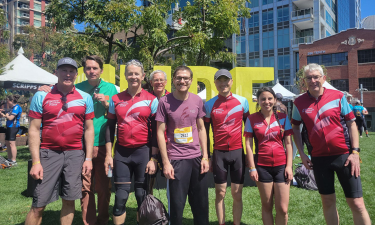
 Mar 17, 2026
 Oct 6, 2025