cancer Archives - Page 7 of 11 - Sanford Burnham Prebys
Institute News

Fleet Science Center cancer series kicks off with lung cancer discussion

AuthorMonica May
Date

August 22, 2019

New lung cancer treatments are making a difference for patients. Pill-based, personalized medicines and immunotherapies are allowing some individuals to survive for years instead of months. Still, lung cancer remains the deadliest cancer—killing more people each year than breast, prostate and colorectal cancer combined. 

To help the public better understand the newly available medicines—and the research advances on the horizon—our Institute teamed up with the Fleet Science Center to host a panel discussion on Sunday, August 18. 

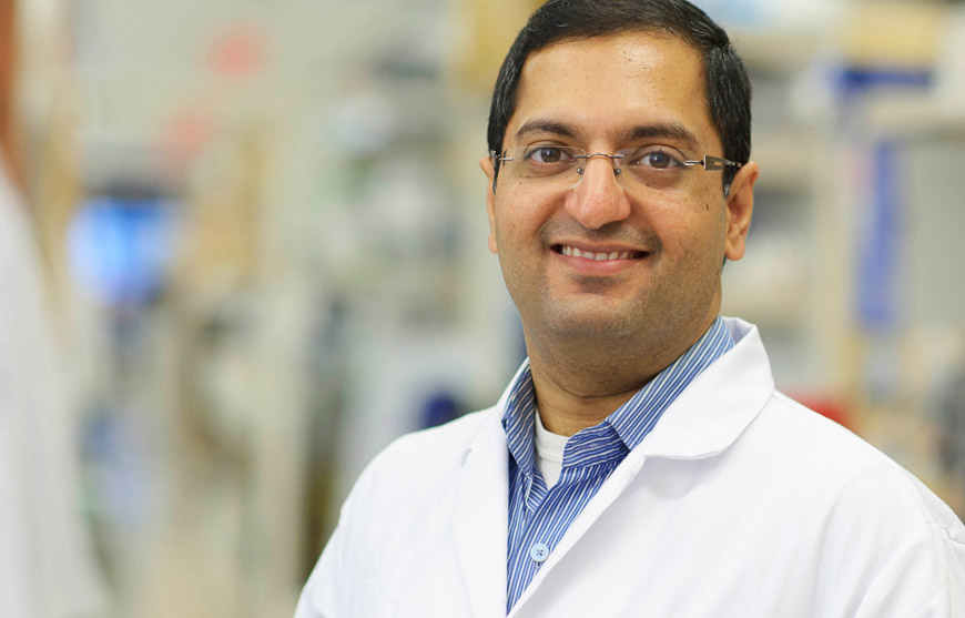
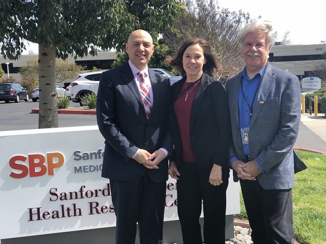
“Many people who live in San Diego aren’t aware of the incredible research advances taking place in their backyard, especially in cancer,” said speaker Garth Powis, D. Phil., professor and director of Sanford Burnham Prebys’ National Cancer Institute (NCI)-designated Cancer Center (on left). “We hope this discussion and future events will help more people understand cancer research and the breakthroughs that might come from their own community.”

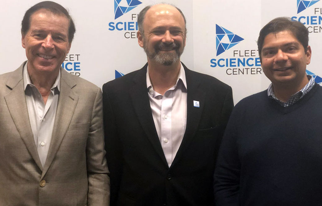
Powis was joined by Hatim Husain, MD, a clinician at UC San Diego (on right); and Steven Snyder, PhD, president and CEO of the Fleet Science Center (center), who moderated the discussion. The speakers described how targeted treatments, which are only prescribed if a patient’s tumor has a specific mutation; and immunotherapies, which harness a patient’s immune system to melt the tumor, are extending survival for lung cancer patients. Husain expressed excitement surrounding new blood tests to detect lung cancer—which he hopes will be more commonplace in five to ten years. The speakers also noted that advances made in lung cancer have the potential to extend to other tumor types. 

“Many of the mutations that drive lung cancers are found in other tumors,” said Husain. “Targeted treatments that shrink lung tumors are being studied broadly in patients with a variety of cancers.” 

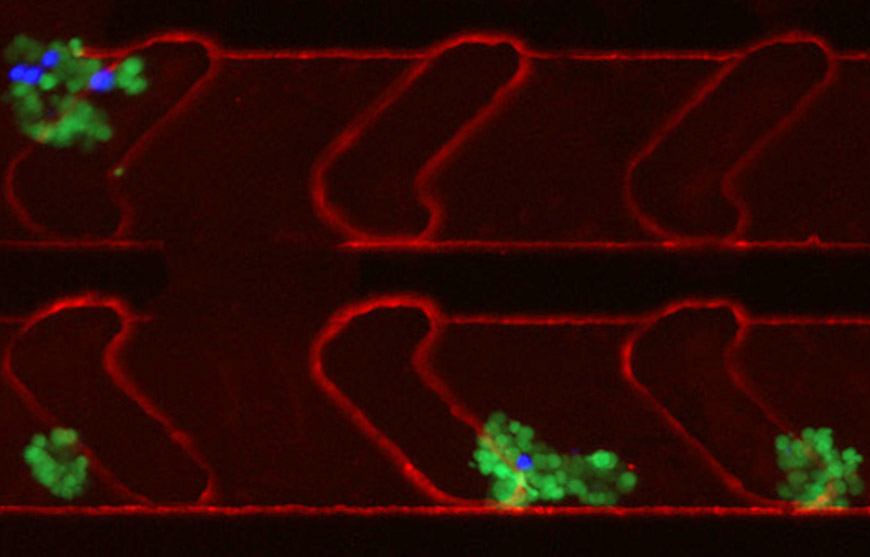
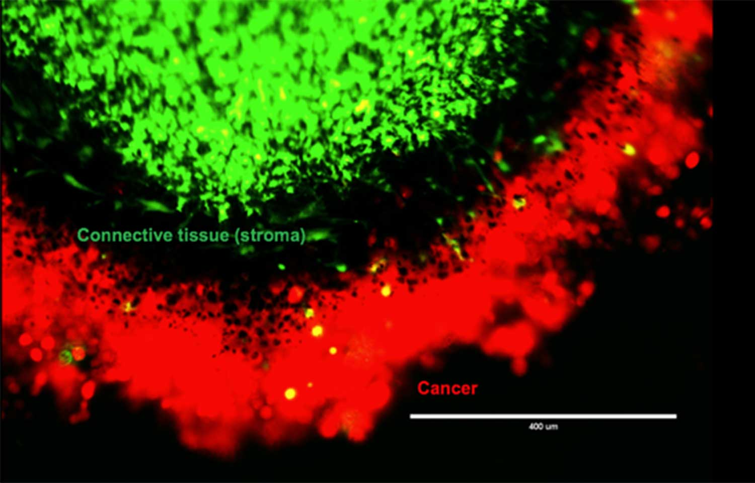
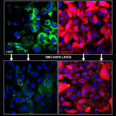
Powis and Husain also touched on their own collaboration to learn how lung cancer becomes resistant to treatment. Fluid buildup in the pleural space, the area between the lungs and chest wall, is often removed during routine checkups to help patients breathe. Working with Husain, Powis’ team is tracking the cellular and molecular makeup of this pleural fluid over the course of the disease. By regularly analyzing this fluid, they hope to gain insights into how lung cancer becomes treatment resistant and how it can be stopped.

“Scientists are getting close to mapping all of the mutations that drive lung cancer growth,” said Powis. “One day, patients may take one pill that contains all the anti-cancer compounds they need to fight the tumor.”

Upcoming topics in the series include breast, brain, and pancreatic cancer and more. The events will take place from 7:00 p.m. to 8:30 p.m. on select Sundays in the Heikoff Giant Dome Theater at the Fleet Science Center in San Diego. Space is limited. Reserve your ticket today. 

Institute News

Solar power gone awry

AuthorZe’ev Ronai, PhD
Date

July 29, 2019

Are you enjoying the summer? Out grilling, swimming and hiking? Beware: those sunny days may come with a cost. 

When the sun’s rays touch your skin, they don’t stop there. Ultraviolet (UV) light enters your cells, and photons—tiny particles of light—landing on the proteins and DNA in your cells. With just the right amount of activation energy, proteins change their shape and function, and your DNA becomes damaged, or as we say—mutated. Under normal circumstances, cells use special proteins to repair mutated DNA, but when the repair proteins are damaged, DNA mutations become permanent.

Certain DNA segments called genes are more vulnerable to mutations than others. The BRAF gene, which normally makes a protein that controls cell growth, is mutated in more than 50% of melanomas—the most dangerous type of skin cancer. Melanoma appears when BRAF mutations crop up with other mutations in the same skin cell. For patients with these tumors, drugs that target BRAF and related proteins are often successful at slowing or stopping melanoma growth—but only for a while.

Unfortunately, patients who initially respond to such targeted therapy often relapse. Some patients relapse because their tumors generate a new mutation, making it resistant to the drug.  Overall, it may be only a small fraction of cells within the original tumor that develop resistance. So although 99.5% of the cancer cells in a tumor may have a mutated BRAF gene, the other 0.5% can harbor different mutations that either evolved during therapy or were present in the first place, but didn’t drive the initial tumor. For these patients, the bulk of BRAF mutant cancer cells are killed with targeted therapy, but another melanoma can evolve from the remaining 0.5%. This is why combination therapy, where drugs aim for multiple targets, are important.

But targeting every single mutation in a tumor may not be feasible. There will always be a fraction of cells with a different mutation that evolves, making patients vulnerable to a relapse. This is where attacking the tumor from another angle comes into play.  

Checkpoint immunotherapies—which have revolutionized the treatment of melanoma—attack tumors independent of their mutational makeup. They work by loosening the brakes of the immune system—brakes that normally prevent immune cells from attacking our own self. Tumors are very good at hiding from the immune system, but with the brakes released, tumors become exposed and are successfully attacked by the immune system, irrespective of their mutational makeup. 

But not everyone responds to immunotherapy—and we don’t yet know why. Is it the tumor? Is it the patient’s immune system? There is even evidence that the gut microbiome plays a role. Once we understand why some patients respond and or stop responding to immunotherapy, we can improve selection of patients for therapy, the effectiveness of these treatments and the possible combinations that work best. 

So where is skin cancer therapy headed? A combination of checkpoint immunotherapy with targeted therapies, as well as some new tricks we are learning, such as coaching tumor cells to be better recognized by the immune system, are moving the needle.

In my lab at Sanford Burnham Prebys we are dissecting the cell signals that drive cancer. Our studies are guided by data derived from patients’ tumors, coupled with advanced bioinformatics. We seek to understand how physiological processes are modified as cancer develops and how they can be exploited for cancer therapy. For example, we recently demonstrated a connection between the composition of the gut microbiome and the response to immunotherapy, establishing new paradigms, but raising important new questions. Can we better predict who will respond to immunotherapy? Can we enhance the response to immunotherapy by manipulating the gut microbiome? Can we make tumors that don’t initially respond start responding to immunotherapy? The bar is always raised, as one discovery opens so many new avenues to explore and advance our understanding, aspects that members of my lab are working hard on to answer.  

Yes—we are making progress. But preventing the initial sun exposure by using protective gear and sunscreens is needed now as much as ever.

Ze’ev Ronai, PhD, professor in Sanford Burnham Prebys’ Tumor Initiation and Maintenance Program, is a world-renowned cancer research expert and recipient of the Lifetime Achievement Award from the Society of Melanoma Research. The award recognizes his major and impactful contributions to melanoma research over the course of his career.

Institute News

Capturing circulating cancer cell clusters using a new microfluidic device

AuthorMonica May
Date

July 16, 2019

Nearly 90 percent of cancer deaths are a result of metastases, when tumors spread to other vital organs. Researchers are learning that cancer metastases are not due to individual cells but rather distinct clusters of cancer cells that circulate and metastasize to other organs. However, obtaining these clusters to learn more about the metastatic process has proved difficult. 

Now, in a study published in AIP Advances, researchers from Sanford Burnham Prebys, San Diego State University and TumorGen MDx™ have described a new microfluidic device that captures circulating cancer cell clusters. 

“The reason for such little research activity on cancer clusters is the overwhelming difficulty of capturing these extremely rare samples from a patient’s blood sample,” says Peter Teriete, PhD, a study author and a research assistant professor at Sanford Burnham Prebys. “But we realized that if we’re ever going to understand the complex process of cancer metastasis, we’d need to develop a tool to easily find these clusters.”

To do so, the researchers first identified the basic requirements essential to collecting useful information from isolated cancer cell clusters. It involves a sample size large enough to likely contain appreciable numbers of cancer cell clusters (about 10 milliliters of whole blood), as well as using whole blood to preserve rare circulating clusters. Whole blood, however, requires special channel-coating procedures that reduce nonspecific binding properties to prevent biofouling. And the device channel dimensions must be of a suitable size to accommodate single cells and cancer cell clusters of varying diameters.

“Our device’s channel design had to generate microfluidic flow characteristics suitable to facilitate cell capture via antibodies within the coated channels,” Teriete explains. “So we introduced microfeatures—herringbone recesses—to produce the desired functionality. We also developed a unique alginate hydrogel coating that can be readily decorated with antibodies or other biomolecules. By connecting bioengineering with materials science and basic cancer biology, we were able to develop a device and prove that it performs as desired.”

The group’s microfluidic device brings a new therapeutic strategy to the fight against cancer metastasis. Capturing viable circulating cancer stem cell clusters directly from cancer patients is a novel approach for the development of new anti-metastatic drug therapies.

“Drug development that specifically targets distant metastases has been greatly restricted due to the lack of adequate tools that can readily access the metastatic cells responsible for cancer’s dissemination,” says Teriete. “Our microfluidic device will provide cancer researchers with actual human cancer cell clusters so they can begin to understand the critical mechanisms involved with metastasis and develop highly effective drugs that ultimately can save more cancer patients’ lives.”

Story materials courtesy of the American Institute of Physics. Content has been edited for style and length. 

Institute News

Padres Pedal the Cause presents record-breaking check for nearly $3 million to fund local cancer research

AuthorMonica May
Date

January 29, 2019

Local cancer research just got a big boost. 

On Thursday, January 24, SBP president Kristiina Vuori, MD, PhD, joined leaders from Moores Cancer Center at UC San Diego Health, Salk Institute for Biological Studies and Rady Children’s Hospital–San Diego to help Padres Pedal the Cause (PPTC) reveal that this year’s event raised a record-breaking $2.9 million for local cancer research. The leaders joined executive director Anne Marbarger onstage to receive the official check. 

This year’s event—which invited participates to cycle, spin, run or walk—had more than 2,500 participants, an increase of 35 percent. Total fundraising grew by 22 percent. SBP has participated in the event since its inception; and this year our team of more than 60 scientists, staff and SBP supporters raised more than $30,000 for the cause. Since the inaugural ride six years ago, PPTC has raised more than $10 million.

Nearly 300 of the event’s participants, including Tony Gwynn Jr., Pedal founders Bill and Amy Koman, San Diego business leaders, and top donors and fundraisers, gathered at the Del Mar racetrack to witness the funding reveal and check presentation in person. 

Gwynn shared a moving story about his father’s battle with salivary cancer, a journey he still finds difficult to recount. “If he saw this progress, he would be smiling today,” he said. 

A full 100 percent of the proceeds fund collaborative research taking place at the four San Diego research institutes. Past PPTC grants have accelerated SBP’s research into cancers of the breast, skin, brain, colon, pancreas and more. This year’s grant announcement will be revealed in the spring. 

In the meantime, make sure to mark your calendars for the 2019 event, which will take place on Saturday, November 16. Registration will open in mid-April.

Interested in keeping up with SBP’s latest discoveries, upcoming events and more? Subscribe to our monthly newsletter, Discoveries. 

Institute News

SBP scientist awarded Susan G. Komen® and NIH grants to advance breast cancer research

AuthorMonica May
Date

October 25, 2018

Breast cancer remains the second most common cancer for American women. While treatment advances are being made, more research is needed. Current treatments don’t work for every woman.
 
Now, breast cancer researcher Svasti Haricharan, PhD, assistant professor at Sanford Burnham Prebys Medical Discovery Institute (SBP), has been awarded more than half a million dollars in combined grants from Susan G. Komen® and the National Institutes of Health (NIH). 

This funding will advance Haricharan’s breast cancer research—including developing a diagnostic test that could guide therapeutic options—and allow her to apply lessons from breast cancer to additional cancers. 

Susan G. Komen grant

The majority of women diagnosed with breast cancer have the estrogen-positive (ER-positive) form, meaning the tumor grows in response to estrogen. Hormone therapies (anti-estrogen drugs) that block estrogen—and thus stop the tumor from growing—are available. However, this treatment doesn’t work for 40 percent of women with ER-positive breast cancer. 

“Currently, doctors are unable to predict which ER-positive patients will respond to treatment—so an estrogen-blocking medicine is given, and a ‘wait and see’ approach is taken to see if the treatment will work,” says Haricharan. “However, if a woman doesn’t respond to treatment, during this time the tumor is instead still growing and may metastasize—when it becomes deadlier and even harder to treat. Knowing upfront if an individual will respond to treatment allows doctors to skip a treatment that won’t work and move immediately to prescribing a medicine that may be effective.” 

Haricharan’s previous work found that about one-third of women with ER-positive breast cancer who were treatment resistant had a mutation in DNA damage-repair genes—providing a potential biomarker that could predict who would respond to treatment. 

Luckily, an FDA-approved test that detects defects in DNA damage repair is currently available for colorectal cancer patients. The grant from Susan G. Komen enables Haricharan to evaluate whether this same test can be used to predict response to anti-estrogen drugs in ER-positive breast cancer patients. 

Additionally, research from Haricharan’s previous lab identified a medicine that is FDA approved for advanced or metastatic breast cancer patients and holds potential as a frontline breast cancer treatment (the first treatment prescribed by a doctor). The grant will allow her to bring these pieces of the puzzle together—developing a predictive test and evaluating a potential alternative treatment. 

“Because an FDA-approved test is already on the market, development of a breast cancer test to predict response to hormone therapy may be accelerated. I’d estimate my work could enable a commercially available test in less than five years—though of course a real-world assessment will be needed to obtain doctor and insurance-company approval,” says Haricharan. “Pairing a new test that can guide therapeutic options with a potential treatment would be an important advance for ER- positive breast cancer. I want to express my greatest thanks to Susan G. Komen for funding this important work.” 

NIH grant

Haricharan was also awarded a K22 grant from the NIH, which helps early-career scientists transition to independent research careers. This grant will allow her to apply insights from her breast cancer research to additional cancers. 

Studies have indicated there are links between the growth of colorectal and bladder tumors and estrogen response. While women are less frequently diagnosed with bladder cancer, they tend to have a greater risk of dying from the disease. In contrast, estrogen may have a protective effect on the development of colorectal cancers. 

The NIH grant will enable Haricharan to work to better understand the role DNA damage-repair mutations may play in response to standard-of-care treatment for ER-positive breast, colorectal and bladder cancers. Once this role has been established, the grant will help fund a search for effective targeted treatments.

“Both bladder and colorectal cancers are often caught at a late stage, when the cancer is harder to treat,” says Haricharan. “I hope that this research will ultimately yield tests that can predict response to treatment and guide treatment options for these deadly cancers.” 

Link to the NIH grant: A pan-cancer role for MUTL loss in inducing treatment resistance 

More information about the Susan G. Komen grant: Susan G. Komen Announces $26 Million Investment in New Research to Find Solutions for Aggressive and Metastatic Breast Cancers, and to Help Communities Most at Risk
 

Interested in keeping up with SBP’s latest discoveries, upcoming events and more? Subscribe to our monthly newsletter, Discoveries.

Institute News

5 things to know about acute myeloid leukemia (AML)

AuthorMonica May
Date

September 26, 2018

It’s no surprise that our blood is important. The cargo it transports—nutrients, infection-fighting cells, clotting factors, waste and more—keeps our body healthy and running smoothly. So when blood cells don’t form properly, serious cancers can occur. 

Scientists divide blood cancers into three broad categories—leukemia, lymphoma and myeloma—based on the cell type affected. Leukemias disrupt white blood cell production; lymphomas affect the lymphatic system, which removes extra fluid from the body; and myelomas affect plasma cells, which produce intruder-fighting antibodies. There are many subsets within each category.

In honor of Blood Cancer Awareness Month, we spoke with Sanford Burnham Prebys Medical Discovery Institute scientist Ani Deshpande, PhD, to learn more about the blood cancer he studies: acute myeloid leukemia (AML). Of the 60,000 American children and adults diagnosed with leukemias each year, nearly 30 percent will have AML. 

  • Most patients receive the same treatment used nearly five decades ago. Drug developers have created medicines for AML patients who have certain changes in their DNA, called mutations. But the majority of AML patients receive the treatments used in the ’70s: chemotherapy, radiation and possibly a bone marrow transplant. This isn’t last-decade science; it’s last-century science.
  • It’s deadly. The five-year survival rate for adults with AML—the number of people who are alive five years after diagnosis—is only 24 percent, according to the American Cancer Society. New medicines and treatment approaches are urgently needed. 
  • Sequencing is making strides. Now, scientists can sequence patients’ genomes to learn the underlying mutation driving their cancer. This technology has advanced our understanding to the point that about 60 to 70 percent of the time, their doctor knows the mutation involved. Our new problem is that we don’t have effective medicines that target most of these mutations. 
  • Speaking of sequencing. Because of DNA sequencing, we also know that a large fraction of the mutations in AML are epigenetic changes—alterations that affect which genes turn on but don’t change the DNA itself.

To better understand how epigenetic changes work, imagine a cookbook. If recipes are DNA,               then epigenetic changes are bookmarks. These bookmarks signal whether the recipe should be made or not, without altering the underlying text of the recipe.

Our laboratory is studying the epigenetic changes that drive AML. Our hope is that once we identify these changes, we can create drugs that restore the epigenome to its normal state. 

  • There is hope. After nearly 50 years of little progress, four new drugs have been approved for AML over the last 18 months. And there are currently more than 330 clinical trials enrolling patients in the U.S., so more treatments may soon follow. 

Resources:

Interested in keeping up with Sanford Burnham Prebys’ latest discoveries, upcoming events and more? Subscribe to our monthly newsletter, Discoveries.
 

Institute News

Cancer cells lure in nutrient-rich neighbors, then mug them

AuthorMonica May
Date

August 9, 2018

If your grocery store was out of food, would you still invite your friends to a dinner party?

Unless you happen to have a flourishing garden, probably not.

Our cells would have the same answer. Food is hard to come by, so cells in our body rarely share nutrients. The only product a cell releases is waste.

But cancer cells don’t follow these rules. Despite living in a low-nutrient environment, cancer cells draw neighboring stroma cells—the glue-like cells holding our body together—toward them. In this setting you’d expect cells to keep to themselves, not bring more people to the party.

Petrus R. de Jong, MD, PhD

This odd behavior fascinates Petrus R. de Jong, MD, PhD, research assistant professor at Sanford Burnham Prebys Medical Discovery Institute (SBP), who is studying cancer cells to try to better understand this strange act.

“Scientists are learning more and more about the importance of the tumor’s surrounding environment, called the tumor microenvironment,” says de Jong. “We are finding there are many interactions between stroma and cancer cells. If we could block this cross talk, we might be able to find a new way to treat cancer.”

Many people could benefit from this research, though it is still in its earliest stages. Breast, prostate, ovarian and colorectal cancers are all known to interact with stroma cells. De Jong’s lab is studying pancreatic cancer, which remains one of the deadliest cancers. Less than 10 percent of patients live more than five years after diagnosis. If surgery is not possible, chemotherapy and radiation are the only remaining treatment options.

Using pancreatic cancer cells, including cells isolated from patients after surgery, de Jong and his team designed an experiment to better understand why the cancer cells draw stroma cells near. They placed the cancer and stroma cells in a special container that separated the two but still allowed them to interact. After two days, they removed the cells and used special dyes to visualize different parts of the cells. DNA shows up bright blue. Cancer cells, a vibrant red. And a nutrient cells crave—fats, called lipids—shows up an intense green. [To learn more about how fluorescence microscopy works, read our primer.]

Peering under the microscope, de Jong and his team saw green lipids emerge from the stroma and taken up by the red cancer cells.

Flourescent cancer cells
    Green lipids emerged from the stroma
and were taken up by the red cancer
cells. 

“Lipids are a valuable source of energy, so it is unusual for the stroma cells to release this nutrient to the cancer cells,” says de Jong. “It appears the cancer cell is sending out a signal that tells the stroma cells to give them this food.”

To try to determine how the cancer cells were taking up these lipids, de Jong used chemicals to halt autophagy—a process that allows cells to destroy and recycle their own guts. Cancer cells are known to use autophagy to break down elements that aren’t useful any longer and reuse the material as building blocks for growth.

“When the autophagy process was halted, the exchange of lipids stopped,” explains de Jong. “This finding indicates the pancreatic cancer cells forced the stroma cells to start eating themselves, then took up the resulting nutrients.”

De Jong’s team is now focused on the next mystery: how the pancreatic cancer cells are taking up these nutrients. If scientists can identify this process, they might be able to find a medicine that halts cancer cells while leaving healthy cells unharmed, the goal for any cancer treatment.

In other words, they could stop cancer cells from mugging their unsuspecting neighbors.

Read the AACR 2017 abstract detailing this research. For the full poster, contact de Jong at pdejong@sbpdiscovery.org. 

Interested in keeping up with SBP’s latest discoveries, upcoming events and more? Subscribe to our monthly newsletter, Discoveries.

Institute News

New study honors SBP scientist Marcia Dawson

AuthorSusan Gammon
Date

June 11, 2018

Professor Marcia Dawson was a fixture at Sanford Burnham Prebys for more than 25 years. The Stanford-educated biochemist was particularly interested in synthesizing compounds that induce apoptosis (programmed cell death) in tumors. Dawson passed away in 2016, but her work has continued.

Recently, researchers investigated several variations of a compound produced in the Dawson laboratory, called DIM-Ph-4-CF3. This molecule is designed to modulate a nuclear receptor protein called NR4A1. In this paper, published in the journal Oncotarget, the multi-lab team investigated whether oxidized versions of the compound could be even more potent against cancer.

“We tested a number of analogs, and I think the most interesting thing is that the oxidation products were more potent than their parent form,” said Marisa Sanchez, a PhD candidate in the lab of Dieter Wolf, PhD, and the first author on the paper. “This potency was exhibited by a significant decrease of cell viability in multiple cancer lines. They kill cancer very well.”

The NR4A1 receptor is usually found in the cell’s nucleus and cytoplasm. When modulated by the DIM-Ph-4-CF3 products, the cytoplasmic fraction appears to trigger the unfolded protein response, a cellular stress mechanism that often leads to apoptosis. The anti-cancer molecules showed particularly strong activity in prostate cancer and exhibited no obvious side effects.

As work continues on these molecules, they could potentially be used to augment the cancer-killing impact of chemotherapy or other treatments. Over time, cancer cells can evolve the ability to resist apoptosis, and this approach might work synergistically with existing therapies to overcome that resistance.

“They target those pathways in a different way to induce cell death,” says Sanchez. “It might be harder for cancers to develop resistance.”

Still, it’s quite early in the discovery process, and much more work needs to be done. Sanchez feels that further investigation could confirm the mechanism of action and perhaps make the molecule more specific.

In addition to being a rewarding effort to develop and test new anticancer molecules, this was a labor of love for the research team, several of whom had worked with Dawson for decades.

“We finished this in Marcia’s memory,” says Sanchez. “We really wanted to honor her.”

Institute News

Cancer immunology symposium highlights hot area in cancer research

AuthorSusan Gammon
Date

March 19, 2018

The Cancer Immunology and Tumor Microenvironment Symposium held at Sanford Burnham Prebys Medical Discovery Institute (SPB) on March 8, 2018 attracted a full house of international attendees.

Its success likely stems from the impressive roster of speakers invited by Carl Ware, PhD, director of the Infectious and Inflammatory Diseases Center and Linda Bradley, PhD, also a professor in that program. The presenters included many thought leaders in the field from such prestigious institutions as University of Pittsburgh, University of Ontario Fred Hutchinson Cancer Research Center, the Mayo Clinic, Moores Cancer Center at UC San Diego and University of Washington School of Medicine.

Today, immunotherapy is one of the most exciting areas of new discoveries and treatments for many types of cancer. Although huge strides have been made—some patients experience complete remission—more breakthroughs are needed. Some patients do not respond at all, some relapse and others experience undesirable, often life-threatening side effects. And some cancers, such a pancreatic, brain, breast and prostate, have shown very limited benefit.

“This symposium brings experts in the fields of cancer and immunology together to promote scientific exchange and collaboration,” says Ware. “It’s meetings like this that will help us accelerate the understanding and development of new immune system-based therapies for cancer patients.”

Institute News

The slow, silent process of “inflammaging” might kill you

AuthorSusan Gammon
Date

October 5, 2017

You may recall from biology classes that most DNA is located in the nucleus, the cell’s command center that dictates cell growth, maturation, division and even cell death.  But occasionally, in aging cells that stop growing and dividing (senescent cells), bits of DNA pinch off and accumulate in the cytoplasm.  Although this may seem like an innocent act, cytoplasmic DNA actually triggers an inflammatory path that contributes to many diseases linked with aging.

“We are studying the mechanics of “inflammaging,” says Peter Adams, PhD, professor at SBP.  “The term refers to the pervasive, chronic inflammation that occurs in aging tissue. Understanding how inflammation occurs in aging tissue opens new avenues to treat a variety of age-related diseases such as rheumatoid arthritis, liver disease, atherosclerosis, muscle wasting (sarcopenia), and even cancer.”

Adams’ most recent study, a collaboration with Shelley Berger, PhD, professor at University of Pennsylvania, studied senescent cells to figure out how cytoplasmic DNA activates inflammation.  Senescent cells can be long-lived and accumulate in aged and damaged organs, attracting inflammatory cells that promote tissue damage.

Their new research, published in Nature, is the first to describe how in senescent cells, cytoplasmic DNA fragments activate the cGAS-STING pathway, a component of the immune system that leads to the secretion of pro-inflammatory cytokines.  

“Pro-inflammatory cytokines, such as interferon and tumor necrosis factor (TNF) promote inflammation, which can be a good thing when you need it,” explains Adams.  “Acute inflammation, for example, is a natural, healthy process that attracts and activates immune cells to heal wounds and fight infections.  And in the right circumstances, when our immune system recognizes cancer cells as foreign, these cytokines can activate powerful anti-tumor immune responses.

“But chronic, uncontrolled inflammation is a potentially harmful process.  It can lead to the destruction of tissue, and a list of diseases that range from skin conditions like psoriasis to deadly liver cancer.  So the inflammatory process must be tightly regulated to avoid excessive tissue damage and spillover to normal tissue—and these risks increase with age.

“Now that we understand how cytoplasmic DNA leads to chronic inflammation in senescent cells—through the cGAS-STING pathway—we have the opportunity to think about therapeutic strategies to intervene to delay or prevent “inflammaging” related diseases.

DOI: 10.1038/nature24050

Related: Cancer biology: Genome jail-break triggers lockdown (Nature Magazine)