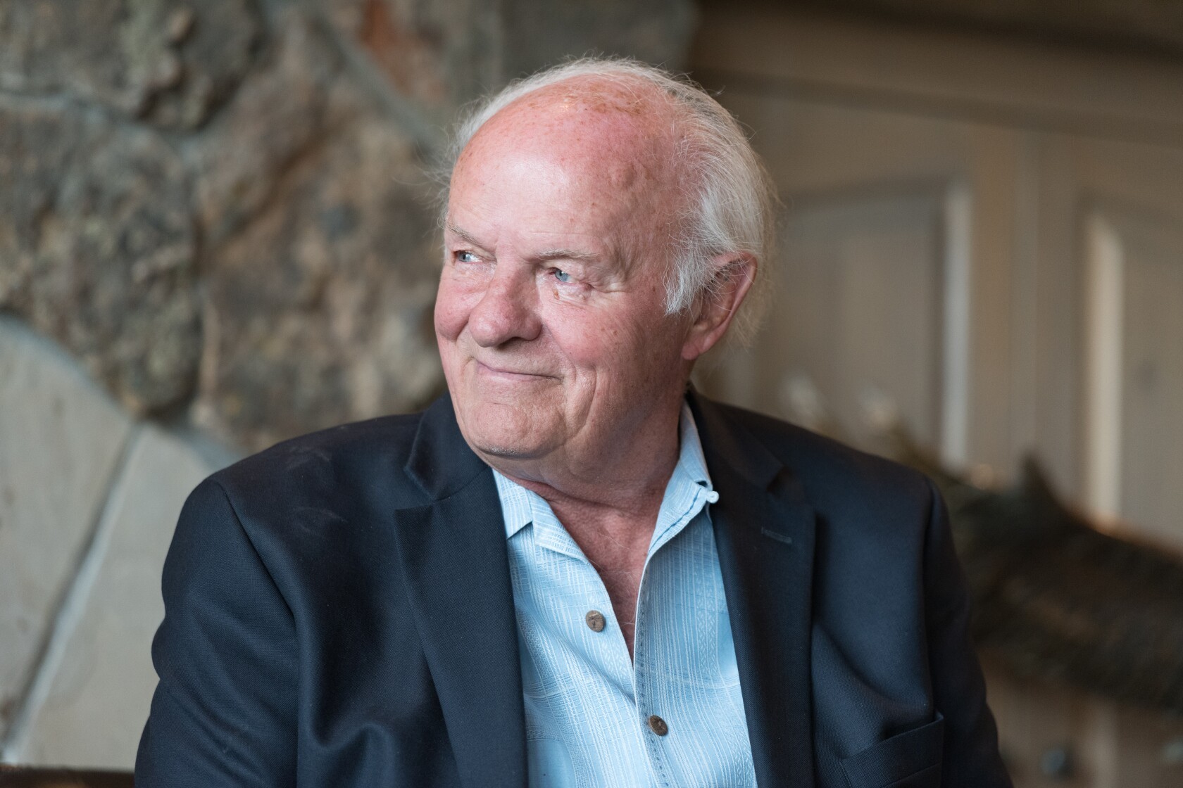
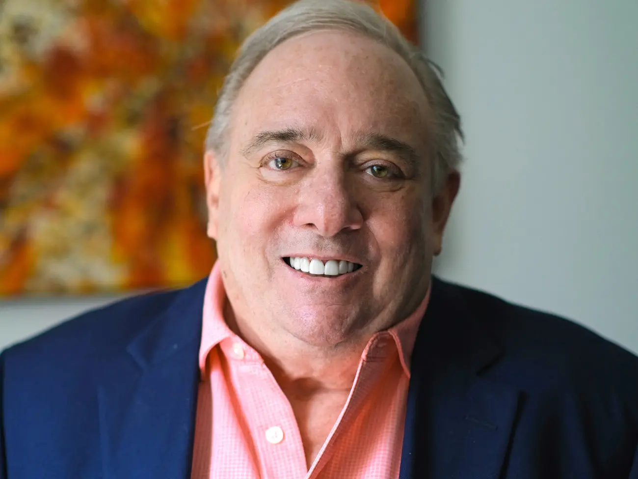
Renowned philanthropist and businessman T. Denny Sanford passed away July 18, 2026 in his hometown of Sioux Falls, South Dakota. He was 90 years old.
Renowned philanthropist and businessman T. Denny Sanford passed away July 18, 2026 in his hometown of Sioux Falls, South Dakota. He was 90 years old.
A self-made billionaire, Sanford had spent recent decades diligently spreading that wealth through philanthropy, primarily focusing on education, health services and biomedical research. His motto: “Aspire to inspire before you expire.”
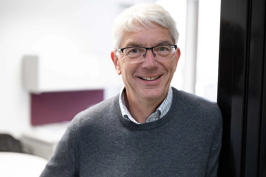
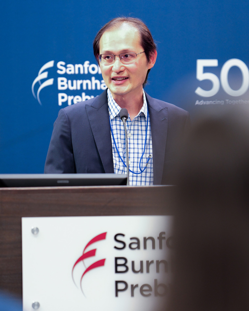
“Denny was larger than life. He loved people, philanthropy, business and golf. He was my supporter and advisor, but more importantly, my confidant and friend. Denny was fascinated by science and education, especially in advancing ideas and efforts that would make lives better, both in terms of health and in well-being,” said David Brenner, CEO and president of Sanford Burnham Prebys Medical Discovery Institute.
“His generosity was manifest, from supporting research and treatment of rare children’s diseases to stem cells to recruiting new generations of bright, young scientists to carry on the work. He loved to hear about how his philanthropic investments have benefited young people.”
Sanford Burnham Prebys was a frequent beneficiary. In 2007, for example, Sanford gave the institute $20 million through Sanford Health, the largest rural health system in the U.S. and a longtime beneficiary of Sanford. The gift helped the then-called Burnham Institute to create the Sanford Children’s Health Research Center. A year later, Sanford donated $30 million to create the Sanford Consortium for Regenerative Medicine, with the Institute as a founding member.
Two years later, Sanford donated $50 million and The Burnham Institute became the Sanford-Burnham Medical Research Institute. In 2023, he gave $70 million to Sanford Burnham Prebys to launch an ambitious plan to recruit up to 20 new scientists.
Sanford has been an honorary trustee at Sanford Burnham Prebys since 2009.
“Long before Denny’s and my names became inextricably linked through the Sanford Burnham Prebys Medical Discovery Institute, we shared a common goal to make an impact in our communities and to serve others,” said Malin Burnham, who has served on its board of directors since 1982. “Denny embodied that purpose throughout his life, generously investing himself and his resources to benefit others, often with truly transformational results.”
“Denny’s commitment to health and medicine was extraordinarily broad and deep,” noted Donald Kearns, MD, chair of the Board of Directors at Sanford Burnham Prebys and president emeritus of Rady Children’s Hospital-San Diego. Indeed he partnered with and supported specialized pediatric care for local families through the hospital, Sanford Children’s Clinic in Oceanside and the Children’s Primary Care Medical Group.
Sanford fundamentally believed that basic research and the benefits derived were driven and sustained by the people who do it. That was most evident in his philanthropy supporting education and in supporting new generations of scientists, said Brenner.
“Denny was a visionary committed to making a better world. His $70 million gift to recruit new faculty—the young investigators who will be future leaders—was a transformational investment in the continued strength of biomedical research at Sanford Burnham Prebys and across the Torrey Pines Mesa.,” said Brenner.
Sanford was born in Saint Paul, Minn. in 1935 during the Great Depression. His mother died of breast cancer when he was four years old. “She had been in the hospital for about a year before she died, so I have no memory of her at all,” Sanford recalled, but her death would inspire significant, subsequent support of cancer research by her son.
His father passed when he was 20.
“We have a lot of heart disease in my family,” he told the Horatio Alger Association, which honored him in 2016. “My father had several heart attacks throughout my childhood. My older brother had a fatal heart attack at the age of 40. I also had a heart attack at the age of 44, but I have had good health since then. But the loss of my father when I was still so young was a major blow to me.
“He was the most caring person I ever met. He never graduated high school because he had to help support his family. He was very giving, and I think my ideas about philanthropy, which is such an important part of my life today, came from him.”
Sanford did graduate from high school—and the University of Minnesota in 1959. In the following years, Sanford would be a sales representative for industrial cork and building materials. He started a distribution company that he transformed into a manufacturing and research firm.
In 1986, he purchased a small bank in Sioux Falls, South Dakota with $80 million in assets, then created a credit card business: United National Corp., with its two subsidiaries, First Premier Bank and Premier Bankcard, soon had a $1 billion portfolio of credit card loans.
The scope of Sanford’s philanthropy is staggering.
He has donated more than $1 billion to Sanford Health, a system of 48 medical centers, 211 clinics and 160 senior living centers in the upper Midwest, California and Florida, including funding to establish the Edith Sanford Breast Center in honor of his mother.
In addition, Sanford has made multi-million dollar gifts to the Sanford Underground Research Facility, the Sioux Falls Development Foundation, the Sioux Falls Zoo and Aquarium and the Children’s Home Society of South Dakota.
A part-time resident of San Diego, Sanford has made similar and substantial gifts to a host of local institutions: UC San Diego, National University, the Salk Institute, San Diego Public Library and the Zoological Society of San Diego.
To date, it’s estimated Sanford’s total philanthropy is approximately $2 billion.
“We are on earth to provide for other people, not just ourselves and our families,” Sanford said before his induction into South Dakota’s Hall of Fame in 2007. “We all have the opportunity to work a little bit harder and help someone else, not just ourselves.”
Remembrances
“One of the greatest gifts of my 20-plus-year friendship with Denny was learning how deeply he cared about helping others. Whether we were talking about life, philanthropy or our Institute, he always brought the conversation back to making a difference. He believed that a truly meaningful life is measured by the impact you have on people’s lives. That philosophy has stayed with me ever since and continues to guide the mission and purpose of our biomedical research.”
Kristiina Vuori, MD, PhD
Professor
Cancer Genome and Epigenetics Program
Pauline and Stanley Foster Distinguished Chair
“I’ve always felt Denny was the older, wiser brother I never had. First time I saw his smile and those blue eyes with shirt to match, I knew I was in friendly territory. But friendly was quite an understatement. Right from the beginning, he wanted to know what we at the institute and me personally could do for kids. He was all about kids, especially those who didn’t get all the normal genes like the rest of us. Those blue eyes were quick to glow when meeting some of the kids we’ve worked with, and especially those few we’ve ‘saved.’ Those eyes teared up easily when he met the kids that neither of us could help. But their smiles would coax out a broad grin on that man’s kind and generous face.
“Whenever we had a big meeting inviting families, scientists and physicians, Denny was right there to welcome everyone and let them know how important they were to him. He showed it all the time. The kids and families wrote him thank you notes and, years later, he’d ask me about some of them.
“They asked about him too. Last year I went to a meeting in Sioux Falls and Denny said, ‘Let’s get together.’ I thought he meant a friendly drink. Not Denny. He’s the surprise man, asking me, “What do you need, pal?” I was caught totally off guard, speechless. He quickly added, ‘Not for you, for the kids.’
“He was generous, but you had to pull your own weight. We mostly got there. Just like 20 years before, being around my ‘big brother’ was all I needed. Of course, seeing that blue-eyed smile always charmed me. In May, he came to my birthday party, maybe he knew it would be our last time. I’ll miss those eyes, that man and his advice.”
Hudson Freeze, PhD
Director, Sanford Children’s Research Center
Professor
William W. Ruch Distinguished Endowed Chair
“Denny Sanford’s passing marks the loss of a truly extraordinary philanthropist whose vision, generosity and unwavering belief in the power of people to make a difference will be felt for generations.
“His support was instrumental in bringing Dr. David Brenner to Sanford Burnham Prebys, setting in motion the recruitment of world-class scientists whose discoveries continue to advance human health.
“Beyond our institute, Denny’s commitment to improving lives touched countless organizations and communities through his remarkable philanthropy. His legacy lives on not only in the buildings that bear his name, but in the scientific breakthroughs, healthier lives and brighter futures made possible by his generosity. As we honor his memory, we do so with deep gratitude and a renewed commitment to carry forward the vision he so passionately believed in.”
Lori Moore
Vice Chair, Sanford Burnham Prebys Board of Directors
“Over the fifteen years I have directed the Prebys Center for Chemical Genomics and the Center for Therapeutics Discovery, I have seen what it means for an institution to be shaped by Denny Sanford’s vision.
“Denny did not merely support biomedical research in the abstract: He embraced, and then championed, the harder and less certain part of our mission — the part that moves beyond understanding the molecular basis of disease into discovering first-in-class drugs for the diseases that have proven so stubbornly difficult to treat.
“That expansion of ambition was not the safe path. It was the visionary one, and it took someone who grasped the big picture: that transformational advances in healthcare for all begin with someone willing to fund the decades-long, uncertain crossing from biology to medicine.
“The pipeline of emerging therapeutics we are advancing today — for pain, for addiction, for cancer, for neurodegeneration — exists in large measure because Denny believed that ‘curing incurable diseases’ was a worthy investment, not a naive one. The tribute he would value most is not words, but the work itself — bringing these drugs to the patients who need them. That is the promise his generosity made possible, and the one we strive to keep.”
Michael Jackson, PhD
Senior Vice President, Drug Discovery and Development
Conrad Prebys Center for Chemical Genomics
Director, Center for Therapeutics Discovery
Associate Director, Translational Initiatives
“As a board member of Sanford Burnham Prebys, what I admired most about Denny was that his generosity didn’t just fund buildings or research. It gave big ideas the chance to become breakthroughs and helped transform our institute into a place capable of changing the future.
“As a San Diegan, I believe our fair city is healthier, stronger and more hopeful because he chose to invest here. His vision lives on in the discoveries we make, the patients we help and the lives we improve.
“As a person, Denny made me think bigger about what philanthropy can accomplish, and to believe that each of us can be a part of something greater than ourselves. His legacy will continue to inspire me—and many others—to do the very best we can, just as he did.”
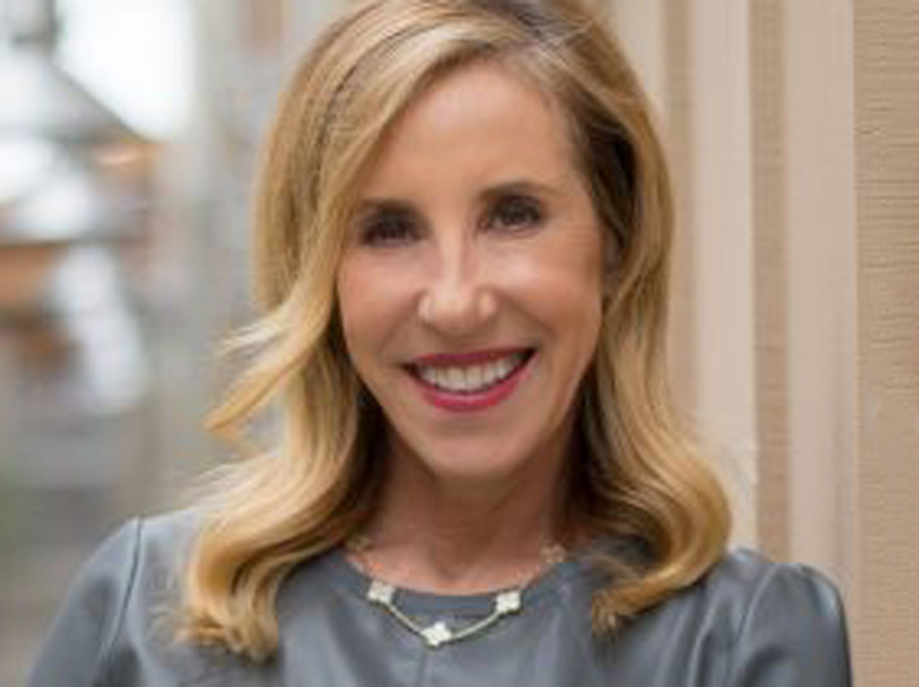
Katherine Chapin
Sanford Burnham Prebys Board of Directors
“Like so many in our Sanford Burnham Prebys family, I am deeply saddened by the passing of Denny Sanford, who was a remarkable philanthropist, visionary and long-time friend. Denny’s unwavering belief in the power of scientific discovery shaped our institute and empowered our scientists to pursue breakthroughs that will bring hope to patients and families for generations to come.
“His legacy extends far beyond his extraordinary philanthropy. He inspired all of us to think bigger, dream bolder and recognize the power of philanthropy to accelerate medical discovery. As we mourn his passing, we also celebrate a life devoted to improving the human condition and making a lasting impact. There is no greater tribute we can offer than to carry his vision forward by cultivating a community of philanthropists who share his passion for discovery and are committed to ensuring that Sanford Burnham Prebys remains a place where bold science continues to change lives.”
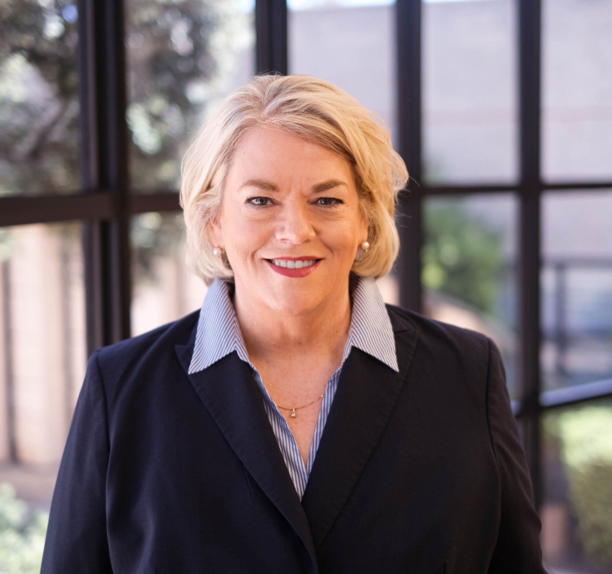
Sandy Liarakos
Vice President, Philanthropy
Institute News
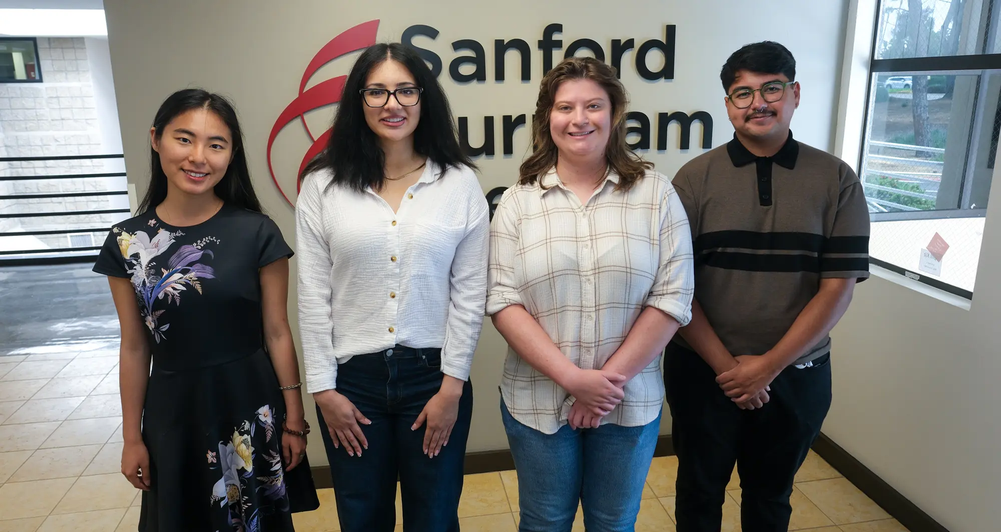
LEAP scholars share scientific and professional development achievements
LEAP program participants recently shared their research at a capstone presentation event. From left to right: Yun Ma, Sarina Safavi, Isabel Sakowicz and Josue Navarrete. Not pictured: Mahek Shah. Image credit: Sanford Burnham Prebys.
Postbaccalaureate training program participants deliver capstone presentations, thank mentors and colleagues
In late June 2026, trainees in the LEAP (Laboratory Experience As Pathway to Graduate School) program at Sanford Burnham Prebys Medical Discovery Institute participated in a capstone presentation event. The scholars shared their research findings following a year of work and mentorship in labs throughout the Institute.
The LEAP program was designed to bridge the gap between college graduation and graduate school. It provides recent graduates with hands-on biomedical research experience, career development opportunities and mentorship to enhance their applications for—and ability to succeed in—highly competitive biomedical PhD programs.
With generous support from the Prebys Foundation, the LEAP program is offered through a collaborative partnership between the Sanford Burnham Prebys NCI-designated Cancer Center and Office of Learning and Development.
“Today we celebrate five of our LEAP program participants who have gained new skills, demonstrated research successes and grown as scientists throughout their journeys here,” said Kevin Yip, PhD, during his introductory remarks.
Following Yip’s welcoming address, the LEAP program mentors introduced the participants and invited them forward for their capstone presentations.
Yun Ma, a trainee in the Feng and Yip labs, discussed her study of proteins that group together in what are called protein complexes to carry out biological functions.
Ma developed a method for comparing data on RNA and proteins to identify priority genes affecting how protein complexes form. These priority genes may help shed new light on how diseases progress and may inform efforts to design new medications.
Her presentation was titled, “Inferring disease-associated changes in protein complex assembly from multimodal data.”
Kevin Yip, PhD, the Andrew and Erna Viterbi Distinguished Chair and director of the Center for Data Science and Artificial Intelligence, provided opening remarks at the event.
Sarina Safavi, a trainee in the Dhar lab, described her work to understand why cardiovascular disease—especially heart failure with preserved ejection fraction—is the leading cause of death among liver disease patients. Safavi developed tools needed to make new liver disease research models to study how the disease affects the heart.
The continuation of this research may lead to the discovery of new drug targets and methods for identifying liver disease patients at greater risk of heart failure to enable earlier treatments.
Her talk was titled, “Mechanistic understanding of MASLD-associated cardiac dysfunction and its reversibility.”
Josue Navarrete, a trainee in the Deshpande and Yip labs, detailed his project focused on small proteins from areas beyond the protein-coding genes that get the most scientific attention. Increasing amounts of evidence supports these microproteins’ biological importance, including in cancer.
Navarette optimized a computational technique for discovering microproteins which enables large-scale studies of thousands of genes. He tested the approach and found microproteins involved in stem cells becoming blood cells and other cells. Studying these microproteins may lead to new diagnostic techniques and potential treatments for blood cancers.
His lecture was titled, “Optimized pipeline enables genome-wide discovery of candidate microproteins in hematopoiesis.” Navarette will be joining the Institute as a doctoral student this fall.
Mahek Shah, a trainee in the Spruck lab, shared her study of a cancer treatment strategy that compels tumor cells to act as if they are infected by a virus. Known as viral mimicry, this therapeutic approach triggers the innate immune system to respond against cancer cells. Shah described drug screening experiments that identified promising novel candidate compounds.
In further tests, these compounds were found to activate viral mimicry, cause an immune response and to be toxic to cancer cells. Continued research may lead to new treatments that can boost the effectiveness of chemotherapy, radiation therapy and targeted therapies.
Her presentation was titled, “Novel compounds targeting histone modifying enzymes induce viral mimicry in breast cancer.”
Isabel Sakowicz, a trainee in the Tharp lab, discussed her research regarding the most common and difficult-to-treat form of ovarian cancer. These tumors can wall themselves off from the immune system with structural collagen proteins that resemble scar tissue.
Yun Ma, a trainee in the Feng and Yip labs, discussed her study of proteins that group together in what are called protein complexes to carry out biological functions.
Sakowicz explored how a potential treatment strategy might reduce levels of a form of collagen known to promote ovarian cancer invasiveness and metastasis. Lowering collagen VI levels may break down the barrier around ovarian cancer cells to make other treatments more effective, including immunotherapies.
Her presentation was titled, “FAK protein loss rewires amino acid metabolism in high-grade serous ovarian cancer.” Sakowicz will be joining the Institute as a doctoral student this fall.
In addition to discussing their respective projects, the trainees credited their mentors, fellow lab members and access to avenues for training and advancing as professionals.
“Beyond my scientific achievements, I have enjoyed many career development opportunities, including attending and presenting at workshops, seminars and conferences,” said Ma.
“Everyone in the lab has contributed to a truly supportive and cooperative environment,” said Safavi. “I would also like to thank the LEAP program coordinators for their efforts and making sure we were set up for success.”
Institute News
Kevin Tharp awarded $450,000 Ovarian Cancer Research Alliance grant to break down tumor defenses
Kevin Tharp, PhD, is an assistant professor in the NCI-designated Cancer Center’s Cancer Metabolism and Microenvironment Program at Sanford Burnham Prebys. Image credit: Sanford Burnham Prebys.
The new award will fund research regarding how to bust through the barrier between tumors and immune cells
Kevin Tharp, PhD, was awarded a three-year, $450,000 Ovarian Cancer Research Alliance grant to study high grade serous ovarian cancer, the most common and deadly form of the disease.
In collaboration with researchers at the University of California San Diego, Tharp recently published findings in Cell Reports demonstrating a treatment approach in mice that allowed more tumor-fighting immune cells to approach tumors, shifted the behavior of other immune cells to work against tumors, and made immunotherapy more effective.
Tharp will use the new funding to follow up on these findings regarding how high grade serous ovarian cancer circumvents the immune system’s anti-tumor defenses. His team will focus on the physical barrier that tumors build to keep immune cells at bay.
“We know that tumors can wall themselves off with structural collagen proteins that resemble scar tissue, and that the presence of this obstacle determines the effectiveness of anti-cancer immunotherapies,” said Tharp.
“We want to know how and why tumors create this obstruction and understanding this will help us find ways to break through these defenses to make immunotherapies more effective.”
Tharp will conduct this research under the mentorship of Cosimo Commisso, PhD, the deputy director of the NCI-Designated Cancer Center at Sanford Burnham Prebys and a professor in the Cancer Metabolism and Microenvironment Program, and David Schlaepfer, PhD, a professor in the department of OBGYN and Reproductive Sciences at the University of California San Diego Moores Cancer Center.
“Too many ovarian cancer patients progress and do not respond to the standard of care, so finding new treatments is an extreme clinical need,” said Tharp.
“There is potential for immunotherapies to treat recurrent and metastatic cancer if we can bridge the divide between tumors and immune cells.”
The Ovarian Cancer Research Alliance is the oldest and largest ovarian and gynecologic cancer charity in the world. Since its founding in 1994, the alliance has grown into the leading non-government funder of ovarian and related gynecologic cancer research by investing more than $140 million in grants to scientists.
Institute News
Women in Science Lecture series spotlights structural biology and immunology leader
From left to right: Sanford Burnham Prebys NCI-Designated Cancer Center scientist Kelly Kersten, PhD, and structural biology and immunology leader Erica Ollmann Saphire, PhD, MBA. Image credit: Sanford Burnham Prebys.
The series highlights the groundbreaking work and unique perspectives of women leaders in the biomedical sciences.
Saphire told the audience about a turning point in 2013 in her field studying how antibodies work against the Ebola virus. Antibodies are especially important treatments for infectious diseases that lack an effective vaccine, as was the case for the Ebola virus until 2019. Saphire described the challenge that emerged when an antibody predicted to be effective based on laboratory results had no effect on survival in animal studies, whereas a cocktail of three antibodies that was ineffective in laboratory cell culture tests actually protected every animal against the infection.
“Collectively, as a body of scientists, it became clear we were missing some information about how to study the neutralizing effects of antibodies,” said Saphire. “The key was seeing the clue that how well the treatment protected was dependent on what kind of cell it was made in and how that effected the antibodies’ ability to recruit the immune system.”
To enable experiments following up on that lead, Saphire organized a large coalition of academic, industrial and government labs from across the world. Each partner sent their antibodies or neutralization assays to a single location where they could be studied side by side under code names to protect intellectual property. The consortium fast-tracked a smaller study that led to the first therapeutic approved for treating Ebola, and the longer-term comprehensive study would go on to develop much better therapeutics for the disease. Due to this initiative’s success, Saphire was asked to lead a Gates Foundation-supported project to evaluate antibody therapeutics against SARS-CoV-2.
Saphire closed her presentation with her reflections on how to improve the scientific system so that it works better for researchers.
Kersten and Saphire with fireside chat moderator and cardiovascular researcher Sanjeev Ranade, PhD. Image credit: Sanford Burnham Prebys.
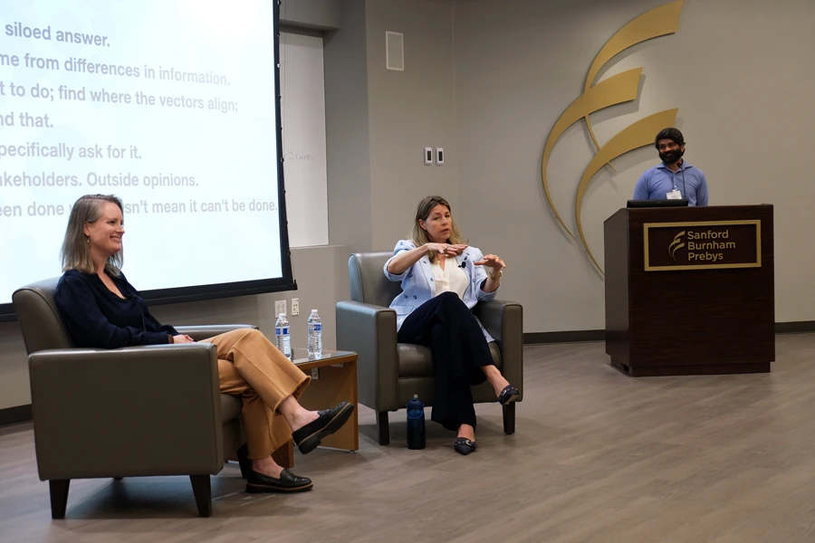
“I’ve learned over 20 years that a siloed approach very much gives a siloed answer,” Saphire said. “It is important to find out how different experts’ interests and motivations align and intersect and then put resources behind that to find success.”
Sanjeev Ranade, PhD, assistant professor in the Center for Cardiovascular and Muscular Diseases at Sanford Burnham Prebys, then moderated a fireside chat and audience question-and-answer session with Saphire and Kelly Kersten, PhD, assistant professor in the Cancer Metabolism and Microenvironment Program at the Sanford Burnham Prebys NCI-Designated Cancer Center. Topics included: the remarkable power and complexity of the immune system; the contrast in needing to mitigate the immune system in allergies and autoimmune disease versus boosting it in the right ways in cancer and infectious disease; how to effectively introduce your work to people outside of science; how roles change as academic and leadership careers progress; and advice for students and trainees beginning their careers in biomedical research.
The Women in Science Lecture Series features events that are free and open to the public. The series is part of broader efforts at Sanford Burnham Prebys to foster an environment that nurtures the success of individuals from all backgrounds. The events are hosted by the Office of Workforce Engagement & Belonging and highlight the groundbreaking work and unique perspectives of women leaders in the biomedical sciences, while fostering mentorship and collaboration across the Torrey Pines Mesa.
Speakers and attendees at the fourth annual SoCal Metabolism Symposium.
The fourth annual SoCal Metabolism Symposium brought together hundreds of experts and trainees to share the latest advances
SoCal Metabolism Symposium co-organizer Brooke Emerling, PhD, opened the meeting held at Sanford Burnham Prebys Medical Discovery Institute on Friday, March 20, 2026, by celebrating the event’s momentum.
“In 2023, when it started, we had about 12 talks, 28 posters, about 120 attendees and three sponsors, and now we’re up to 18 talks, 64 posters, more than 200 attendees and six sponsors,” said Emerling, director of and associate professor in the Sanford Burnham Prebys Cancer Metabolism and Microenvironment Program.
Speakers were mostly postdoctoral researchers and graduate students from Sanford Burnham Prebys, the Salk Institute, the University of California Irvine, the University of Southern California, the University of California Los Angeles and the University of California San Diego.
The event began with a session of scientific talks focused on the theme of cancer metabolism. Aaliyah Balagtas, a graduate student in the lab of Cosimo Commisso, PhD, at Sanford Burnham Prebys, discussed her research on a cellular scavenging process known as macropinocytosis that pancreatic tumors use to survive and grow when resources are scarce. The morning continued with a second thematic session focused on metabolism in aging and cell fate.
Before the event’s lunch break and poster viewing, Emerling introduced the symposium’s first-ever guest speaker from outside Southern California, Navdeep Chandel, PhD, the David W. Cugell, MD, Professor and professor of Medicine (Pulmonary and Critical Care), Biochemistry and Molecular Genetics at Northwestern University.
Chandel began by sharing his delight that the speakers in the morning sessions showed genuine enthusiasm and interest in studying mitochondria and targeting metabolism to improve human health and treat disease. He thinks there is a significant opportunity to use the fundamental knowledge we’re learning about intermediary metabolism in mitochondria and translate it into concrete advances for human health.
Brooke Emerling, PhD, is director of and associate professor in the Cancer Metabolism and Microenvironment Program at Sanford Burnham Prebys. Image credit: Sanford Burnham Prebys.
Chandel focused on one of his lab’s translational projects studying metformin, a longstanding, widely used, cheap and safe drug for treating high blood sugar in prediabetes and type 2 diabetes. Various studies have suggested that metformin also has anti-cancer effects and may reduce inflammation, but it was not clear how the drug worked in our bodies or cells to cause any of this to occur. Chandel shared soon-to-be-published data regarding how metformin builds up in the gut after being taken as a pill, and how it influences mitochondria there to systemically lower blood sugar.
The afternoon opened with a third set of thematic podium presentations centered on the topic of physiological metabolism and new techniques. The fourth and final session of scientific talks were grouped around the theme of immunometabolism.
Cosimo Commisso, PhD, is the deputy director of the institute’s NCI-Designated Cancer Center and a professor in the Cancer Metabolism and Microenvironment Program. Image credit: Sanford Burnham Prebys.
The symposium’s closing podium talk was the Gina Lee Memorial Keynote, a lecture honoring cancer signaling and metabolism expert Gina Lee, PhD, an assistant professor of Microbiology and Molecular Genetics at the University of California Irvine, who passed away on June 23, 2024, at the age of 39.
Cosimo Commisso, PhD, the deputy director of the institute’s NCI-Designated Cancer Center and a professor in the Cancer Metabolism and Microenvironment Program, delivered the 2026 Gina Lee Memorial Keynote and focused on a new direction for his lab. Aging is a major risk factor for pancreatic cancer that also can limit treatment options if a patient is too frail to be safely treated with surgery or other alternatives.
The average age of a patient diagnosed with pancreatic cancer is 70, and nearly two-thirds of cases are in people over the age of 65. Commisso and his lab members are rethinking how therapies in development will work for a frail and aging population that represents the majority of patients.
Following Commisso’s keynote address, the 2026 SoCal Metabolism Symposium concluded with a reception and second poster session. The next SoCal Metabolism Symposium will be held in March 2027 at the University of California Irvine.
Emerling organized the event in partnership with Peter James Mullen, PhD, assistant professor of Microbiology and Immunology in the Keck School of Medicine at the University of Southern California, and Cholsoon Jang, PhD, assistant professor of Biological Chemistry at the University of California Irvine School of Medicine.
Institute News
Shanshan Yin named 2025 recipient of Eric Dudl Endowed Scholarship
Peter Adams, PhD, with scholarship recipient Shanshan Yin, PhD, and Kevin Yip, PhD.
Yin, a postdoctoral associate at Sanford Burnham Prebys, received the honor in recognition of her achievements in research on cancer and aging
Shanshan Yin, PhD, was named the 2025 recipient of The Eric Dudl Endowed Scholarship at Sanford Burnham Prebys Medical Discovery Institute.
The scholarship fund was established at the institute to remember Eric Dudl, a postdoctoral researcher whose life was tragically cut short by cancer at the age of 33. Since 2007, 18 postdoctoral scientists have received support for their research from the endowed scholarship fund.
Yin is a postdoctoral associate in the lab of Peter Adams, PhD, director of the Cancer Genome and Epigenetics Program at Sanford Burnham Prebys. She wants to understand why the incidence of cancer increases with age. Yin studies changes in gene expression and immune system activity in breast cancer tumors as mice age.
Her research has shown that part of the reason that breast cancer is more common with age is because of an impaired immune system. Immune dysfunction due to aging allows the tumors to grow more frequently and more rapidly. Additional research on these findings may guide future preventive treatments.
Yin has garnered recognition throughout her scientific career, including the 2022 Lenka Finci and Erna Viterbi Fishman Fund Award from Sanford Burnham Prebys.
“I’m grateful for the Dudl family’s kindness and generosity,” said Yin. “I am moved by Eric’s determination, bravery and love for life and for science, and I would like to carry his example with me going forward and do my best to honor his legacy.”
“It’s been a real pleasure working with Shanshan over the years,” said Adams. “She truly does embody Eric Dudl’s commitment to and passion for science which is expressed so well by this inspirational award.”
The Eric Dudl Endowed Scholarship at Sanford Burnham Prebys was established at the institute to remember Eric, a postdoctoral researcher whose life was tragicallycut shortby cancerat the age of 33.
For more information on setting up a scholarship or to learn more about our philanthropy program, please contact giving@sbpdiscovery.org.
Institute News
Women in Science Lecture series showcases public health and nutrition policy leader
From left to right: physician-scientist Angela Liou, MD; public health and nutrition policy expert Cheryl A.M. Anderson, PhD, MPH, MS; and researcher Lukas Chavez, PhD, MS.
The series highlights the groundbreaking work and unique perspectives of women leaders in the biomedical sciences
On February 11, 2026, Sanford Burnham Prebys Medical Discovery Institute hosted the second event in the Women in Science Lecture Series. The occasion opened with a presentation by Cheryl A.M. Anderson, PhD, MPH, MS, professor and dean of the Herbert Wertheim School of Public Health and Human Longevity Science at the University of California San Diego and director of the UCSD Center of Excellence in Health Promotion and Equity.
Anderson introduced attendees to some of the pivotal findings of her mentors studying the effects of nutrition on public health, including the landmark dietary approaches to stop hypertension (DASH) clinical trial. Because of the challenges in achieving significant heart disease prevention benefits outside of the controlled environments used in studies such as the DASH trial, Anderson was determined to explore other approaches.
“I put together this concept that instead of asking the individual to figure it all out from our dietary recommendations, maybe we could figure out how to have a healthy, sustainable food system,” said Anderson.
“I see a sustainable food system as one that maintains our ability to get lots and lots of nutrition and where you meet the current population’s needs without compromising what future generations might also need.”
In addition to discussing her scientific journey, Anderson provided insight into her experience serving with other experts to provide input into two different iterations of the federal government’s Dietary Guidelines for Americans. These guidelines from the Department of Health and Human Services and Department of Agriculture set the standards for food in federally funded programs such as public school and day care lunches as well as the Women, Infants and Children (WIC) special supplemental nutrition program. Anderson shared her experience working collaboratively to provide science-based counsel in an ecosystem that also contains political considerations such as the interests of industries involved in agriculture and food production.
Anderson (at right) opened the event discussing her career journey focused on how to develop a healthy, sustainable food system. The event also featured a fireside chat and audience question-and-answer session with Anderson and Liou.
Lukas Chavez, PhD, MS, associate professor in the Cancer Genome and Epigenetics Program at Sanford Burnham Prebys and scientific director of the Pediatric Neuro-Oncology Molecular Tumor Board at Rady Children’s Institute for Genomic Medicine, then moderated a fireside chat and audience question-and-answer session with Anderson and Angela Liou, MD, physician-scientist and pediatric oncologist with a dual appointment at Rady Children’s Health and the Cancer Genome and Epigenetics Program at Sanford Burnham Prebys. Topics included: how new scientific insights are translated to reduce population-level health risks or guide care for children facing serious illnesses; how new technologies change the way you conduct research and deliver patient care; what can be done to ensure that scientific discoveries can be equitably accessed and lead to better outcomes for all; and what do future clinicians and scientists need in terms of skills, mindset and institutional support to succeed as public health researchers and physician-scientists.
The Women in Science Lecture Series, featuring quarterly events that are free and open to the public, is part of broader efforts at Sanford Burnham Prebys to foster an environment that nurtures the success of individuals from all backgrounds. The series is hosted by the Office of Workforce Engagement & Belonging and highlights the groundbreaking work and unique perspectives of women leaders in the biomedical sciences, while fostering mentorship and collaboration across the Torrey Pines Mesa.
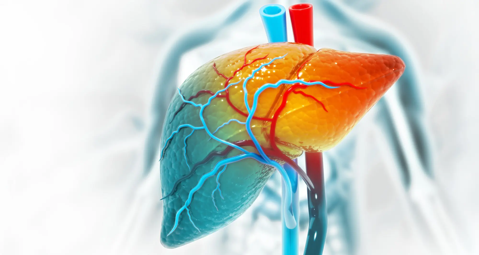
A new study shows that liver cell stress can lead to cancer, yet it also can make tumors less resistant to immunotherapy. The findings may lead to new treatments and help physicians determine which patients will benefit from existing immunotherapies. Image credit: crystal light/Sanford Burnham Prebys.
Cell stress response protein implicated in cancer progression, yet it also weakens resistance to immunotherapies
Metabolic disorders such as obesity and type 2 diabetes place extra stress on the liver. Liver cells try to protect themselves from the accompanying surge in dysfunctional proteins by activating factors that help restore an appropriate protein balance.
One of these factors is a protein called activating transcription factor 6 alpha (ATF6α) that was recently shown to drive the onset of liver cancer if left permanently active. In a Nature study published February 4, 2026, an international team of scientists demonstrated that activating ATF6α in mice caused liver disease that progressed to liver cancer.
In data from human liver cancer patients, ATF6α activation was linked with more aggressive tumors, a suppressed immune system surrounding tumors and reduced patient survival.
The researchers also uncovered ways that ATF6α might be used to advance the treatment of liver cancer. Liver cells with ATF6α switched off developed fewer tumors. While high ATF6α activity levels were associated with cancer progression, they also were found to make tumors more susceptible to certain immunotherapies.
These findings suggest the need for future clinical trials to test drugs that directly target ATF6α to treat the disease. Additionally, it might prove advantageous to screen liver cancer patients for ATF6α activity to find those most likely to benefit from existing immunotherapies.
Randal Kaufman, PhD, is a professor in the Center for Metabolic and Liver Diseases at Sanford Burnham Prebys and a co-corresponding author of the study. Image credit: Sanford Burnham Prebys.
Xin Li, PhD, a postdoctoral fellow at the German Cancer Research Center (DKFZ), shares first authorship of the study with co-corresponding author Cynthia Lebeaupin, PhD, principal scientist at Pfizer and former postdoctoral researcher at Sanford Burnham Prebys Medical Discovery Institute.
The other co-corresponding authors are Dirk Haller, PhD, Technische Universität München; Randal Kaufman, PhD, Sanford Burnham Prebys; and Mathias Heikenwälder, PhD, University of Tübingen and DKFZ.
Institute News
Sanford Burnham Prebys hosts inaugural event in the Women in Science Lecture Series
Sanford Burnham Prebys president and CEO David Brenner, MD, meets with speaker Susan Tousi, MBA, CEO at DELFI Diagnostics, prior to the inaugural event in the Women in Science Lecture Series.
The series highlights the groundbreaking work and unique perspectives of women leaders in the biomedical sciences
Susan Tousi, MBA, CEO at DELFI Diagnostics, opened the event by discussing the lessons she learned throughout her career journey. At DELFI Diagnostics, she is leading a team focused on improving the detection of lung cancer. The company’s goal is to make lung cancer screening more accessible through a blood test that is analyzed by applying machine learning and next-generation sequencing.
Prior to this role, Tousi served as a senior vice president for more than 10 years at Illumina, Inc., including as chief commercial officer for three years. During her tenure, she contributed to making genomic sequencing more affordable as the cost of sequencing a single genome fell from more than $5000 in 2013 to $200 in 2023. Tousi also borrowed from her experience developing consumer printers for Eastman Kodak and Hewlett-Packard, emphasizing the importance of making Illumina’s sequencing machines easy to use for clients in research labs, hospitals and clinics.
“My time at Illumina was amazing,” said Tousi. “I had the absolute privilege of seeing our genomic capabilities installed in 155 countries around the world.”
Tousi concluded with her optimism about how technology is transforming healthcare.
“I think we are on the precipice of major shifts in technology with the advancement of AI and where we’ve come with genomics, multiomics and the access to large-scale molecular data,” said Tousi. “I think you know these new technologies like blood-based liquid biopsy testing are going to allow us to find disease earlier, to treat it more precisely and monitor its recurrence across many disease areas.
“This can be the dawn of a new beginning in science and the advancement of healthy lives.”
From left: Brooke Emerling, PhD, and Susan Tousi, MBA
Image credit: Sanford Burnham Prebys
Kevin Tharp, PhD, assistant professor in the Cancer Metabolism and Microenvironment Program at Sanford Burnham Prebys, then moderated a fireside chat and audience question-and-answer session with Tousi and Brooke Emerling, PhD, director and associate professor in the Cancer Metabolism and Microenvironment Program. Topics included: different gender-based expectations in scientific fields; the importance of mentorship and paying it forward; dealing with the emotional toll of studying diseases more prevalent in women; and programs providing opportunities for future leaders in science and medicine.
The Women in Science Lecture Series features quarterly events and is part of broader efforts at Sanford Burnham Prebys to foster an environment that nurtures the success of individuals from all backgrounds. The series is hosted by the Office of Workforce Engagement & Belonging and highlights the groundbreaking work and unique perspectives of women leaders in the biomedical sciences, while fostering mentorship and collaboration across the Torrey Pines Mesa.
Women in Science lectures are free and open to the public. Registration is open for the next event in the series on February 11, 2026.
Institute News
Sanford Burnham Prebys expert surveys science on how to treat the most common brain cancer
Glioblastoma brain cancer cells under a microscope. Image credit: Anna Durinikova/Shutterstock.
New editorial recommends a multimodal perspective examining glioblastoma from tumor biology through to surgery
Glioblastoma is one of the most aggressive and treatment-resistant forms of brain cancer. It also is the most common form of cancer that originates in the brain, making research into new and better therapies even more imperative.
Physician–scientist Theophilos Tzaridis, MD, a postdoctoral fellow at Sanford Burnham Prebys Medical Discovery Institute in the lab of Peter Adams, PhD, recently surveyed promising glioblastoma studies after being invited to serve as a guest editor for a special issue of Frontiers in Oncology and Frontiers in Neurology.
More exact and safe surgeries
Tzaridis highlighted two studies focused on improving surgery for glioblastoma, as it continues to be the primary treatment for the disease. The recent publications discussed how to enhance the use of MRI to map out tumors and surrounding tissue, as well as other innovative mapping and monitoring techniques. These approaches would enable neurosurgeons to create better and safer plans for reducing risk of recurrence and avoiding side effects before starting surgery.
Targeted treatments and immunotherapies
Scientists have sought to add treatment options for glioblastoma beyond surgery, radiation therapy and chemotherapy. Some other cancers can be treated with targeted therapies that exploit a unique characteristic of certain tumors, but this approach has yet to yield long-term successes for glioblastoma patients. Tzaridis brought forward a case report of a patient whose tumors were nearly completely cleared by a targeted therapy after chemotherapy was unsuccessful. He suggests that future studies are warranted to identify patient subpopulations that can benefit from these treatments.
Theophilos Tzaridis, MD, a postdoctoral fellow at Sanford Burnham Prebys Medical Discovery Institute. Image credit: Sanford Burnham Prebys.
Immunotherapies that supercharge the immune system to better detect and eliminate cancer have transformed the treatment of many blood cancers and solid tumors. It has not, however, yet born fruit as an effective treatment for glioblastoma. Tzaridis spotlights a study discussing the potential use of chimeric antigen receptor (CAR) natural killer (NK) cells in glioblastoma rather than the more common CAR T-cell therapies.
The blood brain barrier and brain cancer biology
In addition to demonstrating how research is contributing to improving existing treatments and finding new potential therapies, Tzaridis emphasized the importance of continued studies of brain cancer cell biology and the obstacle to treatment posed by the blood brain barrier. He highlighted two studies focused on overcoming the blood brain barrier along with another two studies regarding cellular models and the use of extracellular vesicles to package and deliver treatments.
“With a multimodal perspective from addressing challenges in neurosurgery to improving our understanding of tumor biology and achieving therapeutic delivery into the brain, we have the best chance of improving survival of patients with this devastating disease,” said Tzaridis.