cancer therapy Archives - Page 2 of 3 - Sanford Burnham Prebys
Institute News

“We are desperate for new therapies”

AuthorMonica May
Date

September 23, 2019

Experts discuss AML during the Sanford Burnham Prebys community lecture series 

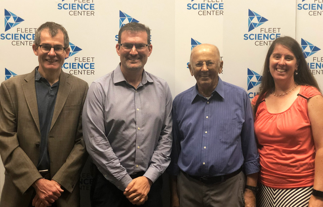
Bill Veljovich had never been sick in his life. “Not even joint pain,” shared the 80-year-old retired engineer at our recent Fleet Science Center discussion about acute myeloid leukemia (AML), a life-threatening type of blood cancer. He was joined by experts from Sanford Burnham Prebys and UC San Diego Health.
 
However, his doctor noticed that his white blood cells counts were off during a routine blood test. He was diagnosed with a blood cancer called myelodysplastic syndrome (MDS), which progressed to AML (this occurs in one out of three people with MDS). Fortunately, Veljovich responded well to a then off-label treatment that only recently was approved for older patients with AML. 

“The truth is, we are desperate for new therapies,” said speaker Rafael Bejar, MD, PhD, a clinician at UC San Diego who specializes in blood cancers. “AML typically occurs in people over the age of 60, who often aren’t able to tolerate intensive chemotherapies.” 

Until two years ago, the treatments for AML remained the same as those used in the 1970s: a chemotherapy combination and perhaps a bone marrow transplant. Only 24% of adults with AML remained alive five years after treatment. 

Now, thanks to foundational research that revealed the underlying genetic drivers of AML, eight new drugs have been approved in the past two years. Several more targeted therapies are nearing potential FDA approval. 

However, AML, which usually arises in cells that turn into white blood cells, is an incredibly complex and fragmented disease. Genome sequencing has revealed that more than 30 genes drive the cancer. Many different treatment types will be needed to truly conquer AML.

Peter Adams, PhD, a professor in Sanford Burnham Prebys’ National Cancer Institute (NCI)-designated Cancer Center, hopes to find a treatment that works for a broader AML population. He focuses on a protein called p53, often called the “guardian of our genome.” This protein senses DNA damage and kills the faulty cell—protecting us from developing cancer. However, to scientists’ surprise, 90% of people with AML have a normal p53 gene. 

“Emerging research suggests that AML inactivates p53 through other means,” said Adams. “My team is working to develop a drug combination that could reactivate the protective powers of p53—and thus fight AML.”

New research advances can’t come soon enough for people living with the cancer. 

“I’ve always taken the approach of learning as much as possible—and then fixing the problem,” said Veljovich, who designed and tested rocket engines before he retired. “I have learned that blood cancers are extremely complex. I wish there was a simple solution, but there isn’t. I’m grateful that we have smart folks like Dr. Bejar and Professor Adams who are working on these tough problems to find better medicines for AML.”

This event was the second of our five-part “Cornering Cancer” series. Join us for discussions on breast cancer (October 20), pancreatic cancer (November 17) and pediatric brain cancer (December 8). Register today.

Institute News

Cancer’s final frontier: the tumor microenvironment

AuthorMonica May
Date

September 3, 2019

Cancer researchers are setting their sights on a new kind of cancer treatment that targets the tumor’s surrounding environment, called the tumor microenvironment, in contrast to targeting the tumor directly. 

To learn more about this approach, we spoke with cancer experts Jorge Moscat, PhD, director and professor in the Cancer Metabolism and Signaling Networks Program at Sanford Burnham Prebys; and Maria Diaz-Meco, PhD, professor in the Cancer Metabolism and Signaling Networks Program at Sanford Burnham Prebys. Both scientists recently authored a review article centered on a family of cancer-linked proteins that regulate the tumor’s microenvironment. The paper was published in Cancer Cell

What is the tumor microenvironment exactly? 
Moscat: Just like every person is surrounded by a supportive community—their friends, family or teachers—every tumor is surrounded by a microenvironment. This ecosystem includes blood vessels that supply the tumor with nutrients; immune cells that the tumor has inactivated to evade detection; and stroma, glue-like connective tissue that holds the cells together and provides the tumor with nutrients.

Diaz-Meco: These elements are similar to the three legs of a stool. If we remove all three legs, we can deliver a deadly blow to the tumor. FDA-approved drugs exist that target blood vessel growth and reactivate the immune system to destroy the tumor. The final frontier is targeting the stroma.

When did scientists realize it’s important to focus on the tumor’s surroundings—not the tumor itself? 
Diaz-Meco: Scientists have known for more than a century that the tumor’s surroundings are different from normal cells. The tissue surrounding a tumor is inflamed—tumors are often called “wounds that never heal”—and their metabolism is radically different from healthy cells. 

Moscat: The discovery of oncogenes—genes that can lead to cancer—in the 1970s shifted the field’s focus to treatments that target the tumor directly. These targeted treatments work incredibly well, but only for a short time. Cancer researchers are realizing that tumors quickly adapt to this roadblock and become treatment resistant. In addition, many oncogenes are difficult to target, earning the title “undruggable.” As a result, cancer researchers are returning their focus to the tumor microenvironment—especially the stroma. Only a handful of stroma-targeting drugs are in development. None are FDA approved.

Which cancers could benefit most from a stroma-targeting drug? 
Moscat: Pancreatic, colorectal and liver cancers stand to benefit most from a stroma-targeting drug. For example, 90% of a pancreatic tumor consists of stroma—not cancer cells. Combined, these cancers are responsible for more than 20% of all cancer deaths in the U.S. each year. 

What is the focus of your lab’s research? 
Diaz-Meco: Our lab studies the cross talk between tumors and their environment. This conversation is very complex. In addition to “talking” with the tumor, the stroma also “speaks” with the immune system. We are working to map these interactions so we can create drugs that silence this conversation—or change it. For example, we recently showed—in a mouse model that faithfully recapitulates the most aggressive form of human colorectal cancer—that by altering the stroma’s interactions with the immune system, we might make tumors vulnerable to immunotherapy. 

What do new insights into the tumor microenvironment mean for cancer drug development? 
Moscat: It’s likely that the ultimate cancer “cure” won’t be just one drug that kills the tumor cells, but a combination of therapies. I expect this will be a three-part combination treatment that stops blood vessel growth, activates the immune system to attack the tumor and targets the stroma. 

Additionally, this research shows that experimental models of cancer drug development need to take the tumor microenvironment into account. Many current models use mice that lack an immune system—in order to get the tumor to grow—or focus on the tumor in isolation. Based on our knowledge of the tumor microenvironment, this isn’t an accurate representation of human disease. 

Diaz-Meco: In our lab, we have created several animal models of cancers that preserve the immune system and mirror tumor progression. In addition to better modeling human disease, this also allows us to study cancer from its earliest beginnings. This work could lead to early interventions—before the cancer has become large and hard to treat.

Anything else you’d like to add? 
Moscat: We are truly in the golden age of cancer biology. We understand more than we ever have before. New technologies are allowing us to obtain an unprecedented amount of information—we can even map every gene that is “turned on” in a single cancer cell. I am incredibly hopeful for the future. 

Learn more about the future of cancer treatment by attending our next “Conquering Cancer” event at the Fleet Science Center. Details

Institute News

Fleet Science Center cancer series kicks off with lung cancer discussion

AuthorMonica May
Date

August 22, 2019

New lung cancer treatments are making a difference for patients. Pill-based, personalized medicines and immunotherapies are allowing some individuals to survive for years instead of months. Still, lung cancer remains the deadliest cancer—killing more people each year than breast, prostate and colorectal cancer combined. 

To help the public better understand the newly available medicines—and the research advances on the horizon—our Institute teamed up with the Fleet Science Center to host a panel discussion on Sunday, August 18. 

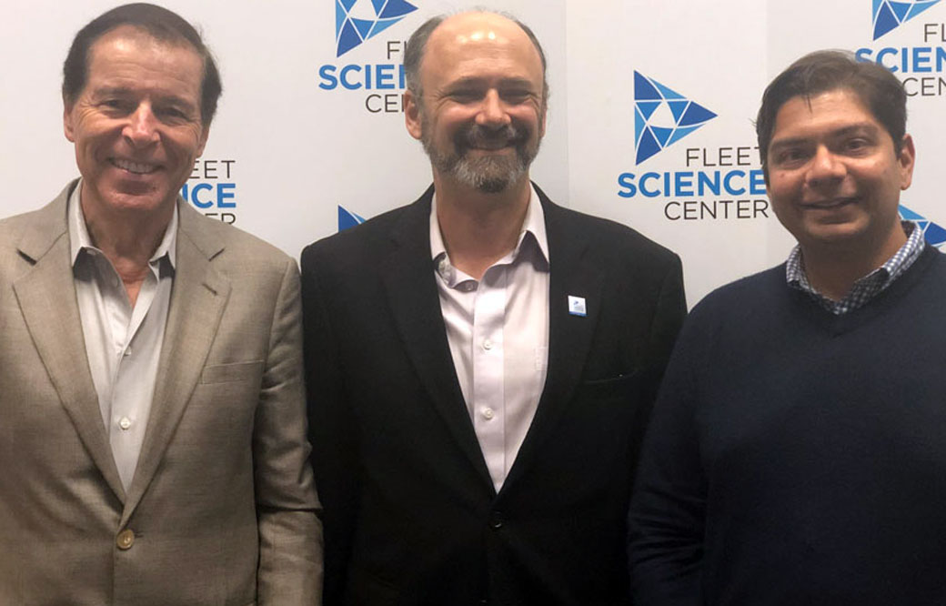
“Many people who live in San Diego aren’t aware of the incredible research advances taking place in their backyard, especially in cancer,” said speaker Garth Powis, D. Phil., professor and director of Sanford Burnham Prebys’ National Cancer Institute (NCI)-designated Cancer Center (on left). “We hope this discussion and future events will help more people understand cancer research and the breakthroughs that might come from their own community.”

Powis was joined by Hatim Husain, MD, a clinician at UC San Diego (on right); and Steven Snyder, PhD, president and CEO of the Fleet Science Center (center), who moderated the discussion. The speakers described how targeted treatments, which are only prescribed if a patient’s tumor has a specific mutation; and immunotherapies, which harness a patient’s immune system to melt the tumor, are extending survival for lung cancer patients. Husain expressed excitement surrounding new blood tests to detect lung cancer—which he hopes will be more commonplace in five to ten years. The speakers also noted that advances made in lung cancer have the potential to extend to other tumor types. 

“Many of the mutations that drive lung cancers are found in other tumors,” said Husain. “Targeted treatments that shrink lung tumors are being studied broadly in patients with a variety of cancers.” 

Powis and Husain also touched on their own collaboration to learn how lung cancer becomes resistant to treatment. Fluid buildup in the pleural space, the area between the lungs and chest wall, is often removed during routine checkups to help patients breathe. Working with Husain, Powis’ team is tracking the cellular and molecular makeup of this pleural fluid over the course of the disease. By regularly analyzing this fluid, they hope to gain insights into how lung cancer becomes treatment resistant and how it can be stopped.

“Scientists are getting close to mapping all of the mutations that drive lung cancer growth,” said Powis. “One day, patients may take one pill that contains all the anti-cancer compounds they need to fight the tumor.”

Upcoming topics in the series include breast, brain, and pancreatic cancer and more. The events will take place from 7:00 p.m. to 8:30 p.m. on select Sundays in the Heikoff Giant Dome Theater at the Fleet Science Center in San Diego. Space is limited. Reserve your ticket today. 

Institute News

What SBP Scientists are Researching to Battle Skin Cancer

AuthorHelen I. Hwang
Date

May 16, 2017

Skin cancer is one of the most common of all cancers, and melanoma accounts for about 1 percent of skin cancers. However, melanoma causes a large majority of deaths from that particular type of cancer. Alarmingly, rates of skin cancer have been on the rise in the last 30 years. Here in Southern California, our everlasting summer comes with a price. Exposure to sun increases our risk to melanoma.

Melanoma occurs when the pigment-producing cells that give color to the skin become cancerous. Symptoms might include a new, unusual growth or a change in an existing mole. Melanomas can occur anywhere on the body.

At Sanford Burnham Prebys Medical Discovery Institute (SBP), we have several researchers working on the causes of melanoma and discovering new ways to treat this deadly disease.

Here is a roundup of SBP’s latest research:

Key findings show how melanoma develops in order to identify potential therapeutic targets

Ze’ev Ronai, PhD
Professor and SBP Chief Scientific Advisor

Ronai’s laboratory has been studying how rewired signaling networks can underlie melanoma development, including resistance to therapy and metastatic propensity. One player in that rewiring is a protein called ATF-2, which can switch from its usual tumor-preventive function to become a tumor promoter when combined with a mutation in the human gene called BRAF.

Ronai’s work on a protein, ubiquitin ligases, led to the identification of RNF125 as an important regulator of melanoma resistance to a common chemotherapy drug. RNF125 impacts melanoma resistance by its regulation of JAK2, an important protein kinase which could play an important role in melanoma resistance to therapy.

Work on the ubiquitin ligase Siah2 identified its important role in melanoma growth and metastasis, and its contribution to melanomagenesis. Melanoma is believed to be a multi-step process (melanomagenesis) of genetic mutations that increase cell proliferation, differentiation, and death.

Work in the lab also concern novel metabolic pathways that are exploited by melanoma for their survival, with the goal of identifying combination drug therapies to combat the spread of melanoma. Earlier work on the enzyme PDK1 showed how it can be a potential therapeutic target for melanoma treatment.

Immunotherapy discovery has led to partnership with Eli Lilly

Linda Bradley, PhD
Professor, Immunity and Pathogenesis Program, Infectious and Inflammatory Diseases Center

Bradley’s group is focused on understanding how anti-tumor T cells can be optimized to kill melanoma tumors. They discovered an important molecule (PSGL-1) that puts the “break” on killer T cells, allowing melanoma tumors to survive and grow. Using animal models, they removed this “break” and T cells were able to destroy melanoma tumors. They have extended their studies and found that in melanoma tumors from patients, T cells also have this PSGL-1 “break”. Bradley’s lab has partnered with Eli Lilly to discover drugs that can modulate PSGL-1 activity in human disease that may offer new therapies for patients.

Knocking out a specific protein can slow melanoma growth 

William Stallcup, PhD
Professor, Tumor Microenvironment and Cancer Immunology Program

The danger of melanomas is their metastasis to organs, such as the brain, in which surgical removal is not effective. By injecting melanoma cells into the brains of mice, we have shown that the NG2 protein found in host tissues makes the brain a much “friendlier” environment for melanoma growth.

Specifically, NG2 is found on blood vessel cells called pericytes and on immune cells called macrophages. The presence of NG2 on both cell types improves the formation of blood vessels in brain melanomas, contributing to delivery of nutrients and thus to accelerated tumor growth. Genetically knocking out NG2 in either pericytes or macrophages greatly impairs blood vessel development and slows melanoma growth.

Mysterious molecule’s function in skin cancer identified

Ranjan Perera, PhD
Associate Professor, Integrative Metabolism Program

Ranjan’s research uncovered the workings of a mysterious molecule called SPRIGHTLY that has been previously implicated in colorectal cancer, breast cancer and melanoma. These findings bolster the case for exploring SPRIGHTLY as a potential therapeutic target or a biological marker that identifies cancer or predicts disease prognosis.

 Drug discovery to help babies has led to a clinical trial at a children’s hospital

Peter D. Adams, PhD
Professor, Tumor Initiation and Maintenance Program

Approximately 1 in 4 cases of melanoma begins with a mole, or nevus. Genetic mutations can cause cells to grow uncontrollably. By investigating how this occurs, we can understand why melanoma develops from some moles, but not others.

Babies born with a giant nevus that covers a large part of the body have especially high risk of melanoma, and the nevus cells can spread into their spine and brain. Adams’ research identified a drug that deters the cells from growing. The drug identified will be used in a clinical trial at Great Ormond Street Children’s Hospital in London, England that may help babies with this debilitating disease.

Discovery of a receptor mutation correlates with longer patient survival

Elena Pasquale, PhD
Professor, Tumor Initiation and Maintenance Program

Pasquale’s work has included whether mutations in the Eph receptor, tyrosine kinases, play a role in melanoma malignancy. Eph receptor mutations occur in approximately half of metastatic melanomas. We found that some melanoma mutations can drastically affect the signaling ability of Eph receptors, but could not detect any obvious effects of the mutations on melanoma cell malignancy.

Bioinformatic analysis of metastatic melanoma samples showed that Eph receptor mutations correlate with longer overall patient survival. In contrast, high expression of some Eph receptors correlates with decreased overall patient survival, suggesting that Eph receptor signaling can promote malignancy.

Institute News

Measuring heart toxicity of cancer drugs in a dish

AuthorJessica Moore
Date

February 22, 2017

A class of cancer drugs known as tyrosine kinase inhibitors (TKIs) are often damaging to the heart, sometimes to the degree that they can’t be used in patients. These toxic effects are not always predictable using current preclinical methods, so they may not be discovered until they make it to clinical trials.

New research could make it possible to tell which TKIs cause heart toxicity without putting any humans at risk. The collaborative study, involving Wesley McKeithan, a PhD student in the Sanford Burnham Prebys Medical Discovery Institute (SBP) graduate program and Mark Mercola, PhD, adjunct professor at SBP and a professor at Stanford University, used lab-grown heart muscle cells to assess the drugs’ potential to cause damaging effects.

“This new method of screening for cardiotoxicity should help pharma companies focus their efforts on TKIs that will be safe,” says Mercola, who collaborated with Joseph Wu, MD, PhD, also a professor at Stanford, on the study published in Science Translational Medicine. “That could mean better new TKIs will make it to the market, since we will be able to predict whether or not they cause heart problems early in the development process.”

TKIs with tolerable cardiac side effects, which include imatinib (Gleevec) and erlotinib (Tarceva), are widely used to treat multiple types of cancer. Because tumors often become resistant to these drugs, new compounds in this class continue to be developed to provide replacement treatments. Other TKIs can harm the heart in a variety of ways, from altering electrical patterns to causing arrhythmias, reducing pumping capacity, or even increasing risk of heart attacks.

Mercola and Wu’s team used heart muscle cells derived from induced pluripotent stem cells (iPSCs), which can be generated from adult skin or blood cells. After treating heart muscle cells with one of 21 TKIs, they assessed their survival, electrical activity, contractions (beating) and communication with adjacent cells. They used a new method for measuring heart cell contraction developed by the lab of Juan Carlos del Álamo, Ph.D., at UC San Diego to create a ‘cardiac safety index’, which correlates in vitro assay results with the drugs’ serum concentrations in humans. Importantly, the safety index values matched nicely with clinical reports on the cardiotoxicity of currently used TKIs.

The study also identified a possible way to protect heart muscle cells from impairment caused by TKIs—treating them with insulin or insulin-like growth factor. Although more research is needed, the findings suggest that it may be possible to alleviate some of the heart damage in patients receiving these chemotherapies.

Mercola adds, “By using cells derived from a broader group of individuals, this screening strategy could easily be adopted by the pharma industry to predict cardiotoxicity.”

This story is based in part on a press release from Stanford University School of Medicine.

Institute News

Beating prostate cancer

Authorjmoore
Date

November 7, 2016

About one in seven men will be diagnosed with prostate cancer during his lifetime. Though it has one of the highest survival rates of any type of cancer—95% make it through the first ten years—diagnosis and treatment could still be improved. Since November is Prostate Cancer Awareness Month, we’re highlighting the work our scientists are doing to address key challenges and unresolved questions in prostate cancer.

Early, accurate detection—The current method of screening for prostate cancer is a blood test for prostate specific antigen, or PSA, which detects cancer early, but isn’t very specific—only one in four men with high PSA levels actually has cancer. In general, high levels of PSA mean the next step is a biopsy. A more specific test would avoid unnecessary biopsies, which are invasive and stressful for patients.

Ranjan Perera, PhD, associate professor in the Integrative Metabolism Program, is looking for biomarkers that would enable just such a test. His lab is making progress—they identified five long noncoding RNAs (RNAs that, instead of carrying genetic information to be translated, regulate the translation of other RNAs) found at higher-than-normal levels in the urine of prostate cancer patients. An RNA-based test is on the market, but could be improved—measuring multiple markers would be more sensitive and specific.

Better therapies—For advanced cases, current treatments are either insufficient or overly toxic. Prostate cancer is usually first treated with drugs that block the actions of androgens, the hormones that drive its growth. If the tumor recurs later, as happens for cancers that are already at a late stage before treatment, it forms from cells that are resistant to those drugs. Then, the only option is chemotherapy or radiation.

Towards therapies that cause less collateral damage, Nicholas Cosford, PhD, associate director of Translational Research in the NCI-designated Cancer Center, is designing new drugs against targets found to be important in prostate cancer. These drugs are intended to block two strategies by which cancer cells survive—avoiding cell death, and generating extra energy by recycling the cell’s own parts.

Understanding why obesity correlates with aggressiveness—Obese men are no more likely than others to get prostate cancer, but if they do, it’s more likely to become advanced quickly. To figure out why this happens, Jorge Moscat, PhD, director, and Maria Diaz-Meco, PhD, professor in the Cancer Metabolism and Signaling Networks Program, are looking at how fat adjacent to the prostate interacts with tumor cells. A detailed picture of how fat cells and tumor cells interact could reveal new ways to treat prostate cancer in overweight men.

Institute News

Leukemia research breakthrough: a new way to trigger cancer cell suicide

AuthorJessica Moore
Date

May 18, 2016

Better therapies for acute myeloid leukemia (AML), a fast-growing cancer of the bone marrow, are urgently needed. Nearly 15,000 people in the United States are diagnosed with AML each year, and it’s the most common acute leukemia in adults. The cause of the disease is unknown, and it is usually fatal within the first five years. Continue reading “Leukemia research breakthrough: a new way to trigger cancer cell suicide”

Institute News

Cancer metabolism 101

Authorsgammon
Date

April 21, 2015

“Feed me!” Cancer is caused by the uncontrolled proliferation of cells. Their rapid growth comes with a voracious appetite to support their nutritional demands. To satisfy these demands, cancer cells rewire their metabolism. Increasingly, scientists are looking to exploit the metabolic differences between normal and cancer cells for the development of new anti-cancer therapies. Continue reading “Cancer metabolism 101”

Institute News

Expanding the options to treat melanoma

Authorsgammon
Date

March 16, 2015

Melanoma is the most deadly form of skin cancer with approximately 10,000 deaths per year in the U.S. and more than 65,000 worldwide. Although there are more and better treatment options available today than in previous years, there is still an urgent need to develop drugs that target the numerous pathways melanoma cells use to multiply, spread, and kill. Continue reading “Expanding the options to treat melanoma”

Institute News

Molecule that fixes “leaky” blood vessels can impact cancer, stroke, and blindness

Authorsgammon
Date

March 13, 2015

In a new study by Masanobu Komatsu, Ph.D., associate professor in the Cardiovascular Pathobiology Program and Tumor Microenvironment and Metastasis Programs, a cellular protein called R-Ras was found to suppress the effects of vascular endothelial growth factor (VEGF), a signaling molecule that helps create new blood vessels and is overexpressed in many tumors. The findings create a new route to treat cancer as well as certain causes of blindness and ischemic diseases. Continue reading “Molecule that fixes “leaky” blood vessels can impact cancer, stroke, and blindness”