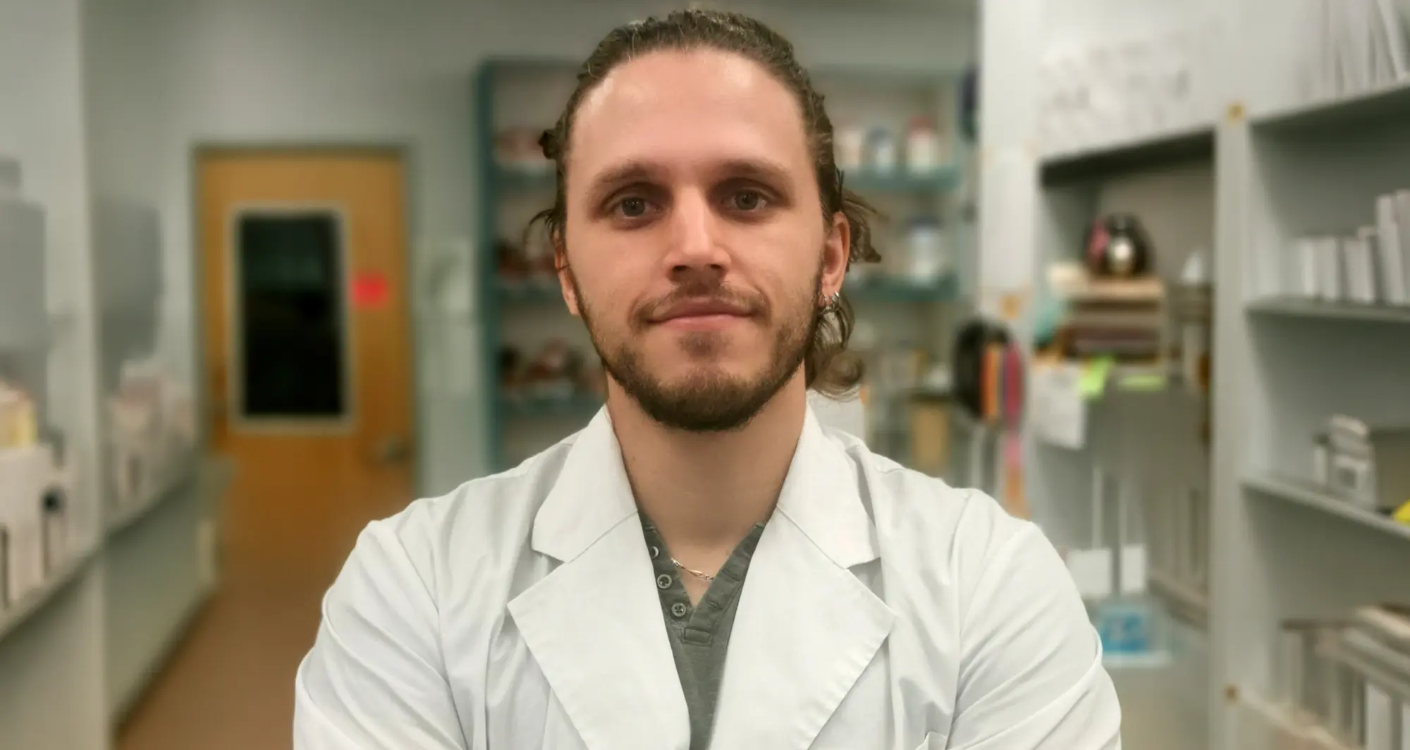
Jimmy Massenet, PhD, is a postdoctoral associate in the lab of Pier Lorenzo Puri, MD, a professor in the Center for Cardiovascular and Muscular Diseases at Sanford Burnham Prebys.
Massenet, a postdoctoral associate in the lab of Pier Lorenzo Puri, MD, was selected to attend the 2026 Muscle Stem Cells and Regeneration Meeting held from July 19-24, 2026, in Victoria, the capital of the Canadian province of British Columbia.
The goal of the Science in Motion Travel Award is to support conference participation for emerging researchers in labs with a primary affiliation in the Center for Cardiovascular and Muscular Diseases.
Applications will open again on July 15, 2026.
Institute News
Two trainees selected for Science in Motion Travel Awards
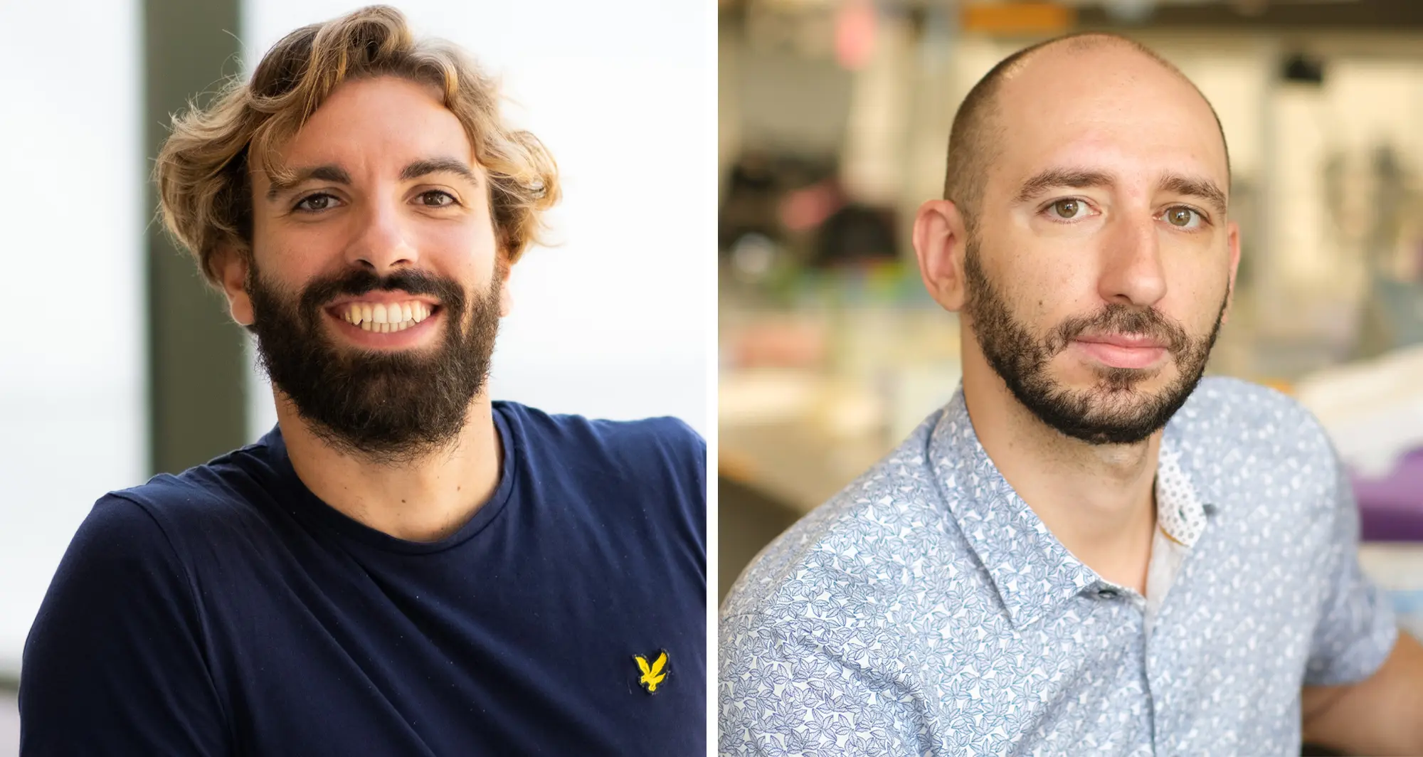
Gabriele Guarnaccia, a graduate student in the lab of Alessandra Sacco, PhD, will use his award to attend the 8th Cancer Cachexia Conference being held September 25–27, 2025, in Turin, Italy.
Luca Caputo, PhD, a postdoctoral associate in the lab of Pier Lorenzo Puri, MD, was selected to attend Frontiers in Myogenesis: Innovations in Myogenesis, From Molecular Mechanisms to Therapeutic Interventions, which will be held October 6–11, 2025, in Sunriver, Oregon.
The goal of the Science in Motion Travel Awards is to support conference participation for emerging researchers in labs with a primary affiliation in the Center for Cardiovascular and Muscular Diseases.
Applications will open again on January 15, 2026.
Institute News
Decades of dedication led to FDA approval of a new treatment for Duchenne Muscular Dystrophy
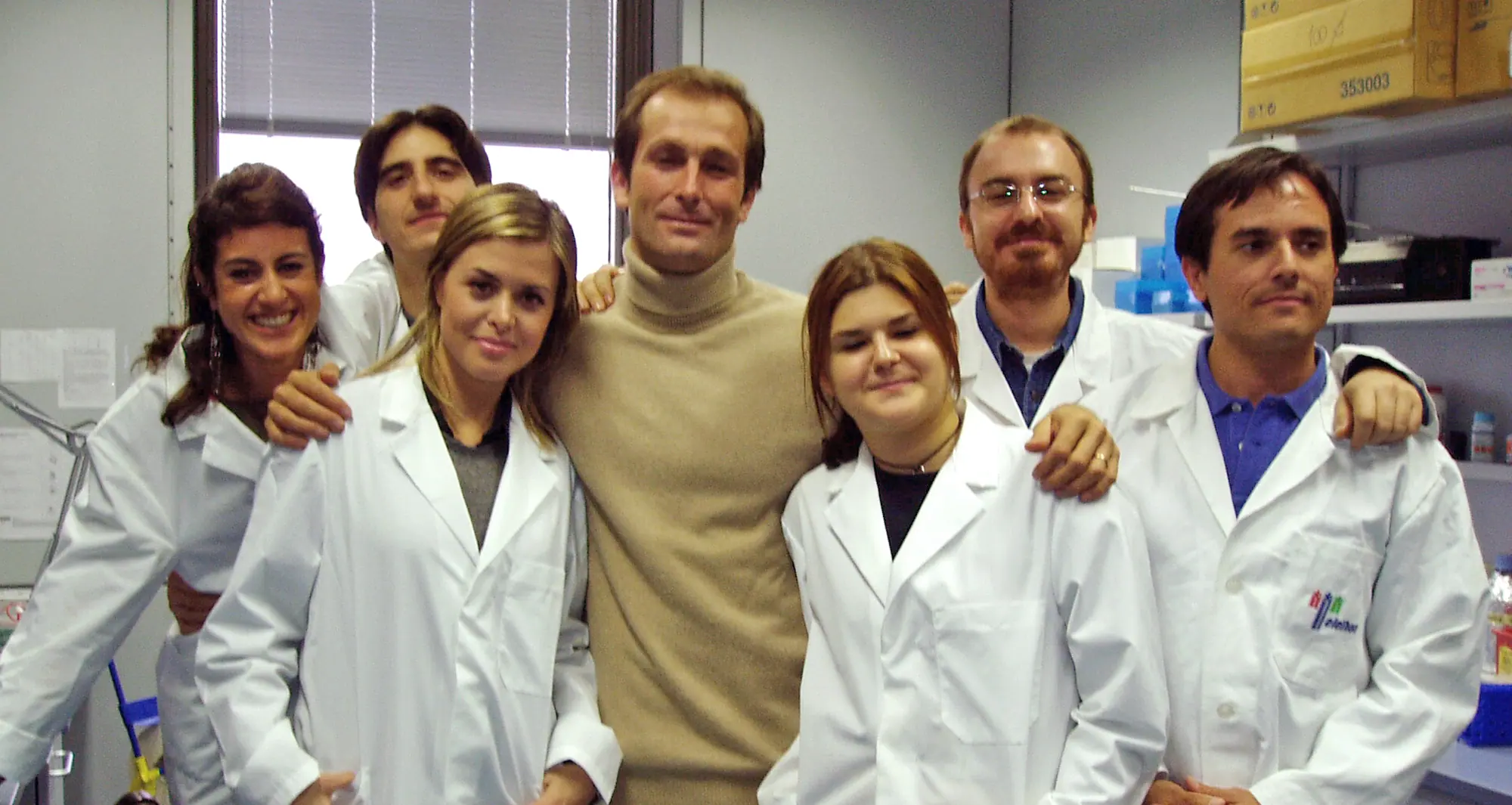
Lorenzo Puri with his lab in Rome in 2004. From left: Lucia Latella, PhD; Silvia Fortuni, PhD; Christian Reale, PhD; Puri; Giulia Minetti, PhD; Cristiano Simone, PhD; and Carlo Serra, MD.
Nearly 30 years of discoveries by a Sanford Burnham Prebys scientist and collaborators lead to federal approval of the first non-steroidal drug to treat Duchenne muscular dystrophy.
For one San Diegan scientist at Sanford Burnham Prebys, the March 2024 federal approval of a new drug to treat Duchenne muscular dystrophy (DMD) marked a milestone in three decades of studying muscle regeneration and muscle-wasting diseases.
The compound, called Givinostat and marketed as DUVYZAT™, is a histone deacetylase inhibitor (HDACi)and was approved by the FDA for the treatment of boys with DMD.
“I have been working from the very beginning of my research career to translate early, basic discoveries into a treatment for DMD,” said Pier Lorenzo Puri, MD, director and professor in the Development, Aging and Regeneration Program at Sanford Burnham Prebys. “The lack of effective treatments for boys with DMD has left families and patients hopeless since the discovery of this disease. Witnessing the progression of such a disease without any option to counter its progression is cruel and I felt the urgency to help these people.”
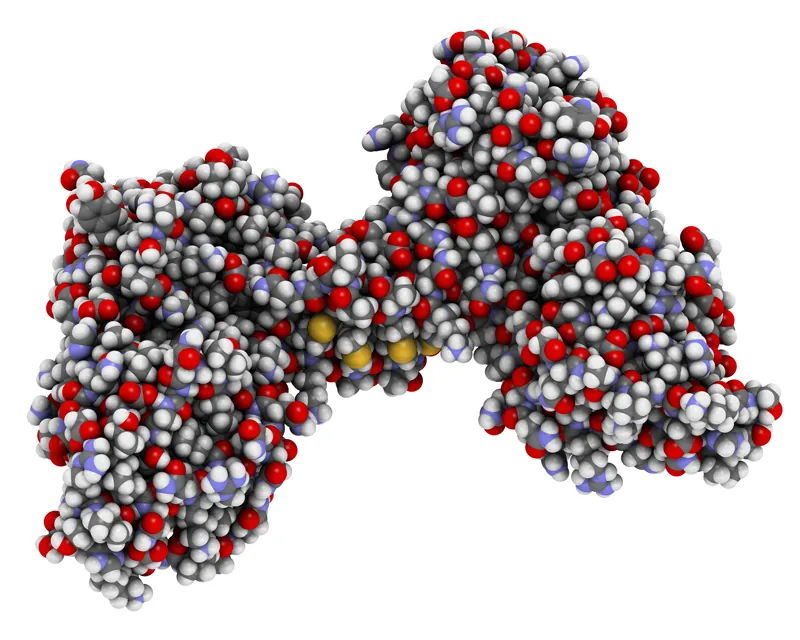
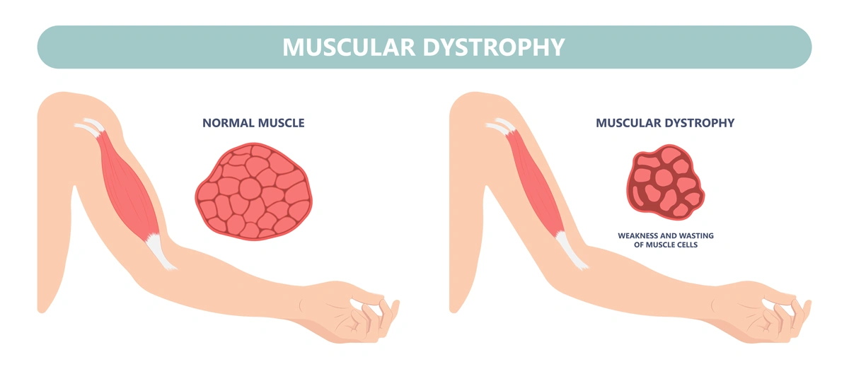
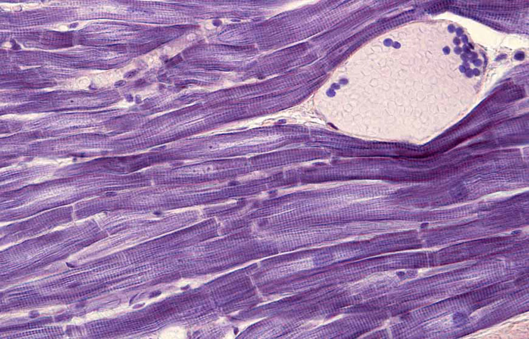
DMD is the most frequent form of muscular dystrophy affecting approximately 1 in 3,500 male births. DMD is linked to the chromosome X, which harbors the gene coding the protein called dystrophin. As such, the disease develops only in males receiving from their mother the X chromosome carrying mutations in the dystrophin gene that impair production of dystrophin. Dystrophin is a protein that protects muscles from degeneration after they contract and relax. In its absence, the muscles of boys with DMD are prone to damage and undergo cycles of contraction and degeneration that eventually lead to muscle wasting and reduced function.
For decades, steroids have provided the standard-of-care for DMD, but steroids represent an empirical and palliative treatment based on their general anti-inflammatory properties, rather than a treatment that targets specific pathological events that contribute to DMD progression. Moreover, the chronic use of steroids is complicated by many side effects, including weight gain, weak bones, high blood pressure and behavior changes.
Puri decided to focus on the potential use of HDACi to treat DMD after he made seminal basic discoveries that revealed how muscle growth and regeneration are regulated by two enzymes with opposing activities: histone acetyltransferases and deacetylases.
“HDACi are not going to cure muscular dystrophy, but they do provide the first pharmacological treatment that can delay DMD progression, regardless of the type of mutation, and do so in a financially affordable way,” said Puri.
The molecular structure of dystrophin, a protein that protects muscles from degeneration after they contract and relax.
“It is important to note that FDA approval of Givinostat is not the end of the journey, but the beginning. The very good news here is that there is room to improve the efficacy of HDACi-based treatment for DMD by using already existing compounds or by developing novel molecules endowed with an improved therapeutic potential. This is because Givinostat has been used at sub-optimal concentrations due to potential adverse effects, and this might have limited its efficacy as a HDACi.
“People ask me whether Givinostat was approved because it is the most effective molecule among existing HDACi. The answer is that Givinostat has been the first, and so far the only, HDACi to be tested in clinical trials for boys with DMD. Givinostat might not entirely express the therapeutic potential of HDACi for DMD. A reasonable and exciting goal of future studies is to identify HDACi that surpass Givinostat in terms of therapeutic efficacy.”
Without dystrophin, the muscles of boys with Duchenne muscular dystrophy are prone to damage and undergo cycles of contraction and degeneration that eventually lead to muscle wasting and reduced function.
Puri noted that, “It is also important to carefully investigate functional interactions between HDACi and steroids, as the clinical trial with Givinostat has been performed while research participants were receiving steroids. However, because the activity of steroids is largely dependent on HDAC, it is very likely that these two treatments could collide, rather than synergize in producing beneficial effects on the muscles of DMD boys.”
He also emphasized that the impact of FDA approval of Givinostat extends way beyond the possibility to offer a treatment for DMD.
“It is the first evidence that it is possible to treat DMD by targeting pathogenic events induced as a consequence of dystrophin deficiency, as an effort parallel to gene and cell therapy, which will hopefully converge into future and more effective combined therapies.”
Building the basic science foundation
Puri’s contributions to the approval of the first non-steroidal drug to treat DMD span nearly 30 years of basic and preclinical research of diseases thought to be incurable — especially pediatric conditions he thought especially cruel.
The puzzle pieces began to take shape as Puri was studying the growth of muscle cells and skeletal muscle tissue, a biological process called skeletal myogenesis. He started working on this project in the 1990s in the laboratory of molecular biology directed by Massimo Levrero, MD, in Rome. Puri had earned his medical degree in 1991 before conducting an internship in Internal Medicine.
“I started there, in a lab inside a hospital in Rome,” said Puri. “I used to see patients until the early afternoon and then I was running to the lab to perform experiments. As a young clinician with a passion for basic research, I was always developing experiments with patients in mind.”
Puri decided to test his first hypothesis by using what was then an innovative technology called microinjection to insert DNA or antibodies inside cultured cells. He decided to spend several months at the Free University of Berlin in the laboratory of Adolf Graessmann, PhD, a pioneer of this technique. Puri later decided to go to the United States to work in the lab of Jean Y.J. Wang, PhD, at the University of California San Diego, to further develop his studies.
After entering the U.S., Puri has worked closely with his long-term collaborator and friend Vittorio Sartorelli, MD, currently the deputy scientific director of the National Institute of Arthritis and Musculoskeletal and Skin Diseases and chief of the institute’s Laboratory of Muscle Stem Cells & Gene Regulation. Together, they uncovered an important role for two groups of enzymes, histone acetyltransferases and deacetylases, that control access to DNA by altering the structure of chromatin.
Histone acetyltransferases control DNA accessibility by adding acetyl groups to histones, which loosens the wrapping of DNA around them, essentially ‘opening’ chromatin and promoting gene expression. Histone deacetylases reverse the process by removing acetyl groups, limiting the activity of genes and the production of key proteins.
“One of our seminal findings was the discovery of associations between myogenesis and an enzymatic activity that could be pharmacologically modulated,” explains Puri.
Puri with current members of hislab at Sanford Burnham Prebys.
Puri and Sartorelli started to explore the possibility that pharmacological modulators of this process by HDACi could affect the growth of muscle cell progenitors and their ability to form contractile muscle tissues.
After finding that HDACs limit muscle cell differentiation, the team’s next step was to find compounds (inhibitors) capable of blocking HDACs from removing acetyl molecules and reducing myogenesis. “We decided see if there is an HDAC inhibitor — an inhibitor of the inhibitor — because this could reduce muscle loss,” notes Puri. “There were a few compounds, and we found a strong effect every time we tested them.”
“I remember vividly the first time I saw the effect of HDACi on cultured muscle cells. I was a postdoc in Jean Wang’s lab at UC San Diego, and one morning I opened the incubator and found that muscle cells treated with HDACi had formed giant myotubes (the contractile muscle). Of course, we started to wonder whether such evidence could provide the rationale we were looking for. If so, this could pave the way toward discovering pharmacological interventions that may promote muscle regeneration.
Puri with Chiara Mozzetta, PhD, faculty at the Institute of Molecular Biology and Pathology at the National Research Council of Italy. Mozzetta previously conducted a postdoctoral fellowship in the Puri lab and made major contributions to the discovery of HDAC inhibitors as therapeutics for DMD.
“But, at that time, it sounded more like a dream. We did not even dare to imagine that the final outcome would have been the identification of a pharmacological treatment for DMD.”
Pursuing the preclinical potential
Puri and Sartorelli pursued their dream, driven by encouraging experimental evidence and discoveries. Still, it took years to provide the rationale for testing HDACi in DMD—years and a few fortunate coincidences. One was the identification of follistatin as a mediator of the action of HDACi on muscle cells. Follistatin is the endogenous inhibitor of myostatin, a potent inhibitor of muscle growth and size, ensuring that muscles do not grow too large.
It was during that time that other groups independently discovered that genetic mutations in the myostatin gene resulted in an abnormal increase in muscle mass in cattle, mice and humans. More importantly, it was published that genetic or pharmacological inactivation of myostatin could exert beneficial effects in a mouse model of DMD.
“That discovery suggested the rationale of testing whether HDACi could exert similar beneficial effect in the same mouse model of DMD,” said Puri. The team hypothesized that this could be achieved by using HDACi to block myostatin activity through the induction of follistatin.
Puri and Sartorelli decided to treat mdx mice — the mouse model of DMD — with a few HDACi. Puri performed these studies with a group of investigators that worked synergistically in his two labs (one in San Diego and the Dulbecco Telethon Institute laboratory in Rome) and in collaboration with Italian scientists Carlo Gaetano, PhD, and Claudia Colussi, PhD.
“The results of the experiment went beyond the most optimistic expectations,” said Puri. “The muscles were bigger. There was no scar tissue, no abnormal fatty deposits and less inflammation. The treated mice were running like normal mice.”
In follow-up studies, Puri and collaborators realized that the therapeutic properties of HDACi extended beyond the targeting of follistatin/myostatin interactions. The pace of discoveries increased along with the improved knowledge on DMD pathogenesis. Key and timely information was gained through identification of a population of muscle interstitial cells, called fibro-adipogenic progenitors (FAPs), from the laboratories of Fabio Rossi, MD, PhD, at the University of British Columbia in Canada and Dr. Kunihiro Tsuchida, MD, PhD, at Fujita Health University in Japan.
FAPs support muscle regeneration of normal muscles, but in dystrophin-deficient muscles these cells turn into the main effectors of fibrotic scars and fat infiltration — the most deleterious events in DMD pathogenesis. Studies from the Puri lab demonstrated that HDACi could restore the ability of FAPs from DMD muscles to promote regeneration, while blocking their pro-fibrotic and adipogenic activity.
Puri with collaborator Vittorio Sartorelli, MD, the deputy scientific director of the National Institute of Arthritis and Musculoskeletal and Skin Diseases.
“The identification of FAPs was a lucky coincidence, as it provided one of the main cell types targeted by HDACi and enabled the identification of the molecular mechanism of action that accounts for the therapeutic properties of HDACi,” said Puri.
Puri (at right) with collaborator Fabio Rossi, MD, PhD, professor of Medical Genetics at the University of British Columbia in Canada, at a muscle research meeting in Montrealin July 2024.
“It also helped to set in our preclinical studies the parameters that have been used in clinical trials. Indeed, I believe that one of the reasons for the success of this journey has been the development of a solid scientific rationale. I was definitely fortunate to have worked with a team of incredibly skilled young scientists that shared with me the wish to help DMD patients and the perseverance to take on a challenge for over 20 years. Overall, discovering the MOA of HDACs has been a fantastic journey.”
“Although the evidence that HDACi could be used to treat DMD was strong and the rationale was very solid, it was hard to convince big pharma to invest on this treatment. There were many excellent HDACi in the market that could have been used, but after knocking on many doors, no one was willing to partner on this task.
Puri felt compelled by these results to keep building support to convince potential industry partners to develop clinical trials. After talking with many businesses, he found Italfarmaco, an Italian pharmaceutical company with an HDAC inhibitor called Givinostat.
“In the end, I had to knock on the door of Italfarmaco, which owned Givinostat — an HDACi that I had never tested in my previous preclincial studies. This was another coincidence since Dr. Christian Steinkulher, a friend and colleague, alerted me about the potential availability of a pan-HDACi called Givinostat from Italfarmaco.
“When I approached Italfarmaco, I immediately realized that they were not very well prepared to take on this type of research. They were not familiar with DMD, and they didn’t have any previous experience on muscular diseases. They also had no background on the epigenetic effects of Givinostat.
“Until that time, they used Givinostat mostly for its anti-inflammatory properties, rather than the epigenetic regulation of gene expression. However, Italfarmaco recognized some potential in this operation and decided to give to me the opportunity to perform preclinical studies with Givinostat for DMD.”
Clinical trials and regulatory approval
After additional preclinical studies to better understand how Givinostat worked, Italfarmaco informed Puri that the company was ready to develop a clinical trial.
The results of the phase II clinical trial were published in Neuromuscular Disorders in 2016. During the trial, the scientists looked at whether the composition of the muscle improved. Muscle biopsies taken from 19 subjects who were treated for more than a year demonstrated that the drug had caused an increase in muscle fiber area and a decrease in fat deposition, scar tissue and other hallmarks of DMD.
“It was remarkable to see that the same positive effects observed in mdx mice treated with HDACi were also observed in DMD boys treated with Givinostat,” noted Puri. “The reproducibility of the outcomes in preclinical studies and clinical trials emphasizes the importance of performing accurate preclinical studies and identifying reliable outcome measures, as we did with HDACi for DMD.”
The trial also helped the team determine what dose of Givinostat was safe and still effective to use in the pivotal phase III trial that was reviewed by the FDA before the agency granted approval.
Italfarmaco established ITF Therapeutics in January 2024 as a new division that is now responsible for marketing DUVYZAT™ in the U.S. ITF Therapeutics announced that the drug was available in the U.S. on July 25. Italfarmaco’s Marketing Authorization Application (MAA) for Givinostat to the European Medicine Agency (EMA) was validated in fall 2023. This means that the drug is eligible to be reviewed by the EMA. If approved, Givinostat can be made available throughout the European Union (EU).
Puri surfs a wave in San Diego. Surfing is his passion outside of the laboratory.
Paving the way forward
Puri is not resting on his laurels and the approval of the first non-steroidal drug to treat DMD.
“Right now, we have steroids, HDAC inhibitors and gene therapy. We are working on the idea that gene therapy and HDAC inhibitors without steroids can perfectly synergize.”
Lorenzo and his dog Mojo.
The researchers are also investigating ways to enhance the effect of HDAC inhibitors through the use of extracellular vesicles (EV) released from FAPs following the exposure to HDACi. EVs are small biological bubbles that the body uses to carry compounds between cells. They are non-immunogenic and therefore suitable for transplantation into dystrophic muscles.
Puri is also investigating whether there are treatment conditions (including dietary supplements or other synergistic molecules) that can improve the therapeutic efficacy of HDACi. The researchers want to test if HDAC inhibitors can treat other forms of muscular dystrophy beyond DMD.
As much as Puri is focused on the future and continuing to find new and better approaches to treat muscular dystrophy, he also appreciates the importance of this vital moment and how the FDA’s decision positions the field for even more innovation.
“While muscular dystrophy was formally described by scientists 40 years ago, it has been a part of the human story since the beginning. People have been chasing something that could help, and for so long there was nothing to offer. Right now, we are paving the way for even better treatments that will be found.”
More information on the development of HDACi as a treatment for DMD is available in the following manuscripts:
Puri P.L., Avantaggiati M.L., Balsano C., San N., Graessmann A., Giordano A., and Levrero M. p300 is required for MyoD-dependant cell cycle arrest and muscle specific gene transcription. EMBO J. 16,369-383 (1997)
Puri P.L., Sartorelli V., Yang X.J., Hamamori Y., Ogrizko , Howard B., Kedes L, Wang J.Y.J., Graessmann A., Nakatani Y., Levrero M. Differential roles of p300 and PCAF acetyltransferases in muscle differentiation. Mol. Cell 1, 35-45 (1997)
Sartorelli V*., Puri P.L.* , Hamamori Y, Ogrizko V., Nakatani Y., Wang J.Y.J., Kedes L. Acetylation of MyoD directed by PCAF is necessary for the execution of the muscle program. Mol. Cell. 4, 725-734 (1999). *equal contribution
Puri P.L., Iezzi, S., Stiegler P., Chen T.T., Shiltz L., Muscat G., Giordano A, Wang J.Y.J. and Sartorelli V. Class I histone deacetylases sequentially interact with MyoD and pRb during skeletal myogenesis. Mol Cell. 8, 885-897 (2001)
Iezzi S., Cossu G., Nervi C. Sartorelli V., and Puri P.L. Stage-specific modulation of skeletal myogenesis by inhibitors of nuclear deacetylases Proc. Natl. Acad. Sci 99, 7757-7762 (2002)
Iezzi S., Di Padova M., Serra C., Caretti G., Simone C., Maklan E., Zhao P., Hoffman E., Puri P.L. and Sartorelli V. Deacetylase Inhibitors Increase Muscle cell Size by Promoting Myoblast Recruitment and Fusion Through Induction of Follistatin. Dev. Cell. 5:673-84. (2004).
Minetti G. C., Colussi c., Adami R., Serra C., Mozzetta C., Parente V., Illi B., Fortuni S., Straino S., Gallinari P., Steinkhuler C., Capogrossi M., Sartorelli V., Bottinelli R., Gaetano C., Puri P.L. Functional and morphological recovery of dystrophic muscles in mice treated with deacetylase inhibitors. Nature Medicine 12 (10): 1147-50 (2006)
Colussi C., Mozzetta C., Gurtner A. , Illi B., Straino S., Ragone G., Pescatori M., Zaccagnini G., Rosati G., Minetti G., Martelli F., Ricci E., Piaggio G., Gallinari P., Steinkulher C., Capogrossi M.C., Puri P.L*, Carlo Gaetano*. A Common Epigenetic Mechanism Underlies Nitric Oxide Donors and Histone Deacetylase Inhibitors Effect in Duchenne Muscular Dystrophy. Proc. Natl. Acad. Sci 105, 19183-7 (2008) *Corresponding authors. PMCID:PMC2614736.
Mozzetta C., Consalvi S., Saccone V., Tierney M., Diamantini A., Mitchel K.J., Marazzi G., Borsellino G., Battistini L., Sassoon D., Sacco A., Puri P.L. Fibroadipogenic progenitors mediate the ability of HDAC inhibitors to promote regeneration in dystrophic muscles of young, but not old mdx mice. EMBO Mol. Med. (2013), Apr;5(4):626-39 doi: 10.1002/emmm.201202096. [Epub ahead of print]. PMC Journal In Process.
Consalvi S, Mozzetta C, Bettica P, Germani M, Fiorentini F, Del Bene F, Rocchetti M, Leoni F, Mascagni P, Puri P.L., Saccone V. Preclinical studies in the mdx mouse model of Duchenne Muscular Dystrophy with the Histone Deacetylase inhibitor Givinostat. Mol Med. 2013 Mar 27. doi: 10.2119/molmed.2013.00011. [Epub ahead of print]. PMCID: PMC3667212.
Saccone V, Consalvi S, Giordani L, Mozzetta C, Barozzi I, Sandoná M, Ryan T, Rojas-Muñoz A, Madaro L, Fasanaro P, Borsellino G, De Bardi M, Frigè G, Termanini A, Sun X, Rossant J, Bruneau BG, Mercola M, Minucci S, Puri P.L. HDAC-regulated myomiRs control BAF60 variant exchange and direct the functional phenotype of fibro-adipogenic progenitors in dystrophic muscles. Genes & Development. 2014 Apr 15;28(8):841-57. doi: 10.1101/gad.234468.113. Epub 2014 Mar 28.
Bettica P, Petrini S, D’Oria V, D’Amico A, Catteruccia M, Pane M, Sivo S, Magri F, Brajkovic S, Messina S, Vita GL, Gatti B, Moggio M, Puri P.L., Rocchetti M, De Nicolao G, Vita G, Comi GP, Bertini E, Mercuri E. Histological effects of givinostat in boys with Duchenne muscular dystrophy.Neuromuscul Disord. 2016 Jul 11. pii: S0960-8966(16)30069-4. doi: 10.1016/j.nmd.2016.07.002. [Epub ahead of print]
Consalvi S, Tucciarone L, Macrì E, De Bardi M, Picozza M, Salvatori I, Renzini A, Valente S, Mai A, Moresi V, Puri P.L.. Determinants of epigenetic resistance to HDAC inhibitors in dystrophic fibro-adipogenic progenitors. EMBO Rep. 2022 Jun 7;23(6):e54721. doi: 10.15252/embr.202254721. Epub 2022 Apr 4. PMID: 35383427
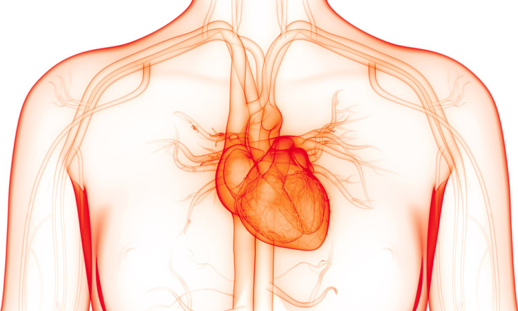
A collaborative study led by researchers at Sanford Burnham Prebys is paving the way to identifying gene networks that cause atrial fibrillation (AFib), the most common age-related cardiac arrhythmia.
The findings, published in Disease Models & Mechanisms, validate an approach that combines multiple experimental platforms to identify genes linked to an abnormal heart rhythm.
“One of the biggest challenges to solving the AFib genetic puzzle has been the lack of experimental models that are relevant to humans,” says Alex Colas, PhD, co-senior author and assistant professor in the Development, Aging and Regeneration Program at Sanford Burnham Prebys. “By working with colleagues who focus on AFib but in different systems, we have created a robust multiplatform model that can accurately pinpoint genes associated with this condition.”
AFib is characterized by an irregular, rapid heartbeat that causes a quivering of the upper chambers of the heart, called the atria. This condition is the result of a malfunction in the heart’s electrical system that can lead to heart failure and other heart-related complications, which include stroke-inducing blood clots.
AFib impacts more than 5.1 million people in the United States, with expectations of 15.9 million by 2050. It is more common in individuals over the age of 60 but can also occur in teenagers and young adults.
“There will never be a one-size-fits-all solution to AFib, since it can be caused by many different genes—and the genes that do cause it vary from person to person,” says Karen Ocorr, PhD, also a co-senior author and assistant professor in the Development, Aging and Regeneration Program at Sanford Burnham Prebys. “A better understanding of the gene network(s) that contribute to AFib will help us design tests to predict a person’s risk, and develop individualized approaches to treat this dangerous heart condition.”
To overcome the limitations of current AFib research models, Colas, Ocorr and researchers from UC Davis and Johns Hopkins University combined forces to assemble a multi-model platform that combines:
A high-throughput screen using atrial-like cells (derived from human-induced pluripotent stem cells) to measure how a gene mutation alters the strength and duration of a heartbeat.
A Drosophila (fruit fly) model—with heart genetics and development remarkably similar to human hearts—that permits analysis of gene mutations in a functioning organ.
A well-established computational model that uses computers to simulate the effects of gene mutations on the electrical activity in human atrial cells.
The accuracy of the multi-model platform was confirmed when each screened 20 genes, and all three platforms identified phospholamban, a protein found in the heart muscle with known links to AFib.
“This collaboration has greatly expanded our ability to understand AFib at the genetic level,” says Colas. “Importantly, the high-throughput screening component of the model will also allow us to rapidly and effectively screen for drugs that can restore a heart to its normal rhythm.”
He adds, “Hopefully this is just the beginning. There are many more cardiac diseases to which our system can be applied.”
Institute News
José Luis Millán joins international initiative to study calcification in aging
Sanford Burnham Prebys professor José Luis Millán, PhD, has joined a five-year, $13 million program that will study misplaced calcification in the eyes and brains of patients suffering from age-related macular degeneration (AMD) and Alzheimer’s disease (AD).
The initiative is funded by the National Institute on Aging and will be led by Francesca Marassi, PhD, an adjunct professor at Sanford Burnham Prebys and chair of biophysics at the Medical College of Wisconsin.
AMD affects nearly 20 million adults in the U.S. and is the leading cause of central vision loss and legal blindness. AD affects more than 6 million people in the U.S., and it is the top cause of dementia across the globe. Age is a prominent risk factor for both diseases. However, how AMD and AD progress over time is not well understood, and research is needed to drive the development of effective pharmaceutical treatments.
Both diseases are associated with the progressive accumulation of mineralized deposits under the retina and in the brain. Healthy calcification processes are needed to grow and repair bones, but these same processes can cause misplaced deposits in the eye and the brain that contribute to disease. Scientists do not yet know what causes these deposits to form, and answering this question may provide clues to better understand AMD and AD, as well as aid the development of new ways to diagnose and treat these diseases.
The international research team, which also includes scientists from UC San Diego, University of Maryland School of Medicine, and Queen’s University Belfast, will explore the characteristics of misplaced calcifications in both the eye and the brain. They have devised four projects to examine calcifications at varying scales, from their atomic structure up to their accumulation in cells and animals.
Millan will direct the fourth project, which will study how cells and tissues maintain their balance of phosphorus. In human adults, approximately 90 percent of the body’s total phosphorus is crystalized in bone, and these same crystals also are part of the calcified deposits that form in AMD and AD. Dr. Millan’s team will study mice to determine how cells control phosphorus levels and how these biochemical pathways contribute to the formation of calcified deposits in the eye.
The grant, funded by the National Institute on Aging, is titled “Molecular mechanisms of calcification: roles and opportunities in diseases of aging.”
Katya Marchetti has had her heart set on research since childhood. Today, she’s a bright, confident scientist making her dream a reality at Sanford Burnham Prebys.
Katya Marchetti, a first-year PhD student in the lab of Karen Ocorr, PhD, was recently awarded an Association for Women in Science (AWIS) scholarship. This competitive award encourages outstanding women pursuing degrees in science, technology, engineering and mathematics (STEM) fields at San Diego colleges and universities.
“Receiving this recognition highlights the importance of advocating for women’s empowerment in STEM and fostering an inclusive and diverse scientific community,” says Marchetti.
Marchetti grew up in Bakersfield California and finished her undergraduate degree from UC San Diego in just three years. Last year, she enrolled as a graduate student at 21 years old, making her one of the youngest PhD students to ever join the Institute. For her, the AWIS award is a culmination of a lifelong enthusiasm for science, inspired and encouraged by her family.
“I’m a very curious person,” says Marchetti. “I just inherently have to know how everything works, and my dad is the one got me inspired and interested in exploring things. I am so grateful for the opportunities that he fought for me to have, because he gave me everything that he didn’t.”
With the enthusiastic support of her family, Marchetti began her research career at the ripe age of nine years old.
“My first-ever science project was heart research,” she says. “My favorite song was “Kickstart My Heart” by Mötley Crüe, and I wanted to see if it would raise blood pressure. I tested myself and my family, and we actually found that it did, obviously.”
Today, Marchetti’s heart research is a bit more sophisticated. She studies hypoplastic left heart syndrome (HLHS), a rare disease in which the left side of the heart is underdeveloped and unable to effectively pump oxygenated blood to the rest of the body. HLHS is a congenital disease that is nearly always fatal without heart surgery. Marchetti’s research focuses on uncovering the genetics that underpin this disease to find new ways to prevent and treat it.
“Researching heart disease is very rewarding in and of itself, but it’s also really motivating to work on a disease that occurs in one of the most vulnerable populations,” says Marchetti.
Marchetti is also heavily involved on campus at the Institute, as one of just two graduate students to serve on the Institute’s Education and Training committee, part of the Institute’s Diversity Equity and Inclusion Council. She has also mentored interns for the Institute’s CIRM-sponsored SPARK program, which provides research experiences to high school students from underrepresented backgrounds.
“I really love mentoring people who don’t have a lot of lab experience,” says Marchetti. “It’s my favorite thing I’ve done in graduate school so far. I think that’s kind of my way of paying forward the opportunities that I’ve had.”
Marchetti will use the funds from the AWIS scholarship to further support her HLHS research. She also maintains that even after finishing her PhD, her long-term goal is to continue working in the San Diego research community.
“If were to describe myself as a city, it would be San Diego,” she says. “It’s really the perfect place for me.”
The National Institutes of Health recognizes April as National Stress Awareness Month, with the goal of bringing awareness to the health impact of stress.
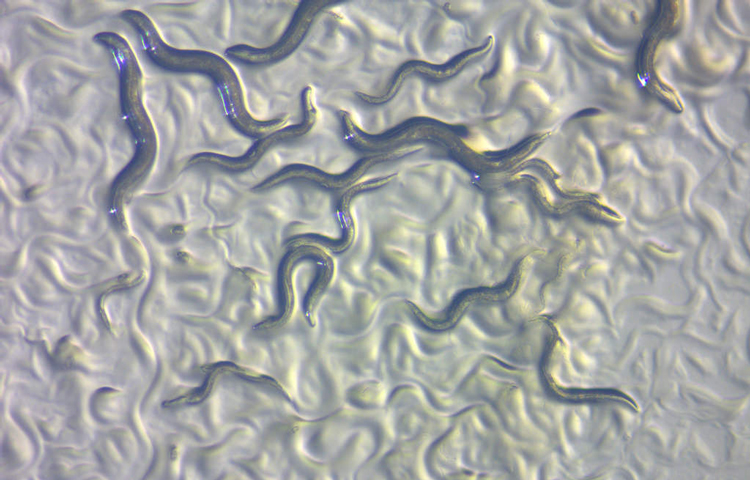
Stress comes in many forms—from the psychological stress we experience during difficult moments to the biological stress in our smallest cells. At Sanford Burnham Prebys, Assistant Professor Caroline Kumsta, PhD, uses small worms (nematodes) to study the negative relationship between cellular stress and aging (yes, aging can be stressful!). On the flip side, she’s also exploring how we can use small amounts of stress to improve health and potentially treat neurodegenerative diseases.
We spoke to Kumsta about her research to learn more about how nematodes, stress and neurodegenerative diseases are all related.
Why do you work with nematodes? Nematodes are very good for aging research because they have a short life span—only a few weeks—so we can measure the effects of aging within a reasonable amount of time. Another reason why we like these worms is because we can measure stress responses in more than individual cells. We can use nematodes to study broader, more systemic responses, as well as how stress responses are communicated from tissue to tissue. We can only see these effects if we look at how stress responses are orchestrated across the entire organism. Even though worms don’t look like us, a lot of the basic biological machinery we study is the same as in humans because stress responses evolved a long, long time ago.
How do worms help you study aging and cellular stress responses? “What doesn’t kill you makes you stronger” is true in biology—in other words, small amounts of stress can actually be beneficial for organisms, including humans. We’re studying this idea in nematodes by giving them a small heat shock early in their lives, almost like giving them a few minutes in a sauna. The heat triggers stress responses in the worms at the cellular level, and one of these responses is that the worms’ cells induce a cellular recycling process, called autophagy. Autophagy recycles cellular components and helps keep cells healthy and free from debris. This is a beneficial process that helps increase the life span of the worms. My team is exploring how this process works and figuring out how we can use it to fight diseases.
How can your work in nematodes help us study human diseases? Our main target is neurodegenerative diseases. One of the drivers of diseases like Alzheimer’s, Parkinson’s or Huntington’s disease is that proteins accumulate in the brain in aggregates or clumps. We’ve seen that nematodes that have had a heat shock early in their lives have reduced clumping of disease-relevant proteins. This is because when autophagy kicks in as a stress response, it helps slow the accumulation of these clumpy proteins. We’re ultimately looking for ways to boost the cellular recycling process in humans as a way to treat degenerative diseases. We can imagine heat therapy as a treatment intervention, and we are currently developing methods of measuring autophagy status in humans so that we will be able to test potential interventions.
Institute News
How your DNA takes shape makes a big difference in your health
The more we learn about our genome, the more mysteries arise. For example, how can people with the same disease-causing mutation have different disease progression and symptoms? And despite the fact that it’s been more than 15 years since the human genome was sequenced, why can’t we explain the significance of the vast majority of genomic variations that occur in noncoding, or “junk,” elements of the genome?
Now, Pier Lorenzo Puri, MD, a professor in the Development, Aging, and Regeneration Program at Sanford Burnham Prebys, has used a cutting-edge technique called Hi-C, which maps millions of interactions between proteins and tightly coiled DNA, called chromatin, to shed light on this mystery. The study, published in Molecular Cell, shows that a specific protein called MyoD—a master regulator of muscle development—reshapes chromatin’s architecture to alter gene expression—revealing fundamental insights into how genetic variations may affect our health.
“One of the greatest mysteries of medicine is how people with the same mutation can have different symptoms,” says Puri. “Our study indicates that some genetic variations may affect our health by altering how DNA coils and interacts in its 3D shape. This alteration may be helpful or detrimental—and could even explain why some people seem to be naturally athletic.”
In the study, the scientists used several genomic technologies to map the interactions between MyoD and chromatin as cells turned into skeletal muscle upon MyoD expression. Among other findings, the scientists determined that MyoD rearranged chromatin’s shape during this process—similar to the retying of a tangled shoelace. Importantly, the researchers found that MyoD-driven reconfiguration of 3D chromatin architecture is mediated by interactions between noncoding elements of the genome—where most disease-associated genetic variants occur. These findings demonstrate that the noncoding genome can act as a structural element that defines the chromatin architecture—key information that will help predict the functional outcomes of these variants.
Puri is already applying this insight to help solve other genomic mysteries. He plans to review a worldwide database of gene variations with unknown significance—meaning that scientists are unsure if the change is harmless or a risk factor for disease. Then, he aims to create models that help us better understand the impact of these genetic variations on an individual’s ability to respond to environmental changes and eventually develop disease.
“It’s possible that many genetic variations alter chromatin folding. Instead of directly causing disease, the changes may increase or decrease our disease risk,” explains Puri. “I hope that my next studies will shed light on these genomic mysteries and help more people get definitive answers about what lies in their DNA.”
The co-first authors of the study are Alessandra Dall’Agnese, PhD, and Luca Caputo, PhD, of Sanford Burnham Prebys.
Additional study authors include Chiara Nicoletti, PhD, of Sanford Burnham Prebys and University of Modena and Reggio Emilia; Sole Gatto and Ranjan Perera of Sanford Burnham Prebys; Julia di Iulio, PhD, and Amalio Telenti, MD, PhD, of Scripps Research; Anthony Schmitt, PhD, Yarui Diao, PhD, and Zhen Ye of the Ludwig Institute for Cancer Research; Mattia Forcato, PhD, and Silvio Bicciato, PhD, of University of Modena and Reggio Emilia; and Bing Ren, PhD, of the Ludwig Institute for Cancer Research and UC San Diego School of Medicine. The study’s DOI is 10.1016/j.molcel.2019.07.036.
Research reported in this article was supported by the U.S. National Institutes of Health (NIH) (R01AR056712, R01AR052779, AR061303), Epigen, Ellison Medical Foundation (AG-NS-0843-11), AFAR (G16294AD), Ludwig Institute for Cancer Research, Human Frontier Program and San Diego Muscle Research Center. The content is solely the responsibility of the authors and does not necessarily represent the official views of the NIH.
Heart disease is the number one killer of Americans. Now, the National Institutes of Health (NIH) has awarded a four-year grant totaling nearly half a million dollars to Sanford Burnham Prebys to find medicines that could help people repair damaged heart muscle—and potentially reduce the risk of heart attack or other cardiovascular events.
“Each year we lose far too many loved ones to heart attacks and other heart conditions,” says grant recipient Chris Larson, PhD, adjunct associate professor in the Development, Aging and Regeneration Program at Sanford Burnham Prebys. “Now, we have the opportunity to find medicines that may help more people live long, active lives by strengthening their heart muscles.”
Nearly half of American adults—approximately 120 million people—have cardiovascular disease, according to the American Heart Association and NIH. The condition occurs when blood vessels that supply the heart with oxygen and nutrients become narrowed or blocked, increasing risk of a heart attack, chest pain (angina) or stroke. Current medications for cardiovascular disease can lower blood pressure or thin the blood to minimize risk. Still, five years after a heart attack, 47% of women and 36% of men will die, develop heart failure or experience a stroke. No medicines that repair heart muscle exist.
To identify drugs that may stimulate heart muscle growth, Larson and his team will screen hundreds of thousands of compounds against human heart muscle cells, called cardiomyocytes. The work will be done in collaboration with Alexandre Colas, PhD, assistant professor in the Development, Aging and Regeneration Program at Sanford Burnham Prebys, who developed the high-throughput screening system that will be employed.
Once the scientists identify drug candidates that promote heart muscle growth, they will study these compounds in additional cellular and animal models of heart disease in the hopes of uncovering insights into the biology behind the repair process.
“After experiencing a heart attack or other cardiovascular event, many people live in fear that it will happen again,” says Colas. “Today we embark on a journey toward a future where people living with cardiovascular disease don’t have to be afraid of a second heart attack.”
Armed with wiggly worms and striped zebrafish, on Saturday, March 2, more than 20 volunteers from Sanford Burnham Prebys helped kids and their families learn about the power of DNA at the San Diego Festival of Science & Engineering’s EXPO Day.
One of the largest STEM (Science, Technology, Engineering and Math) festivals in the U.S., this year’s event featured more than 130 interactive exhibits designed to ignite a passion for science in K–12 students. Despite an uncharacteristically rainy morning, an estimated 17,000 people attended.
For Joseph Lancman, PhD, a postdoctoral researcher at our Institute who was the first in his family to graduate from college, the festival was an opportunity to provide children with the experience he wishes he’d had as a kid.
“Growing up, I knew I was interested in human health, but I had no idea that research was an option,” Lancman says. “Like many kids, I thought I wanted to be a doctor. But in college, I quickly learned that I wanted to know more. I wanted to know what causes disease and how scientists go about finding cures.”
Dr. Lancman and his son
At our booth, postdoctoral researchers, graduate students and staff helped children don paper lab coats and explore DNA-themed activities.
Children were able to see live worms with DNA mutations that affect their movement, courtesy of the lab of Malene Hansen, PhD, professor in the Development, Aging and Regeneration Program. Compared to normal worms, some mutant worms moved mindlessly in circles, and others remained relatively immobile—illustrating how changes in a DNA sequence can dramatically affect life.
At the adjacent station, provided by the lab of Duc Dong, PhD, assistant professor in the Human Genetics Program, children squinted through microscopes and peered into fish tanks to observe how DNA changes can dramatically affect the heartbeat of zebra fish—one of the most powerful model organisms used to study vertebrate biology.
Lancman, who works in Dong’s lab, took care to explain the exhibit in child-friendly language (he credits his four-year-old son for helping him develop this skill).
“I want kids to know that science is like a puzzle,” he explains. “It takes time to put all the pieces together, but when you’re done, you can see the big picture—and that big picture can lead to improving human health.”