children's health Archives - Page 2 of 4 - Sanford Burnham Prebys
Institute News

How community collaboration shapes leukemia research at Sanford Burnham Prebys

AuthorMiles Martin
Date

October 4, 2022

Since 2020, Todd and Rena Johnson, co-founders of the Luke Tatsu Johnson Foundation (LTJF), have helped fund the research of Associate Professor Ani Deshpande, PhD

But it all started with their son Luke. He had a very rare subtype of acute myeloid leukemia, one of the most difficult-to-treat cancers, and, sadly, he passed away from the disease in 2016. This inspired the Johnsons to become involved with fundraising and advocacy for cancer research.

“Our foundation started with a fundraising golf tournament to honor Luke, and that was about taking something so horrific and so horrible and finding a way to turn it into something positive,” says Rena. “If you can take that tragedy and put a positive spin on it, then everything around Luke and his name and his memory becomes positive.”

How “the stars and planets aligned” to bring the Johnsons to the Institute

In a remarkable coincidence, the Johnsons discovered on their first visit to the Institute that Deshpande’s research focuses on AF10 fusion AML, an extremely rare subtype of the disease that accounts for about 5 percent of cases. It’s also the subtype of AML that Luke had.

“It was a goosebumps-raising moment,” says Todd. “Once we visited Ani and saw his lab, we realized there was a lot more in common with our story and his research than we had realized before.”

“The stars and planets aligned and brought us to Ani,” adds Rena. 

Luke Tatsu Johnson

Luke Tatsu Johnson

As well as helping fund Deshpande’s research through LTJF and their partnership with the Rally! Foundation, the Johnsons are also on the Community Advisory Board (CAB) for the Institute’s Cancer Center, which advocates for cancer research by engaging the community. 

“The CAB does such a wonderful job of connecting the community with the scientists, and we’re so excited to be involved in that,” says Todd. “That’s fundamentally what we do as a foundation—we support the folks doing this work so that children and families down the road can have a different outcome from Luke’s.”
 

AML research “needs more support and needs more funding”

The Johnsons’ support helped the AML research team discover a new potential treatment for AML, which is currently in preclinical studies, after which they hope it will advance to clinical trials. The research team maintains that it would have been impossible to secure the NIH grants necessary to do these studies without the jump start given by the LTJF and the Rally! Foundation.

“We couldn’t do what we do without the Johnsons’ support,” says Deshpande. “We are so grateful to have them in our corner, and we’re confident that our work will help improve outcomes for kids like Luke down the line.”

Despite this progress, more research into AML and other leukemias is still needed. Leukemia is the most common cancer in children and teens. About 4,000 children are diagnosed with leukemia each year, and AML accounts for about a third of these cases.
 

Studying AML from all angles

To tackle this pressing problem, the Institute has established an AML disease team composed of researchers across labs and clinician partners. The team’s research falls into several large categories, including studying the genetics of AML, studying how the disease works in animal models and working to develop drugs that can target specific mutations associated with the disease, which are numerous. 

“AML has many different subtypes, so it’s been difficult for researchers to make major advances to treat all cases of AML,” says Deshpande, who co-leads the AML team with Professor Peter D. Adams, PhD “Most patients with AML are given the same treatments that have been used since the ’70s, which is why we want to look at AML from as many angles as possible.”

In addition to being difficult to treat, it is also challenging to get funding for AML research, particularly for the rarer subtypes. This makes the support of foundations such as LTJF even more vital to researchers like Deshpande. 

“This is exactly why AML research needs more support and needs more funding, because this is a much more difficult disease than other forms of leukemia,” says Todd. “Many patients don’t have positive outcomes, and the only way to turn that pendulum is to intensify our efforts and increase the amount of research being done.”

Institute News

Heating up cold brain tumors: An emerging approach to medulloblastoma

AuthorMiles Martin
Date

July 6, 2022

Immunotherapy has revolutionized cancer treatment, but it doesn’t work on many childhood brain tumors. Researchers from Sanford Burnham Prebys are working to change that.

Brain tumors account for about a quarter of all cancer cases in children. Medulloblastoma, a particularly aggressive form of childhood brain cancer, often develops resistance to radiation and chemotherapy. Researchers from Sanford Burnham Prebys are working to solve this problem by harnessing the power of the immune system.

They describe the potential of this approach in their recently published paper in Genes & Development

“The brain’s location makes it very difficult to target medulloblastoma tumors with current therapies,” says first author Tanja Eisemann, PhD, a postdoctoral associate in the lab of Robert Wechsler-Reya, PhD “They’re also immunologically cold, which means they’re good at evading the immune system.” 

The researchers hypothesize that it may be possible to enhance the body’s immune response to medulloblastoma and help the body’s immune cells enter the brain, making treatment with immunotherapy possible.

“Immunotherapy has so much potential as a  cancer treatment, but its scope is limited right now,” says Eisemann. “We want to bring the benefits of this therapy to medulloblastoma patients and their families.”

Eisemann has been studying this approach in mice, and although the research is still at an early stage, she and her colleagues are highly optimistic about its potential.

“The brain has long been considered immune privileged, hidden from immune-system surveillance and immune responses. But we’re starting to see that this isn’t the case,” says Eisemann. “This is a rapidly evolving field, and I’m excited to be working in a lab on the forefront of that research.”

Institute News

Rare Disease Day gathers scientists, doctors and families

AuthorMiles Martin
Date

March 3, 2022

The 2022 Rare Disease Day Symposium took place last weekend at the Dana On Mission Bay Resort in San Diego. The event, sponsored by Sanford Burnham Prebys and CDG CARE, brought together researchers, clinicians and families from around the world to discuss new medical breakthroughs and meet other families living with rare diseases.

The 2022 Rare Disease Day Symposium took place last weekend at the Dana On Mission Bay Resort in San Diego. The event, sponsored by Sanford Burnham Prebys and CDG CARE, brought together researchers, clinicians and families from around the world to discuss new medical breakthroughs and meet other families living with rare diseases.

Rare Disease Day is celebrated on the last day of February to raise awareness for rare diseases, defined by the United States government as those that affect fewer than 20,000 people. Although there are more than 7,000 individual types of rare diseases that affect more than 30 million people in the United States, this year’s conference gathered more than 200 people focused on CDG, an extremely rare group of genetic disorders that affect children. 

CDG, which stands for congenital disorders of glycosylation, occurs when sugar molecules on many of our proteins are absent or incomplete. CDG causes serious, often fatal, malfunctions in various organ systems throughout the body.

“This is a chance for the global CDG community to come together, support one another and continue to try to find treatments,” says Hudson Freeze, PhD, director of the Human Genetics Program at Sanford Burnham Prebys. “It’s always my favorite weekend of the year, and I’m thrilled that we’re able to do it again safely.” Freeze’s primary research focus is CDG, and he has personally worked with more than 300 patients. 

Exchanging knowledge
The three-day symposium opened Friday morning with introductory comments from three important figures and philanthropists in Sanford Burnham Prebys’ history: T. Denny Sanford, Malin Burnham and Debra Turner. Congressman Scott Peterson also spoke on the importance of funding medical discoveries. 

“Our job is to make a positive difference. We do that best when we all work together,” said Sanford in his video introduction. “Congratulations on all your work. You make me very proud.”

This year, 19 scientists and clinicians in total spoke on the latest research in modeling, treating and understanding CDG. The full program of presentations can be found here.

Connecting families
Although Rare Disease Day is an important opportunity to share the latest scientific research, one of the highlights of the event doesn’t involve science at all. To provide space for families to take a break from the presentations and socialize, staff and volunteers transformed the Bayside Conference Room of the Dana resort into a child care and respite area packed full of toys and games.

In addition to giving families space to play, Rare Disease Day hosted several group activities for families, including a magic show on Saturday and a surprise visit on Sunday morning from Disney’s Anna and Olaf.

​Longtime friend of the institute Damian Omler, a thirteen-year-old who is the only person living with his rare genetic mutation, had a great time dancing along to “Let it Go” and playing catch with his father, Donnie.

And while the joy in the respite conference room was palpable, there was something else, less tangible, in the air as well: hope.

“Meetings like this bring us hope and help us raise awareness for CDG,” says Donnie. “That gives us a sense of purpose each and every time we attend the conference. And we won’t stop, even 20 years from now.” 

Omler family

Damian Omler and his family, parents Donnie and Gracie and brother DJ, had a great time at Rare Disease Day the year (image credit: CDG CARE)

Institute News

Rare disease in the time of COVID: Damian Omler’s story

AuthorMiles Martin
Date

February 25, 2022

How a one-of-a-kind kid and his family stay connected during the pandemic

Thirteen-year-old Damian Omler is the only person in the world with his rare genetic mutation, which presents him and his parents (Donnie and Gracie) and 11-year-old brother, DJ, with major challenges every day. Damian’s condition—a congenital disorder of glycosylation, or CDG—causes him to have seizures, and requires him to have help with routine tasks such as using the restroom and dressing. And, he must use a wheelchair for mobility.

Despite these obstacles, Damian lives a rich, fulfilling life. But protecting his health during the COVID-19 pandemic threw a major wrench into the Omlers’ routine.

“In the early days of the pandemic, we didn’t know what kind of effect COVID would have on Damian, so we had to take a lot of precautions, including not seeing a lot of family and friends, which was very isolating,” says Donnie. 

“Damian is also very sociable—we call him the hot potato because he just goes from person to person, so the pandemic was hard for him in that way as well,” adds Gracie. “We were so glad when we were finally able to get our family vaccinated so we could be more a part of the community.”

Staying at home had its ups and downs for the Omlers
Although most of us can relate to the isolation of the pandemic, there are unique challenges that come with being a family living with a rare disease during this time. 

“Appointments were so much more difficult for Damian over Zoom,” says Gracie. “I had to help him through his physical therapy, and I was nervous that I might be doing it wrong or even hurting him.”

Despite these complications to Damian’s care, there were some unexpected silver linings to spending more time at home.

“Damian does choir and dance for his electives at school,” says Gracie. “I love that with remote learning I was able to interact with him and the class and learn the dances with him.”

“She definitely got a lot of accolades from the teachers for being one of the parents who participates,” adds Donnie, jovially. 

Returning to Sanford Burnham Prebys’ Rare Disease Day
The Omlers are longtime friends of Sanford Burnham Prebys. They first visited the Institute in 2012, when Damian was 5. Before then, they’d been struggling to find a diagnosis for their son, who’d been missing developmental milestones since he was born. 

With the help of Institute professor Hudson Freeze, PhD, who has dedicated his career to CDG research, doctors were finally able to diagnose Damian’s specific case in 2015. 

“After the diagnosis, we sat and smiled for a long time,” says Donnie. “Just knowing was such a relief.”

Since 2016, the Omlers have also been regular participants in the Institute’s Rare Disease Symposiums, which help patients, researchers and clinicians from around the world connect in order to support one another and learn about the latest advances in rare disease research.

The most recent Rare Disease Day the Omlers attended was in 2020, just before the pandemic took hold. And although the event didn’t take place last year, this year it’s back stronger than ever. And the Omlers can’t wait to be back too.

“Meetings like this bring us hope and help us raise awareness for CDG,” says Donnie. “That gives us a sense of purpose each and every time we go. And we won’t stop, even 20 years from now.” 

The 2022 Rare Disease Day Symposium & CDG/NGLY1 Family Conference will take place February 25–27 at the Dana Hotel on Mission Bay in San Diego. Scientific sessions will be held on the 25th and 26th, and the Family Conference will take place on the 27th.

And if you see a young man acting like a social “hot potato” on the 27th, that’s Damian. He’ll probably say hi to you.

Institute News

One at a time: How a Sanford Burnham Prebys professor changes patient lives

AuthorMiles Martin
Date

February 22, 2022

Having worked for decades to improve the lives of children with rare diseases, Hudson Freeze is still on the case.

Hudson Freeze, PhD is not your average researcher. His work focuses on congenital disorders of glycosylation, or CDG, a severe group of diseases that affect fewer than 2,000 children worldwide. Those conditions occur when sugar molecules on many of our proteins are absent or incomplete. That can lead to serious, often fatal, malfunctions in various organ systems throughout the body.

Although Freeze is not a clinician, he is deeply involved in identifying these rare CDG mutations, and providing families with answers to what is often a challenging diagnosis. Because CDG is a group of incurable diseases, families of children with CDG reach out to Freeze almost weekly, seeking help.

“If someone asks for help, I say, ‘Let me try,’” says Freeze. “Any glimmer of hope is a path worth pursuing, anything to make life easier for children with CDG.”

Freeze has been working on CDG for more than 25 years and has worked with more than 300 patients, and he has kept in touch with many of them over the years.

“Not a day goes by when I don’t think of them and their struggles—but mostly their smiles,” says Freeze. “It’s the reason we won’t give up on trying to understand them and maybe even finding treatments.”

Treating disease with sugar
Although CDG presents as permanent and irreversible mutations, Freeze’s research has been instrumental in discovering an approach to alleviate severe symptoms of CDG—such as seizures—in certain patients. The answer: sugar. Thanks to Freeze and others, there are about 30 patients worldwide who are now taking mannose, a simple sugar molecule, to help alleviate their CDG symptoms.

Today, the strategy of treating diseases with simple sugar molecules is being explored in other glycosylation disorders, as well as less-rare diseases such as multiple sclerosis, cancer and diabetes.

Hudson Freeze, PhD poses with Damian Omler, who has CDG.

Hudson Freeze, PhD poses with Damian Omler, who has CDG.

Rare Disease Day at Sanford Burnham Prebys
Freeze’s impact on the lives of families living with CDG extends well beyond the walls of his lab. Since 2010, he has organized an annual Rare Disease Day Symposium each February, where scientists, doctors and families gather from around the world to discuss the latest research and meet other families coping with rare diseases. Last year, the pandemic forced the Institute to press pause on the event, but this year, Rare Disease Day is back in San Diego and stronger than ever.

“It’s a chance for the global CDG community to come together, support one another and continue to put our heads together to find treatments,” says Freeze. “It’s always my favorite weekend of the year, and I’m thrilled that we’re able to do it again safely.”

The 2022 Rare Disease Day Symposium & CDG/NGLY1 Family Conference will take place February 25–27 at the Dana on Mission Bay Resort in San Diego. Scientific sessions will be held on the 25th and 26th, and the Family Conference will take place on the 27th.

Register Here

Institute News

From child neurology to stem cells: An interview with Evan Snyder

AuthorMiles Martin
Date

November 18, 2021

What do Evan Snyder and Sigmund Freud have in common? Both radically changed how we see the human brain.

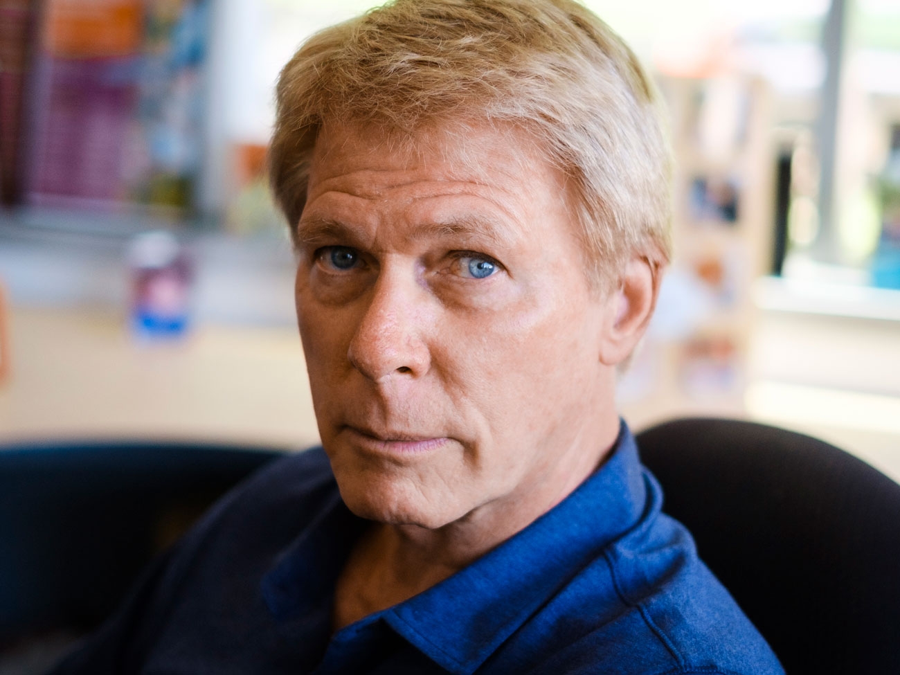
In a first for San Diego, Sanford Burnham Prebys Professor Evan Y. Snyder, MD, PhD has been featured in the second edition of Child Neurology: Its Origins, Founders, Growth and Evolution, a collection of biographies detailing the lives of innovators in child neurology throughout history. 

To celebrate this honor, we caught up with Snyder to discuss how his work in child neurology led him to make foundational discoveries in the fields of stem cell biology and regenerative medicine, as well as where these fields are headed in the future.

“Plasticity, both at the “macro” sociological level and at the “micro” clinical level, always fascinated me. And that fascination with resilience and my curiosity about its source led to my early discoveries surrounding stem cells in the brain.”

How does it feel to be included in this book?
Snyder: Ever since I was a student, I thumbed through the first edition and was inspired by all these old images of people from the early days of the field. To see my chapter in there along with the likes of Sigmund Freud is mind-blowing. It means a lot to be included. 

How did your work in child neurology lead you to work on stem cells?
Snyder: I was always interested in resilience. As a kid, I worked at a camp for children with various challenges: poverty, behavioral issues, disrupted home lives. These children were being raised in terribly deprived environments with challenging family situations, but they adjusted and bore up without complaint. When we took them out of that environment, even for a short time, they flourished. I never forgot that lesson. 

Later, doing my pediatric and neurology residencies and neonatology fellowship at Boston Children’s Hospital, I would care for babies in the newborn intensive care unit with seemingly devastating brain injuries. As a neurologist, I would often follow up with those kids months or even years later.

Despite the horrible injuries they had sustained as newborns, by the time they were children, some had recovered to the point where it was hard to notice a deficit—they were achieving their developmental milestones, were seizure-free off medications, and playing and interacting like normal kids.

It astounded me that some children could have that resilience while I knew that adults with those same injuries would likely be incapacitated. Plasticity, both at the “macro” sociological level and at the “micro” clinical level, always fascinated me. And that fascination with resilience and my curiosity about its source led to my early discoveries surrounding stem cells in the brain.

“There was no notion that there could be such regenerative cells in a solid organ, particularly one that was thought to be as static as the brain.”

How did your early work shape the stem cell field?
Snyder: Before I, and a few others, began examining the molecular biology of cell types that composed the brain, there really was no research area called the “stem cell field”—at least not for solid organs like the brain. What was known clinically about stem cells in the early ’80s was confined to the making of blood cells for bone-marrow transplants.

The goal of hematologists back then was to identify the youngest cell in the bone marrow that could give rise to other types of blood cells. That cell, called the hematopoietic stem cell, was difficult to isolate and identify, but it was always assumed to exist because a healthy person’s blood is always turning over.

There was no notion that there could be such regenerative cells in a solid organ, particularly one that was thought to be as static as the brain. ​My postdoctoral project at Harvard unexpectedly forced me to challenge that notion. I set out to study how the brain is put together by isolating all of its different cell types into separate containers, then building a miniature brain in a dish, cell by cell.

But from the very beginning, I had difficulty making containers of cells with a single specific identity, even if I started with what seemed to be a single cell and its identical sibling cells. The young cells I isolated always seemed to be changing their identities, even though they started out looking the same.

Colleagues observing my initial results assumed it was just an odd tissue culture artifact, but then I thought back to those kids with brain injuries for whom I’d cared, and I started thinking that maybe what I was observing wasn’t an artifact at all; maybe these cell types were one of the repositories of plasticity in the brain I had puzzled over—a “stem cell” of sorts.

To prove that notion, I quietly transplanted those stem cells into a mouse brain and waited two years to examine the brain. The cells I’d transplanted were not only still there but they had integrated into the fabric of the brain and taken on different identities depending on which part of the brain they took up residence.

This observation—which I made alone, in the middle of the night under a microscope—gave me chills that I still recall today.

I shuddered with excitement at the implication of what I was seeing. The central nervous system was always thought to be a part of the body that could not regenerate at all, yet here I had isolated cells from one mouse brain and used them to populate another brain with multiple cell types in multiple locations. I could make pots of those donor cells to be transplanted whenever and wherever I liked, like a bone-marrow transplant in the brain.

The potential seemed enormous, and my curiosity to see where that potential might lead in terms of understanding and healing the human brain became the focus of my lab—first at Harvard and then at Sanford Burnham Prebys.

“Finding how we could address unmet medical needs became somewhat like looking for a lock for which we already had a key.”

How did regenerative medicine enter the picture?
Snyder: Other people started finding stem cells, not only in the brain but everywhere in the body. In the mid-1990s, the focus then became figuring out how to exploit them. Those efforts helped give rise to the new field of regenerative medicine, where the emphasis is on repairing and regrowing tissues rather than just treating the symptoms of a disease. 

We found that we could put these neural stem cells almost anywhere in the nervous system, particularly if that region was diseased or injured—and hit the “reset button” for that region. I was more interested in studying the overall biology of this new cell type rather than focusing on any one illness. So, while learning about this cell, we found many different ways that its biology could be exploited therapeutically.

Finding how we could address unmet medical needs became somewhat like looking for a lock for which we already had a key. Looking for those locks—the therapeutic obstacles that stem cell biology might help circumvent—is where my clinical experience proved handy. 

One example was brain tumors. I’m not an oncologist, but I’d learned from my studies that stem cells will naturally go to regions where pathology exists in the brain, even over long distances from their point of implantation. So, by putting a therapeutic gene into a neural stem cell, I could use that cell like a “heat-seeking missile” to deliver that gene and its product to where it might be needed.

Brain tumors are often so difficult to cure because we can’t safely access all the brain areas where the tumor has infiltrated. The neural stem cell seemed a perfect way to deliver a tumor-killing gene to those disseminated tumor cells simply by harnessing their natural powers. That strategy has now moved into clinical trials, and the use of stem cells to deliver therapeutic genes is being used for other conditions as well.

What came next in the history of stem cells?
Snyder: If we fast-forward a few decades from the beginnings of regenerative medicine, we now know a lot more about the different “flavors” of stem cells, how they work, and how they can be therapeutic.

One of the new flavors of stem cells is induced pluripotent stem cells, or iPSCs. One can take a mature skin or blood cell and push it back to a state where it loses all of its organ identity, and then force it to become a completely different mature cell type.

In other words, plasticity can go in both directions: an immature cell type can take on multiple identities going forward in development, and a mature cell type can be pushed back in developmental time to the point where it can now make different choices. 

Importantly, that induced stem cell retains many of the characteristics of the person from which it originally came, including diseases and genetic defects that individual may have had. This realization gave rise to the idea of modeling diseases in a dish. 

This approach of using iPSCs works best for diseases where we know the specific cell type, pathway, gene, or protein that causes the disease, but we wondered if iPSCs could help for diseases where we have no clue which gene, protein, cell type, or pathway is causing a particular disease. 

Psychiatric disorders are the poster children for such complex and mysterious diseases. To date, there’s no psychiatric disease that we can attribute to a specific defect in a particular gene or protein. Nevertheless, I felt confident that we could use iPSCs to discover the underlying cause of challenging diseases where we have no prior knowledge of what has gone wrong.

I decided to zero in on bipolar disorder because, as a physician, I knew that this condition did have an effective treatment—lithium—which had been accidentally discovered many years ago without knowing why it seemed to help some patients.

I reasoned that If we knew lithium’s target, we could work backward, recognizing that whatever lithium was changing was the underlying cause of bipolar disorder. That strategy worked. We used the iPSCs to map the lithium-response pathway, and we learned that errors in the regulation of that pathway were the likely cause of bipolar disorder. With that in mind, we could discover drugs better than lithium.

In this branch of regenerative medicine, it’s not the stem cell that goes into the patient, but rather the drug discovered by the stem cell that goes into the patient, but this would not be possible if we could not use stem cells to give us those patient-specific disease models.

“Organs and their diseases never involve just one type of cell. If we want to improve the way we model diseases, we need to re-create and target that complexity.”

Where is the field going from here?
Snyder: Our models for diseases are only going to get better. We’re moving away from looking at small numbers of homogenous cells in a flat dish toward creating more complex three-dimensional mini-organs from a patient’s stem cells, directing those cells to become the many different interacting cell types that make an organ.

Organs and their diseases never involve just one type of cell. If we want to improve the way we model diseases, we need to re-create and target that complexity.

As far as using stem cells directly for therapy, we’re becoming much more sophisticated in deriving the cell type we need and directing it to the specific regions in need of repair. Furthermore, as our three-dimensional representations of organs in a dish become more sophisticated, these mini-organs may themselves become the material we transplant.

I’m also certain that there are applications for stem cells we haven’t conceived of yet.

Every generation of scientists needs to be reminded that one is constantly going to be surprised; one’s initial hypotheses, based on old understandings, are likely going to be wrong. One must be prepared to revise those hypotheses when the data—even if counter to expectations—leads one in an unconventional or inconvenient direction.

Although we’ve made much progress in the stem cell field over the past 30 years, there’s so much we don’t know. And, even when we think we know something, we probably don’t. None of my work would have been possible if I’d kept my blinders on and not been able to see the connections between different areas in which I was working—including allowing my work in the clinic and as a social worker to inform my scientific vision. That mindset is critical to the future of science.

Institute News

How microbes shape human health: an interview with Andrei Osterman

AuthorMiles Martin
Date

October 7, 2021

In his work on the human microbiome, Sanford Burnham Prebys professor Andrei Osterman, PhD, has shown how the organisms living within us can be leveraged to boost human health for a humanitarian cause – the plight of malnourished children. 

Describe your research aimed to improve the gut microbiome in malnourished children.
In infants, it’s been well-established that conditions of severe poverty and food insecurity cause a delayed development of gut microbes, and that this results in stunted growth, numerous syndromes, and even death. My team has been collaborating with researchers at Washington University in St. Louis to develop foods that are designed to enhance the microbiome, and we’ve found that these can actually work to correct some of these pathologies. 

One study involved introducing microbes from undernourished Bangladeshi children into the guts of mice. When these mice were then fed a typical Bangladeshi diet, they exhibited a weaker immune response to the oral cholera vaccine. More importantly, this poor response could be repaired by establishing a more normal gut microbiome in the mice and providing them supplements to boost these microbes’ propagation.

What else can we learn from this research that could be applied more broadly?
From a humanitarian perspective, the progress we’ve made is so valuable that there is no question we will continue the work. But studying the microbiome in infants, regardless of their food security, can also provide us with new insights into the importance of the microbiome in human health. 

In a more recent study with collaborators from University of California San Diego and University of Southern California, we found that adding corn syrup to infant formula can enhance the populations of beneficial microbes they might otherwise have gotten from breast milk. 

We hope this is the first of many studies we work on with this team, because the transition of infants from breast milk or formula to conventional foods is thought to be the most drastic example of how the microbiome changes with diet. Studying infants and their diets at this early point in life could help reveal fundamental truths that we’ll be able to translate to other syndromes related to the microbiome in children and adults worldwide, regardless of food security. 

And this isn’t just speculation. Another study with the team in St. Louis used the same methods as the malnutrition study to develop supplementary foods, called “fiber snacks,” to correct microbiome imbalances in people with obesity. One might think that obesity would be the total opposite of malnutrition, but the microbiome is a key player in both. 

More broadly, gut microbes are already the most well-studied part of the human microbiome, and the list of health associations with these microbes extend well beyond the digestive tract, even into the immune system, affecting the risk for diseases like cancer or diabetes. There’s also a growing body of evidence that suggests that gut microbes can have a direct effect on the brain. For example, the microbiome is being studied closely in connection to autism spectrum disorder, since many people on the spectrum experience concurrent gastrointestinal syndromes.

What would you say is important to know for people not familiar with the subject?
We need to acknowledge that our body and many of its problems have a huge microbiome component. The human body is a complex organism, and we are still learning how the microbiome influences and is influenced by different health conditions. The next step is to incorporate the role of the microbiome into the design of new diagnostics and therapeutics—because this undoubtedly influences their effectiveness. We can’t ignore this aspect of our biology, and the time is ripe to improve our understanding of it and leverage it to our advantage. Moving forward, this is going to help us solve so many problems—from issues we’ve already started looking at like obesity and malnutrition, all the way through to problems we aren’t even aware of yet. 

What are the next steps for you and your team?
What we’re really interested in now is exploring new genomic technologies that are starting to revolutionize the field. The latest development is something called MAG genomics, short for metagenomically assembled genomes. This involves looking at the big picture, sequencing DNA from the whole microbiome at once in a way that is much faster and of much better quality than we’ve ever been capable of before. It’s like the difference between watching a movie on a clunky pixelated monitor from the 80’s and seeing that same movie on an HD monitor. Methods like this are moving us into a new paradigm in biomedical research, one that may be more complex, but also one that has the potential to substantially improve health outcomes for people around the world.

Institute News

Conrad Prebys Foundation provides $3 million for pediatric brain cancer research

AuthorSusan Gammon
Date

April 7, 2021

Conrad Prebys was an extraordinary man and a passionate philanthropist. Today, his generosity extends beyond his life through the Conrad Prebys Foundation.

This year, the Foundation provided $3 million to Robert Wechsler-Reya, PhD, and his team of researchers to advance a potential drug to treat medulloblastoma—the most common malignant brain tumor in children.

Children with medulloblastoma often receive aggressive treatment (surgery, radiation and chemotherapy), but many still die of their disease, and survivors suffer long-term effects from therapy. Safer and more effective therapies are desperately needed.

Wechsler-Reya recently combined forces with Michael Jackson, PhD, senior vice president of Drug Discovery and Development, to find a drug(s) that would inhibit the growth of Group 3 medulloblastoma, the most aggressive form of the disease. Using high-throughput screening technology, they identified a compound that reduces levels of a protein called MYC, which is found at exceptionally high levels in Group 3 medulloblastoma, as well as in cancers of the blood, breast, lung and prostate.

“An effective MYC inhibitor could have a major impact on the survival and quality of life of patients with medulloblastoma,” says Wechsler-Reya. “We identified a compound that reduces levels of MYC in medulloblastoma cells, but now we need to learn how it works to optimize it as an anti-cancer drug and advance studies toward the clinic.

“Historically, pharmaceutical companies and funding agencies have under-invested in childhood cancers, and the majority of drugs currently used to treat these cancers were originally developed for adult cancer,” adds Wechsler-Reya. “We believe that effective drugs for pediatric brain tumors must be developed—and this award from the Foundation will help us achieve this goal.”

“We are profoundly grateful to Conrad for his generosity over the years,” says President Kristiina Vuori, MD, PhD “He has a special legacy at our Institute, which was renamed Sanford Burnham Prebys in 2015 to honor him. We are now thankful to his Foundation for including us in their inaugural grant cycle, and for supporting the critical work we do to benefit children and others suffering from cancer.”

The Conrad Prebys Foundation allocated $78 million in its inaugural grant cycle to fund 121 projects. The awards reflect areas of personal interest to Conrad Prebys—including visual and performing arts, higher education, health care, youth development and animal conservation.

Sanford Burnham Prebys joins a long list of recipients, which included other prominent San Diego institutions such as Rady Children’s Hospital, KPBS, San Diego State University, Scripps Research, Museum of Contemporary Art San Diego and the La Jolla Music Society.

Institute News

Fighting rare diseases: Finding treatments and bringing hope to families

AuthorMonica May
Date

March 23, 2021

The majority of rare diseases affect children, most of whom have an underlying genetic cause for their condition that is incurable.

The majority of rare diseases affect children, most of whom have an underlying genetic cause for their condition that is incurable.

Often, their own doctors have never heard of their disease, let alone know how to treat it.

But there is someplace they can turn to for help. The Human Genetics Program at Sanford Burnham Prebys provides insights into the genes and environmental factors that play a role in the development of childhood diseases. Their work often leads to better ways to diagnose, treat, and sometimes, even cure children.

On March 18, 2021, two patients whose lives were saved by discoveries made by Hudson Freeze, PhD, and José Luis Millán, PhD, joined the scientists for a conversation about what this work means to them and how their lives have been impacted. Watch the full discussion below.

Institute News

Meet molecular biologist Jonatan Matalonga-Borrel

AuthorMonica May
Date

February 3, 2021

Matalonga-Borrel is on the hunt for a treatment that could help children born with a rare, life-threatening condition

Thanks to the sequencing of the human genome, scientists have helped parents get answers to the cause of mysterious conditions that have affected their children. Now, researchers are tackling a new challenge: translating this knowledge into life-altering medicines.

Molecular biologist Jonatan Matalonga-Borrel, PhD, a postdoctoral researcher in the Dong lab at Sanford Burnham Prebys, is at the forefront of this effort. We caught up with Matalonga-Borrel as he prepares to take the virtual stage at DASL (the Diversity and Science Lecture Series at UC San Diego) to learn more about his work and his interests outside of the lab.

Did you always know you wanted to be a scientist?
I actually wanted to be an airplane pilot until my senior year of high school. But during the application process, I learned that I have very mild color-blindness, so I had to quickly decide what I wanted to do next. I pivoted to biology, a topic where I had some interest, thinking I would become a teacher. Then, when I was in college, I got the opportunity to complete a lab internship, which is where I discovered my passion for research. I would have never guessed that I would be where I am today, leading a project that might directly help families and children.

What do you study, and what is your greatest hope for your research?
I study Alagille syndrome, a rare disease that affects kids from the day they are born. Many organs are affected, especially the heart and the liver, and almost half of these children die before the age of 19.

Luckily, Alagille syndrome is associated with mutations in only two genes, both belonging to the same pathway, called Notch. This makes our goal easier to achieve: identify drugs that target Notch, which currently don’t exist. I’m excited that we’ve identified a promising option. My greatest hope is to create a medicine that truly helps these children and their families, who currently live without any treatment.

When you aren’t working in the lab, where can you be found?
You will likely find me playing golf at Torrey Pines! There is nothing like playing a twilight round, feeling a slight breeze and looking at the immensity of the Pacific Ocean. With that said, since I became a father, my golfing time has been severely impaired. Now it’s most likely that you’ll find me at home, entertained by the early stages of development of my son…and changing a lot of diapers!

What do you wish people knew about science?
How patient one has to be to move science forward. It can take weeks—or months—of trial and error until a big breakthrough happens.

We live in a world that seems to spin faster and faster. It is critical for our society to understand that proper science is not about rushing experiments. It is about setting the right ones.

How do you think your lab colleagues would describe you?
Upbeat, reliable and organized (hopefully!).

How has the pandemic affected your life?
I had my first baby last June, and the pandemic prevented any relatives to come from our home country, Spain, and meet their first grandchild. Thankfully, we had Skype to get in touch. Looking on the bright side, daycares have never been so clean, and the rate of sickness around kids has dropped significantly!

What is the best career advice you have ever received?

“Have fun and make friends,” from Dr. Eduardo Chini of the Mayo Clinic. It is possible to do great science and have fun—don’t feel guilty about it. My best collaborations came from my greatest friendships among colleagues.

What do you wish people knew about Sanford Burnham Prebys?
It’s an amazing community. Science moves forward thanks to communication and collaboration and it wouldn’t happen without a strong sense of community. This includes wise faculty members who train graduate students and postdocs, an Office of Education and International Services that offers year-round seminars and workshops, and a group I am part of, called SBP-Social Network (SBP-SN), which organizes fun social and scientific events. All of this creates a place where scientific excellence thrives.