immunotherapy Archives - Sanford Burnham Prebys
Institute News

Perkins Fellow Trains Immune System Against Melanoma

AuthorGreg Calhoun
Date

May 15, 2024

Sanford Burnham Prebys scientist works on new methods to boost the body’s natural defenses against melanoma and other cancers

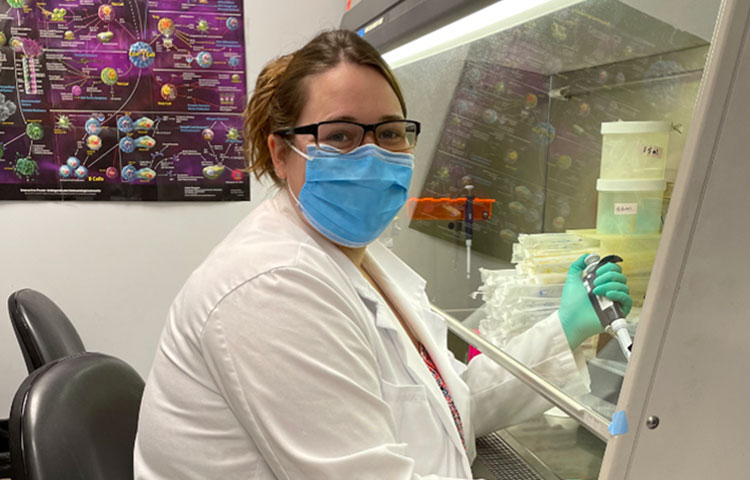
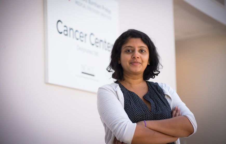
When she was growing up in India, Sreeja Roy, PhD, looked up to her father as he applied his scientific knowledge to care for the patients in his medical practice.

“He was my inspiration,” says Roy. “I found that I was particularly good at biology, and I liked learning about the mechanisms of how things work. Along the way, I realized I didn’t want to be a medical doctor and began focusing on biomedical research—and I fell in love with it.”

After earning her undergraduate degree in biotechnology from The Australian National University in Canberra, Roy obtained a master’s degree in infection biology from the Universität zu Lübeck in Germany. She returned to The Australian National University for her doctoral degree in immunology with an emphasis on viral vector-based vaccine immunology. After completing graduate school, Roy worked as a postdoctoral researcher at Albany Medical Center in New York before joining Sanford Burnham Prebys in September 2021 as a postdoctoral associate in the Immunity and Pathogenesis Program.

“I had been working on basic science in Albany,” notes Roy. “I really wanted to do translational research so I could work on things that would benefit people much sooner. That is why I chose to move to Sanford Burnham Prebys and focus on cancer immunotherapy.”

Roy’s background in immunology prepared her to enter the emerging field of cancer immunotherapy. This discipline involves developing treatments that enhance the human body’s innate immune response to better locate and dispose of cancer cells. She learned about an opportunity to support her interest in translational immunotherapy through the Jean Perkins Foundation Fellowship.

Roy received one of two prestigious fellowships designed to support postdoctoral researchers in the lab of Carl Ware, PhD, director of the Infectious and Inflammatory Diseases Center and professor in the Immunity and Pathogenesis Program.

“The Jean Perkins Foundation Fellowship has been fantastic,” says Roy. “I can work without the pressure of writing an academic grant, which allows me to focus on the science and be more productive.”

Roy’s project at the Ware lab involves making immunotherapies more effective in treating melanoma, the deadliest form of skin cancer.

“Unfortunately, some tumors never respond to immunotherapy treatments,” explains Roy. “Also, tumors can initially begin to shrink before becoming resistant to a treatment.”

Under Ware’s direction, Roy is testing ways to enhance existing immunotherapies through the lymphotoxin-β receptor, which is found on some types of immune cells.

“When the immune system encounters a foreign substance that may cause an infection, a sample of the invader can be shuttled to the lymph nodes as a way of learning about the threat and generating a better immune response,” explains Roy. “Depending on which tissues are being infected, the lymph nodes cannot always be involved, so an active lymphotoxin-β receptor is able to approximate their effect by organizing immune cells in something akin to training centers so that a better attack can be launched.”

Roy and the Ware lab are developing ways to take advantage of the lymphotoxin-β receptor’s ability to recruit and train immune cells as an approach to making immunotherapies more effective.

“If I can target the lymphotoxin-β receptor signaling against tumors, does that enhance the anti-tumor immunity?” asks Roy. “Do the tumors become more responsive to the treatments now? That is what we’re trying to find out.”

With the help of the Jean Perkins Foundation Fellowship, Roy is determined to continue developing her translational science expertise and find ways to improve the effectiveness of immunotherapies for melanoma and other cancers.

“We’ve made quite a bit of progress,” says Roy. “I look forward to sharing our results and seeing how this project advances from the bench to the bedside.”
Institute News

Scientists uncover protein that empowers antibodies

AuthorMonica May
Date

January 11, 2021

Discovery may lead to better vaccine strategies and improve treatments for cancer and autoimmune disorders

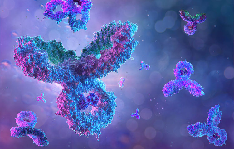
Antibodies are the heroes of our immune system. They protect us from viruses, like SARS-CoV-2 (which can lead to COVID-19), as well as bacteria and other pathogens. They can provide lifelong protection from future infections—if they are strong enough. But, like any hero, they are fallible, and certain cancers or autoimmune disorders can arise when things go wrong.

Now, Sanford Burnham Prebys scientists have revealed that a protein called cyclin D3 tells antibody-producing B cells to start dividing—opening new research avenues that could improve vaccine development or the treatment of B cell lymphoma and autoimmune disorders. The discovery was published in Cell Reports.

Parham Ramezani-Rad, PhD
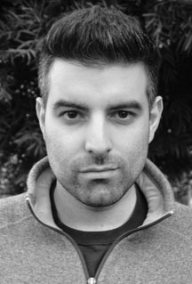
Parham Ramezani-Rad, a
postdoctoral researcher at
Sanford Burnham Prebys and
lead author of the study. 

Antibodies get their power from a complicated process. When an “intruder” is detected in the body, B cells—which produce antibodies—are activated. Each B cell is unique—they contain slight genetic variations to produce a diverse set of antibodies to attack the “intruder.” Later, they undergo optimization through a “survival of the fittest” process to identify the most protective versions.

“Our findings reveal that cyclin D3 is the ‘go’ signal for B cells to start rapidly dividing and producing a set of diverse antibodies,” says Parham Ramezani-Rad, PhD, a postdoctoral researcher in the Tumor Microenvironment and Cancer Immunology Program at Sanford Burnham Prebys and the lead author of the study. “This information might help scientists create better vaccine strategies in the future. On the flip side, researchers may be able to develop better weapons against B cell lymphoma and autoimmune disorders by removing malignant B cells.”
 

Diving into the “dark zone”

After infection, B cells grow and divide in special structures called germinal centers that form in our spleen and lymph nodes. In this structure, a “dark zone”—referring to what scientists saw under the microscope in the 1930s—and a “light zone” are visible. Now researchers know the dark zone is where B cells are rapidly expanding, and this cell density appeared darker in the original microscope studies. After proliferating in the dark zone, B cells head to the light zone where the best potential antibody options are selected—while less desirable options are eliminated.

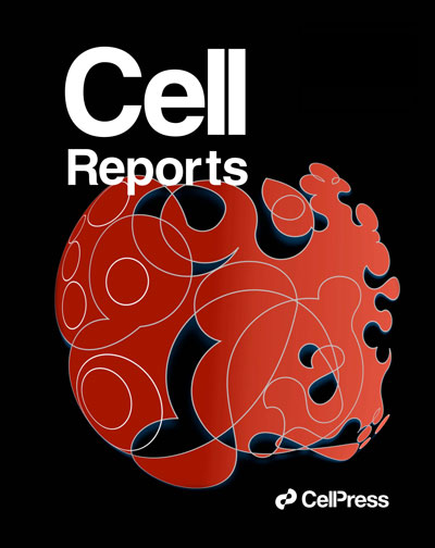
Cell Reports cover
Parham Ramezani-Rad
designed the image that
was featured on the cover of
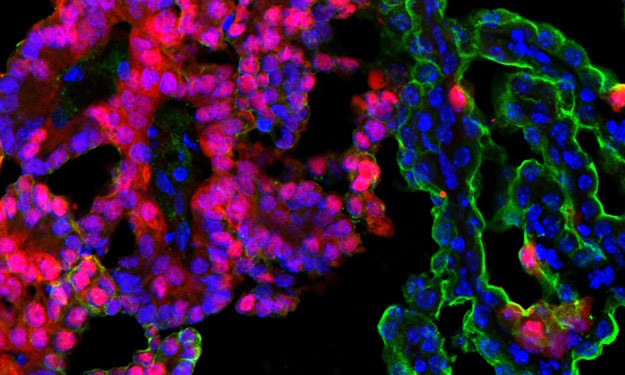
Cell Reports. The image is
an artistic impression of the
dynamics occurring inside of
the germinal center, where
antibody-producing B cells
undergo a “survival of the
fittest” selection process. 

Ramezani-Rad made the discovery when studying B cell lymphoma, a blood cancer that often contains a mutation that leads to hyper-stable cyclin D3. Using mice and sophisticated CRISPR gene editing technology, he discovered that cyclin D3 regulates the expansion or contraction of B cells specifically in the dark zone of germinal centers—and not the light zone. He also identified other regulatory aspects involved in this process that scientists might be able to harness for the benefit of human health.

“B cell lymphoma is often treated with an intensive chemo and immunotherapy combination. The side effects of this treatment can be immense, and relapses may occur,” says Ramezani-Rad. “Our findings about cyclin D3 could form the basis for a more tailored medicine that targets exactly what goes wrong during B cell lymphoma, and is potentially less toxic and more effective.”

Ramezani-Rad also designed the image that was selected for the journal cover, which is his artistic impression of the dynamics occurring inside the germinal center. He finds many parallels between scientists and artists.

“As a scientist, I see myself describing what already exists in nature,” explains Ramezani-Rad. “Musicians and painters are also describing the world. They are just using instruments or paint strokes to express emotions, whereas scientists use data to express knowledge.”

Institute News

5 things to know about immunotherapy and breast cancer

AuthorSusan Gammon
Date

September 30, 2019

If you follow news about medical breakthroughs, you have undoubtedly heard about immunotherapy to treat cancer.

This form of therapy is designed to prime the body’s own immune system to fight the disease head-on. For some cancers, such as melanoma and lung cancer, immunotherapy has helped patients who once had only a life expectancy of months now live for years. But does it work for other cancers?

We sat down with Svasti Haricharan, PhD, assistant professor at Sanford Burnham Prebys and recipient of a Susan G. Komen Career Catalyst Award to discuss where we are with immunotherapy and breast cancer. Here are five things she wants us to know.
 

  1. As scientists, our job is to understand the biology of why immunotherapy works for some cancers but not others. Our goal is to develop approaches to expand the benefits of immunotherapy to as many patients as possible. With breast cancer, we are still in the early days, but there has been some success. Earlier this year a type of immunotherapy called an “immune checkpoint inhibitor” was approved to treat certain types of metastatic breast cancer. But immunotherapy doesn’t work—yet—for all breast cancers.
     
  2. No two breast cancers are alike. Even though two women with breast cancer may have the same size tumor, the individual characteristics of the tumor—the receptors, the genetics, even the way the tumor cells gather fuel to grow, can differ. Just as importantly, the way each woman’s body reacts to the growing cancer is predicated by her immune history: her exposure to immunological challenges, the strength of the immune response her body is capable of mounting, and how long she can sustain an immune response. These factors strongly influence the likelihood that a patient will respond to a specific therapy. The more we drill down on breast tumors, and the tricks they use to evade the immune system, the closer we get to outsmarting them.
     
  3. Today, immunotherapy seems to work best for triple negative breast cancer. Triple negative means three types of receptors—estrogen receptor, progesterone receptor and HER2—are not expressed on the cancer cells. Cancers that express these receptors are easier to treat because these receptors can be targeted directly. We believe part of the reason why immunotherapy is effective for triple negative breast cancer is because these cells can grow rapidly and produce more neoantigens—altered tumor proteins that have not previously been recognized by the immune system. So, these tumors may already have immune cells infiltrating the tumor, and when unleashed via immunotherapy, they can readily attack the cancer. 
     
  4. Immunotherapy—at least the immune checkpoint agents that are used today—target a protein called PD-1 found on T cells, which are the immune cells that roam the body looking for disease. PD-L1 is another protein found on some normal and some cancer cells. When PD-1 attaches to PD-L1, T cells are queued to leave the cell alone and not attack it. We believe cancer cells use PD-L1 to protect themselves from the immune system, and that cancers with large amounts of PD-L1 are the most likely to respond to checkpoint inhibitors. It’s possible that testing breast tumors for PD-L1 levels will help identify more women likely to benefit from these drugs. 
     
  5. Collaboration is key. Although we like to think of scientists as having “Eureka” moments, the reality is that much of the progress we make is incremental. We painstakingly plan, control and execute experiments—gathering and analyzing data to open new avenues that can be tested in the clinic. Working alongside professionals who are responsible for patient outcomes is an important part of the research spectrum. Their input provides direction for our goal of achieving cures—and a means to evaluate if what started in the lab will work in the clinic. There are nearly 300 clinical trials currently ongoing that are testing immunotherapeutic approaches for breast cancer. The information we gather from these trials helps guide the future of what we do next in the laboratory. Advances will be made, and progress is on the horizon.
     
Institute News

Rebooting the immune system after a bone marrow transplant

AuthorJessica Moore
Date

August 30, 2016

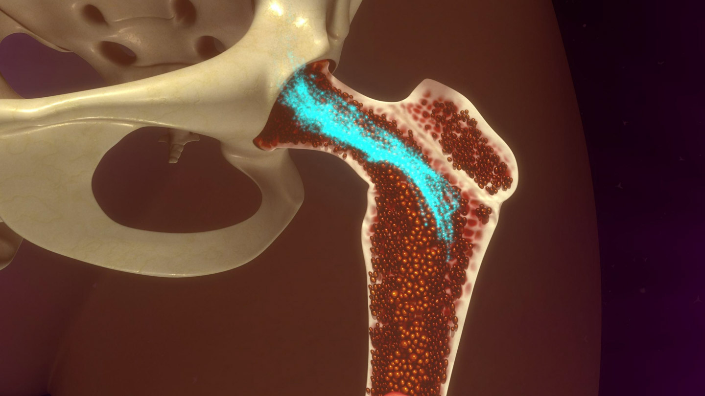
After a bone marrow transplant, it can take months for the number of T cells to reach healthy levels. Because T cells are crucial for launching an effective immune response, this leaves patients—usually cancer survivors whose immune systems were knocked out by chemotherapy—vulnerable to infections for longer. However, new research, to which Carl Ware, PhD, professor and director of the Infectious and Inflammatory Disease Center, contributed, identifies a novel target for immunotherapeutics to shorten this recovery time.

“This study shows that the lymphotoxin β receptor controls the entry of T cell progenitors into the thymus, the organ where T cells mature,” said Ware. “Future compounds that activate this receptor may help transplants give rise to functional T cells faster.”

Within the overall immune response against invading bacteria, viruses, and other pathogens, T cells are the field officers and special forces. Helper T cells send chemical signals to get other parts of the immune system involved, and cytotoxic T cells recognize and kill infected cells directly. They’re ‘trained’ to distinguish threats from the cells of the body in the thymus, where T cell progenitors that react to normal, uninfected cells are eliminated.

Publishing in the Journal of Immunology, the team, led by William Jenkinson, PhD, and Graham Anderson, PhD, of the University of Birmingham, looked at the importance of various receptors in letting T cell progenitors into the thymus, and found that only the lymphotoxin β receptor was required.

Significantly, the researchers also showed that stimulating the lymphotoxin β receptor boosted the number of transplant-derived T cells.

“Post-transplantation, T cell progenitors can struggle to enter the thymus, as if the doorway to the thymus is closed,” said Anderson. “Our work points to a way to ‘prop open’ the door and allow these cells to enter and mature.”

Ware and his lab have made many contributions to understanding how the lymphotoxin β receptor, as well as other related receptors, affect immunity and inflammation.

“The lymphotoxin β receptor is important not only in the thymus, but also at sites of inflammation and infection,” Ware added. “Further investigation of the effects of activating it throughout the body will determine whether this treatment approach is feasible, or perhaps should be targeted to the thymus.”

The paper is available online here.

Institute News

Genetic drivers of immune response to cancer discovered through ‘big data’ analysis

AuthorJessica Moore
Date

July 18, 2016

Scientists at the Sanford Burnham Prebys Medical Discovery Institute (SBP) have identified over 100 new genetic regions that affect the immune response to cancer. The findings, published in Cancer Immunology Research, could inform the development of future immunotherapies—treatments that enhance the immune system’s ability to kill tumors.

“By analyzing a large public genomic database, we found 122 potential immune response drivers—genetic regions in which mutations correlate with the presence or absence of immune cells infiltrating the tumors,” said lead author Eduard Porta-Pardo, PhD, a postdoctoral fellow at SBP. “While several of these correspond to proteins with known roles in immune response, many others offer new directions for cancer immunology research, which could point to new targets for immunotherapy.”

Immunotherapy has been heralded as a turning point in cancer because it can treat even advanced cases that have spread to other organs. Several drugs in this class are now widely used and often lead to remarkable success, eradicating or dramatically shrinking tumors and preventing recurrence.

Most current immunotherapies rely on a similar strategy—releasing the brakes on the immune system. These treatments are powerful if the tumor is recognized by the immune system as a threat and allows immune cell infiltration, but some cancers remain undercover or block immune cell entry into the tumor in as yet unknown ways.

“To develop immunotherapies that are relevant to a wide range of cancers, we need to know a lot more about how the immune system interacts with tumors,” said Adam Godzik, PhD, professor and director of the Bioinformatics and Structural Biology Program and senior author of the study. “Our study provides many new leads for this endeavor.”

“We are exploring cancer mutations at fine resolution by accounting for the fact that mutations can affect the encoded protein in different ways depending on where the resulting change is located,” commented Porta-Pardo. “Our algorithm, domainXplorer, identifies correlations between a phenotype, in this case the amount of immune cells in the tumor, and mutations in individual protein domains—parts of a protein with distinct functions.

“This work emphasizes the value of open data,” Godzik added. “Because we could access genomic data from over 5,000 tumor samples from The Cancer Genome Atlas (TCGA), we could jump straight to analysis without having to set up a big collaborative network to gather and sequence so many samples.”

“Our plan for the next phase of this research is to use this algorithm to search for genetic regions correlating with the levels of specific immune cell types within the tumor, which will reveal further details of cancer immunology.”


Institute News

Upcoming symposium: Cancer Immunology and the Tumor Microenvironment

Authorjmoore
Date

March 10, 2016

On March 17, SBP La Jolla is hosting a symposium on the interactions between the immune system and tumors, including how they can be leveraged for cancer treatment. The symposium is organized by Carl Ware, PhD and Robert Rickert, PhD, the directors of the Inflammatory and Infectious Disease Center and the Tumor Microenvironment and Cancer Immunology Program, respectively, and features presentations by leaders in the field:

Crystal Mackall, MDStanford University

Yang-Xin Fu, MD, PhDUT Southwestern

Mikala Egeblad, PhDCold Spring Harbor Laboratory

Linda Bradley, PhDSanford Burnham Prebys Medical Discovery Institute

Jose Conejo-Garcia, MD, PhDWistar Institute

Jonathan Powell, MDJohns Hopkins School of Medicine

Shannon Turley, PhDGenentech

Karen Willard-Gallo, PhDInstitut Jules Bordet – Belgium

Sandip Patel, MDUC San Diego

Adam Godzik, PhDSanford Burnham Prebys Medical Discovery Institute

The symposium will be held from 9-4:30 in Fishman Auditorium (overflow seating in the Building 12 auditorium), with a reception to follow. If you plan to attend, please register here.