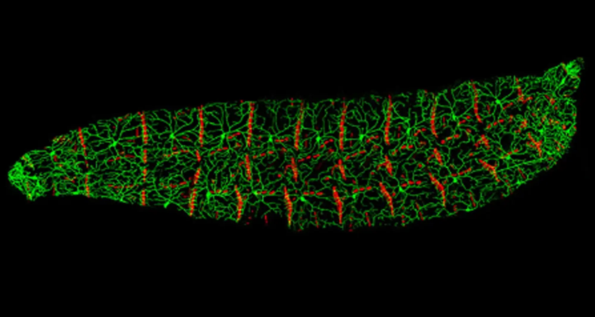
The genus Drosophila (fruit flies) are among science’s most used animal models, employed by researchers at all stages of the insects’ life. The larval stage depicted covers the first five days after an egg is laid. Three growth phases later, the larva will pupate.
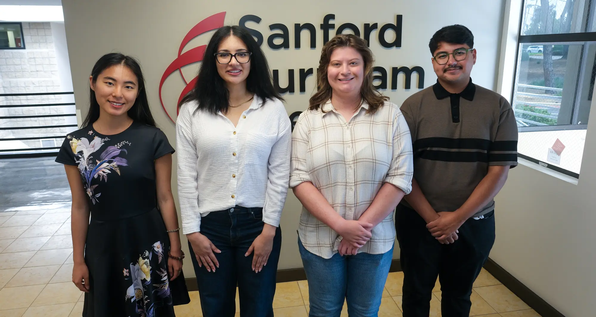
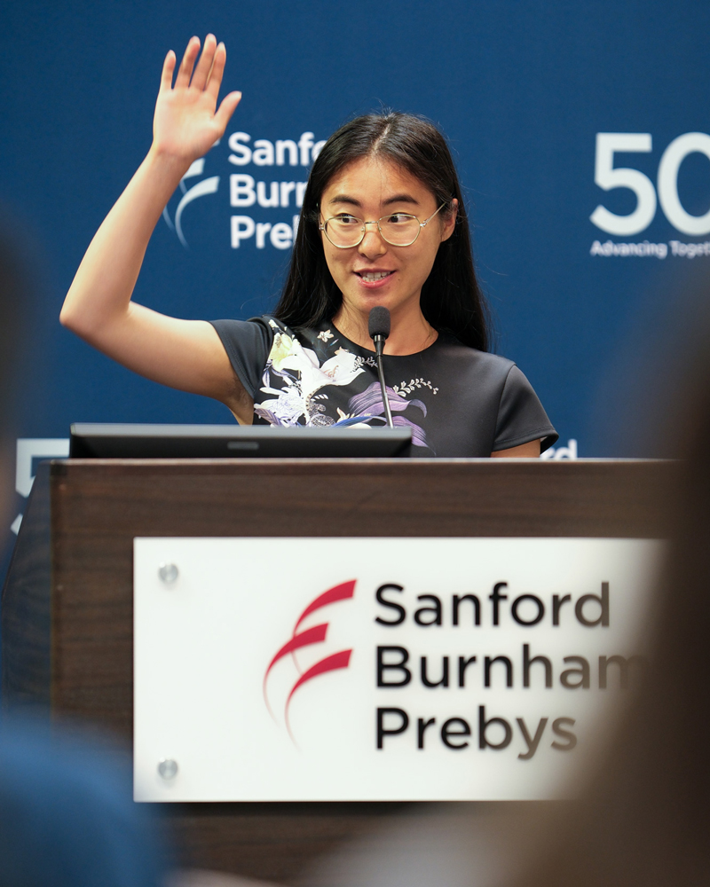
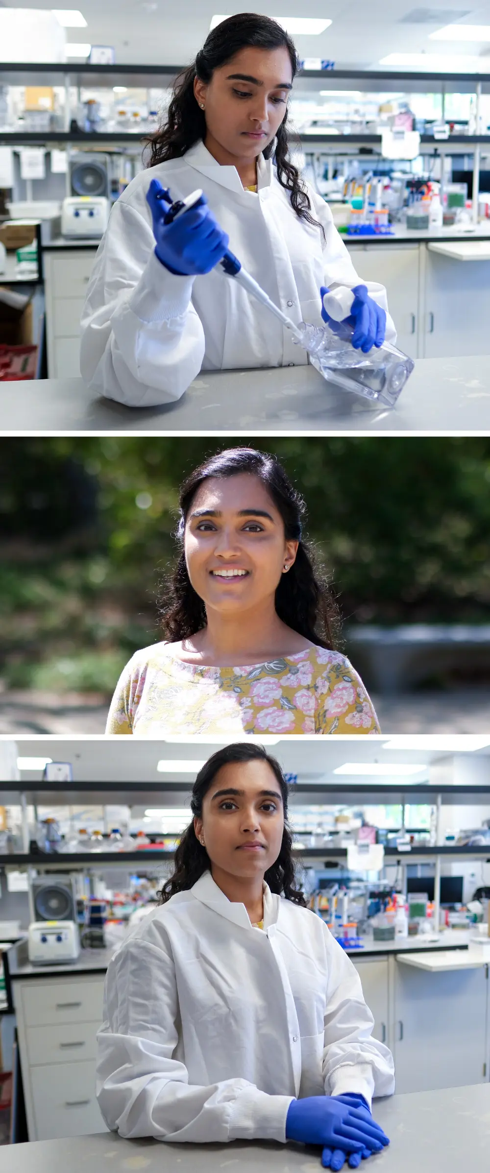
LEAP program participants recently shared their research at a capstone presentation event. From left to right: Yun Ma, Sarina Safavi, Isabel Sakowicz and Josue Navarrete. Not pictured: Mahek Shah. Image credit: Sanford Burnham Prebys.
Postbaccalaureate training program participants deliver capstone presentations, thank mentors and colleagues
In late June 2026, trainees in the LEAP (Laboratory Experience As Pathway to Graduate School) program at Sanford Burnham Prebys Medical Discovery Institute participated in a capstone presentation event. The scholars shared their research findings following a year of work and mentorship in labs throughout the Institute.
The LEAP program was designed to bridge the gap between college graduation and graduate school. It provides recent graduates with hands-on biomedical research experience, career development opportunities and mentorship to enhance their applications for—and ability to succeed in—highly competitive biomedical PhD programs.
With generous support from the Prebys Foundation, the LEAP program is offered through a collaborative partnership between the Sanford Burnham Prebys NCI-designated Cancer Center and Office of Learning and Development.
“Today we celebrate five of our LEAP program participants who have gained new skills, demonstrated research successes and grown as scientists throughout their journeys here,” said Kevin Yip, PhD, during his introductory remarks.
Following Yip’s welcoming address, the LEAP program mentors introduced the participants and invited them forward for their capstone presentations.
Yun Ma, a trainee in the Feng and Yip labs, discussed her study of proteins that group together in what are called protein complexes to carry out biological functions.
Ma developed a method for comparing data on RNA and proteins to identify priority genes affecting how protein complexes form. These priority genes may help shed new light on how diseases progress and may inform efforts to design new medications.
Her presentation was titled, “Inferring disease-associated changes in protein complex assembly from multimodal data.”
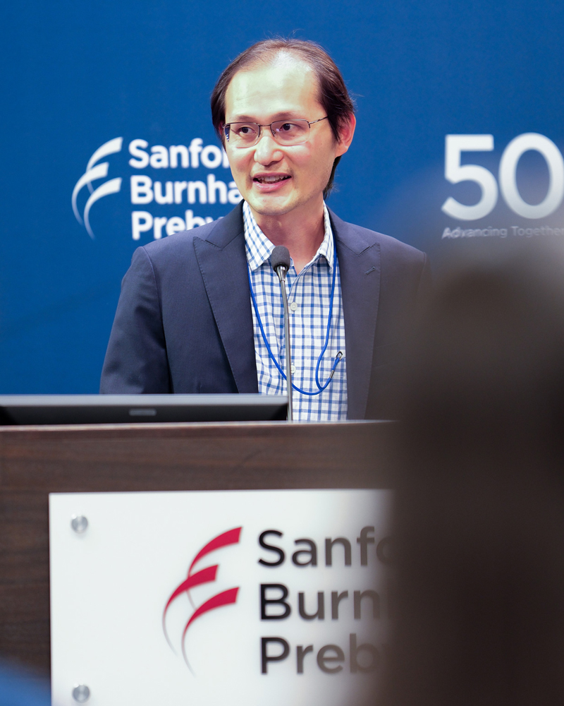
Kevin Yip, PhD, the Andrew and Erna Viterbi Distinguished Chair and director of the Center for Data Science and Artificial Intelligence, provided opening remarks at the event.
Sarina Safavi, a trainee in the Dhar lab, described her work to understand why cardiovascular disease—especially heart failure with preserved ejection fraction—is the leading cause of death among liver disease patients. Safavi developed tools needed to make new liver disease research models to study how the disease affects the heart.
The continuation of this research may lead to the discovery of new drug targets and methods for identifying liver disease patients at greater risk of heart failure to enable earlier treatments.
Her talk was titled, “Mechanistic understanding of MASLD-associated cardiac dysfunction and its reversibility.”
Josue Navarrete, a trainee in the Deshpande and Yip labs, detailed his project focused on small proteins from areas beyond the protein-coding genes that get the most scientific attention. Increasing amounts of evidence supports these microproteins’ biological importance, including in cancer.
Navarette optimized a computational technique for discovering microproteins which enables large-scale studies of thousands of genes. He tested the approach and found microproteins involved in stem cells becoming blood cells and other cells. Studying these microproteins may lead to new diagnostic techniques and potential treatments for blood cancers.
His lecture was titled, “Optimized pipeline enables genome-wide discovery of candidate microproteins in hematopoiesis.” Navarette will be joining the Institute as a doctoral student this fall.
Mahek Shah, a trainee in the Spruck lab, shared her study of a cancer treatment strategy that compels tumor cells to act as if they are infected by a virus. Known as viral mimicry, this therapeutic approach triggers the innate immune system to respond against cancer cells. Shah described drug screening experiments that identified promising novel candidate compounds.
In further tests, these compounds were found to activate viral mimicry, cause an immune response and to be toxic to cancer cells. Continued research may lead to new treatments that can boost the effectiveness of chemotherapy, radiation therapy and targeted therapies.
Her presentation was titled, “Novel compounds targeting histone modifying enzymes induce viral mimicry in breast cancer.”
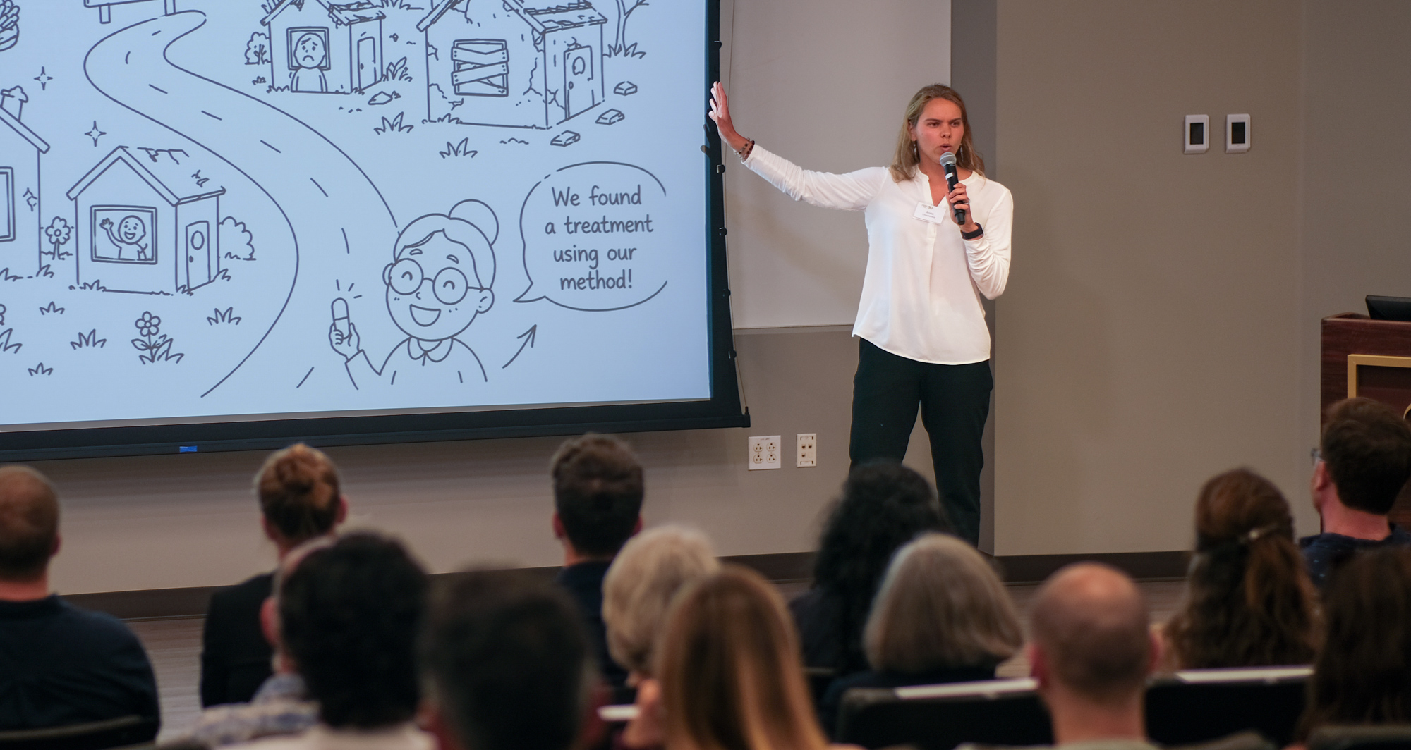
Isabel Sakowicz, a trainee in the Tharp lab, discussed her research regarding the most common and difficult-to-treat form of ovarian cancer. These tumors can wall themselves off from the immune system with structural collagen proteins that resemble scar tissue.
Yun Ma, a trainee in the Feng and Yip labs, discussed her study of proteins that group together in what are called protein complexes to carry out biological functions.
Sakowicz explored how a potential treatment strategy might reduce levels of a form of collagen known to promote ovarian cancer invasiveness and metastasis. Lowering collagen VI levels may break down the barrier around ovarian cancer cells to make other treatments more effective, including immunotherapies.
Her presentation was titled, “FAK protein loss rewires amino acid metabolism in high-grade serous ovarian cancer.” Sakowicz will be joining the Institute as a doctoral student this fall.
In addition to discussing their respective projects, the trainees credited their mentors, fellow lab members and access to avenues for training and advancing as professionals.
“Beyond my scientific achievements, I have enjoyed many career development opportunities, including attending and presenting at workshops, seminars and conferences,” said Ma.
“Everyone in the lab has contributed to a truly supportive and cooperative environment,” said Safavi. “I would also like to thank the LEAP program coordinators for their efforts and making sure we were set up for success.”
A ring nematode (Mesocriconema sp.). While the nematode C. elegans is a broadly used and useful model organism in research, ring nematodes are plant-parasites that feed on agricultural crops.
A thin-melt specimen of hippuric acid, a natural metabolite found in human urine. It acts as a biomarker for dietary polyphenol intake (fruits/vegetables) and gut microbiome activity, with levels rising after consuming tea, wine and fruit.
Image courtesy of Brian Johnston.
Institute News
Sanford Burnham Prebys Science Network hosts Community Outreach Symposium showcasing early-career research
Graduate students and postdoctoral researchers from across Sanford Burnham Prebys took center stage at the 2026 Community Outreach Symposium, sharing their research with colleagues and mentors from across the Institute, as well as members of the broader community.
Organized by the Sanford Burnham Prebys Science Network (SBP-SN) and the Office of Learning and Development, the event aims to spark excitement about biomedical research and build trust with the public, while providing trainees with an opportunity to communicate their science to a diverse audience in short 3-minute talks.
The symposium included a Postdoc Pitch Competition, where postdoctoral researchers competed for the opportunity to represent Sanford Burnham Prebys in a mesa-wide competition this fall against peers from Scripps Research, Salk Institute for Biological Studies and the University of California San Diego.
The event also featured a Science Communication Competition, where seven graduate students presented their research and competed for audience votes. The friendly competition highlighted the importance of effective science communication and public engagement.
Postdoc Pitch speakers:
Jimmy Massenet, PhD, Puri lab — CTCF: The Pillars That Hold Our Genome
Julia Schoenfeld, PhD, Cosford lab — One STEP Forward in the Fight Against Alzheimer’s Disease
Ryan Loughran, PhD, Emerling lab — PIPKs: Sorting Out Your Cell’s Delivery System
Kokila Shankar, PhD, Sheffler lab — Don’t Stress: Targeting Stress Signaling to Treat Brain Disorders- (First place Judges’ Selection and People’s Choice Award)
Jessica Proulx, PhD, Adams lab — Game of Genes: A Song of Activation & Silencing – (Second place Judges’ Selection)
Erik Hultenius, Tian lab — Anti-Aging Interventions for the Treatment of Alzheimer’s Disease
Patrick Hagan, Cosford lab — ATG Attack: Developing New Drugs for Pancreatic Cancer
The symposium also featured a keynote presentation by Sanjeev Ranade, PhD, assistant professor in the Center for Cardiovascular and Muscular Diseases. Ranade shared highlights from his research on congenital heart disease and developmental biology, while also reflecting on the mentors, experiences and scientific communication opportunities that shaped his career.
Drawing on his own career journey, Ranade encouraged trainees to take chances and remain open to unexpected opportunities. He shared how a seemingly small decision to submit an abstract ultimately set in motion a series of events that led to new collaborations, funding opportunities and eventually a faculty position at Sanford Burnham Prebys. Success, he noted, often comes not from following a carefully mapped path, but from being willing to step forward when opportunities arise.
The event concluded with award announcements, followed by a reception hosted by SBP-SN that provided additional opportunities for networking and scientific discussion.
By bringing together members of the Sanford Burnham Prebys community and guests from the broader community, the symposium fostered collaboration, scientific exchange and public engagement while helping early-career researchers strengthen the communication skills needed to share their discoveries beyond the laboratory.
Special thanks to our judges for their time and participation:
Nan Eastman Fishman Community Advisory Board Member
Mitchell Furumoto Associate Director, International Services, Sanford Burnham Prebys
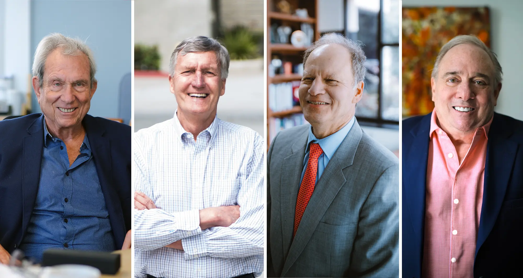
Four researchers at Sanford Burnham Prebys Medical Discovery Institute are among Research.com’s latest ranking of the “1,000 best scientists in the world” for 2025/26.
They are:
Michael Karin, PhD
Erkki Ruoslahti, MD, PhD
David Brenner, MD
Randal J. Kaufman, PhD
Research.com is an academic and higher education platform that produces data-driven rankings and guides. The latest listing of best scientists is primarily based on data derived from OpenAlex, an open-source catalog of the global research system with more than 480 million entries, and CrossRef, a not-for-profit digital registry of published scientific literature.
Additionally, researchers’ H-index is taken into account, which is a measure of the productivity and citation impact of their published work, an indicator of its importance and broader influence. H-index scores vary substantially by academic and scientific discipline. In biomedical research, an H-index of 40 or higher is deemed notable.
More on the four
Michael Karin, PhD, is among the world’s leading authorities on inflammation as a driver of disease, particularly cancer. He joined Sanford Burnham Prebys as director of the Center for Metabolic and Liver Diseases in 2025. Research.com identified Karin as ranking 23rd in the world among best scientists and 17th among U.S. researchers. His H-index was 279, with 348,118 citations in 834 publications.
Erkki Ruoslahti, PhD, joined Sanford Burnham Prebys in 1979 and served as president from 1989-2002. He is perhaps best known for co-discovering integrins—proteins that act as vital structural and chemical middlemen, connecting the inside of a cell to the surrounding extracellular matrix. He continues to conduct active studies. Research.com ranked Ruoslahti 341st in the world and 222nd in the U.S, with a 194 H-index, 153,152 citations in 704 publications.
David Brenner, MD, is president and CEO of Sanford Burnham Prebys and a leader in the field of gastroenterology research, specifically liver fibrosis (scarring). Research.com listed Brenner 872nd in the world and 505th in the U.S., with a 168 H-index, with 86,042 citations in 658 publications.
Randal J. Kaufman, PhD, is a professor in the Center for Metabolic and Liver Diseases. His research focuses on the fundamental mechanisms that regulate protein folding, why proteins misfold and the biological consequences. Research.com ranked Kaufman 890th in the world and 517th in the United States, with a 167 H-index on 119,194 citations in 566 publications.
For the full listing and explanations of methodology, visit Research.com.
Institute News
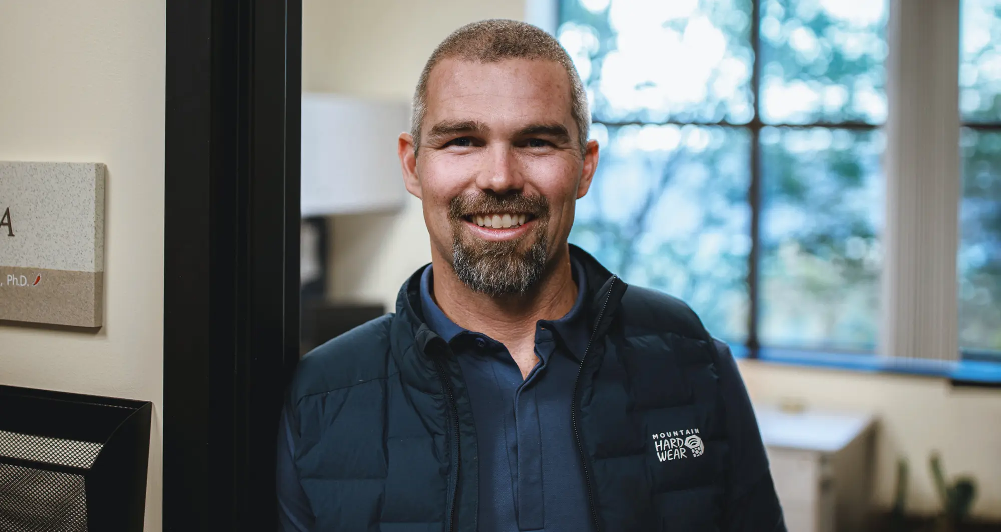
Kevin Tharp awarded $450,000 Ovarian Cancer Research Alliance grant to break down tumor defenses
Kevin Tharp, PhD, is an assistant professor in the NCI-designated Cancer Center’s Cancer Metabolism and Microenvironment Program at Sanford Burnham Prebys. Image credit: Sanford Burnham Prebys.
The new award will fund research regarding how to bust through the barrier between tumors and immune cells
Kevin Tharp, PhD, was awarded a three-year, $450,000 Ovarian Cancer Research Alliance grant to study high grade serous ovarian cancer, the most common and deadly form of the disease.
In collaboration with researchers at the University of California San Diego, Tharp recently published findings in Cell Reports demonstrating a treatment approach in mice that allowed more tumor-fighting immune cells to approach tumors, shifted the behavior of other immune cells to work against tumors, and made immunotherapy more effective.
Tharp will use the new funding to follow up on these findings regarding how high grade serous ovarian cancer circumvents the immune system’s anti-tumor defenses. His team will focus on the physical barrier that tumors build to keep immune cells at bay.
“We know that tumors can wall themselves off with structural collagen proteins that resemble scar tissue, and that the presence of this obstacle determines the effectiveness of anti-cancer immunotherapies,” said Tharp.
“We want to know how and why tumors create this obstruction and understanding this will help us find ways to break through these defenses to make immunotherapies more effective.”
Tharp will conduct this research under the mentorship of Cosimo Commisso, PhD, the deputy director of the NCI-Designated Cancer Center at Sanford Burnham Prebys and a professor in the Cancer Metabolism and Microenvironment Program, and David Schlaepfer, PhD, a professor in the department of OBGYN and Reproductive Sciences at the University of California San Diego Moores Cancer Center.
“Too many ovarian cancer patients progress and do not respond to the standard of care, so finding new treatments is an extreme clinical need,” said Tharp.
“There is potential for immunotherapies to treat recurrent and metastatic cancer if we can bridge the divide between tumors and immune cells.”
The Ovarian Cancer Research Alliance is the oldest and largest ovarian and gynecologic cancer charity in the world. Since its founding in 1994, the alliance has grown into the leading non-government funder of ovarian and related gynecologic cancer research by investing more than $140 million in grants to scientists.
Institute News
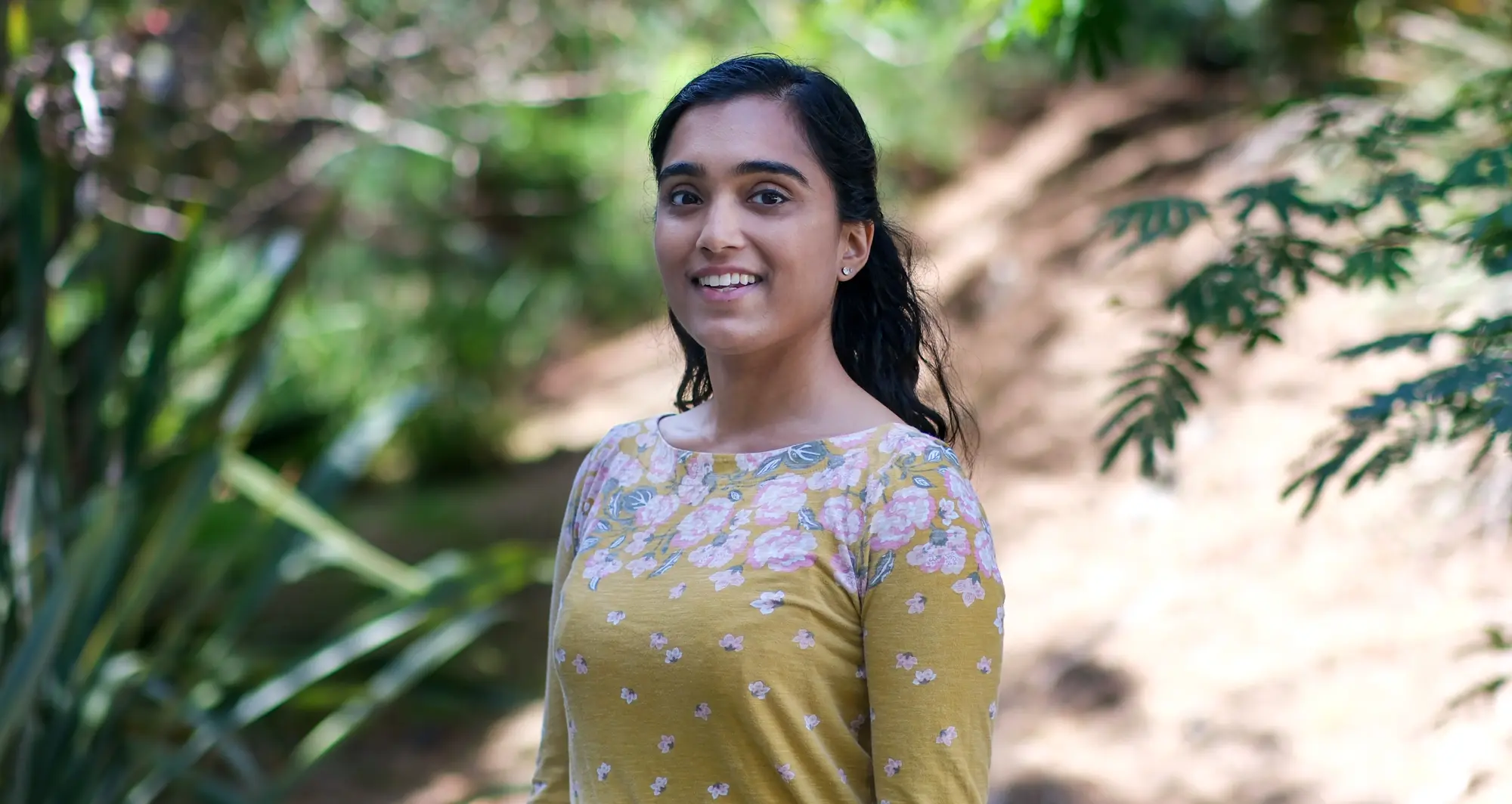
Q & A with Postdoctoral Researcher Kokila Shankar, from the Sheffler Lab
Meet one of our early-career scientists at Sanford Burnham Prebys Medical Discovery Institute: Kokila Shankar, PhD, a postdoctoral researcher in the lab of Douglas Sheffler, PhD. Shankar studies the brain stress signaling system to develop potential treatments for drug addiction.
When and how did you become interested in science? My dad works in science, so I grew up seeing a little bit of that world. Within science, he works on the industry side which provided me with a perspective on how drug discovery works.
While I always liked science growing up, what really hooked me is when I learned about the field of neuroscience. You have to incorporate a lot of different types of knowledge to really understand neuroscience and neurobiology. And it’s one of the fields where I think that no matter how much we study it, there’s always going to be room to learn more and more.
What are the key areas of research you focus on? I am really excited about the fact that I get to work in small molecule drug discovery for neurological and psychiatric disease. Currently, my focus is on treating drug addiction.
We use a drug discovery workflow to be able to take biological targets we’ve identified and develop small molecule chemicals that modify these targets. Essentially, we’re working on ways to either increase or decrease the activity of these targets as a strategy to potentially mitigate drug addiction symptoms.
More specifically, my main project is targeting the brain stress signaling system. By developing a small molecule that modulates a stress signaling receptor and some of its associated proteins, our goal is to change stress signaling in the brain. This is important because stress signaling is very involved in addiction. It can play a large role in people becoming addicted to substances and making their addiction worse and harder to quit.
What motivates you about your research? I think in the field of drug discovery, people tend to portray making new medicines as a linear path. We have a small molecule identified through a screen that we know has some effects on a biological target. Then we just have to make it better, put it in cells, then in animals and finally we get it approved to put into people.
The reality is that there are so many things that can affect every single stage of that workflow. And there’s a lot of troubleshooting and interdisciplinary collaboration involved in keeping things on track. So even when things aren’t going smoothly, I remember that what I signed up to do as a scientist is solve problems. Each time we solve one, we also learn something new and that keeps me very motivated.
What do you like about working here? Sanford Burnham Prebys feels like a very focused place to do research. Everyone’s goal is to work together to drive research forward. And you really feel that through the resources that are available, whether that’s the core facilities or the number of people that are available to collaborate or share reagents.
On top of that, the community here is very welcoming. Everyone is collaborative and wants to see each other succeed. And that’s rare to find in an academic research environment.
What are your career goals? My goal is to transition to a role in biotech or the pharmaceutical industry. And it would be even better if I was able to work in neuroscience therapeutics development.
Beyond that, I think a special aspect of being in science, earning a PhD and completing postdoctoral training is that it all prepares you for so many different types of careers. That could mean staying at the bench in R&D or moving into areas such as business, communications and marketing or science policy.
I feel like I’m very fortunate that there’s a lot of opportunities, and I don’t want to close the door on any of them.
What do you enjoy doing when you’re not in the lab? I am an active volunteer with the Fleet Science Center, so I love doing their K-12 and their adult outreach programs to get more people excited about science.
Also, I used to be a competitive ballroom dancer. Right now, I’m enjoying the opportunity to work with students at the University of California San Diego to help them improve. And hopefully one day I’ll step back into competition.
Postdocs at Sanford Burnham Prebys are pushing the boundaries of science every day through curiosity, collaboration, and innovation. This series highlights their unique journeys, what inspires their work, and the impact they’re making across our labs.