

- Our Machine Learning Resources A Complete Guide to Studying Machine Learning from YouTube | X
- Almost All The Big Data Resources Available In Cancer Research 10 Resources to Learn Almost All the Big Data Resources Available in Cancer Research | X
- Computational Resources To Study Immune System 11 Resources (Videos And Papers) Covering the Fundamentals and Computational Tools Available to Study the Immune System | X
Faculty Type: Faculty
Sanjeev S. Ranade studies how transcription factors specifically control the development and function of cardiac cells — and what happens when things go wrong.
Transcription factors (TF) are proteins that initiate and regulate the transcription of genes, essentially turning genes on and off, boosting or repressing their activity. At last count, there were over 1,500 known TFs, but the contribution of most of the TFs to life and health is unknown.
In particular, Ranade focuses on how disrupted cell-to-cell signaling caused by mutations in TFs can cause congenital heart defects or CHDs.
“My research is focused on understanding why young children are born with heart defects. What are the principles and rules that allow our hearts to develop in the first place and why do these rules get broken in some cases? This is really important because nearly 1 in 100 children are born with some form of heart defect and many of these children will suffer from heart disease at much earlier stages in life compared to the general population.”
For his doctorate in molecular biology at Scripps Research in San Diego, Ranade studied ion channels — proteins that span cell membranes, allowing passage of ions or charged molecules from one side of the membrane to the other. The channels serve many critical functions, including transmitting signals involved in cell-cell communications and muscle contraction.
Working as a post-doctoral fellow and staff research scientist in the lab of Deepak Srivastava, M.D. at Gladstone Institutes, Ranade looked at how genetics and cell biology were connected and how disruptions to these connections led to children with heart defects.
Related Disease
Congenital Diseases
Phenomena or Processes
Transcription Factors
Anatomical Systems and Sites
Heart
 Apr 21, 2026
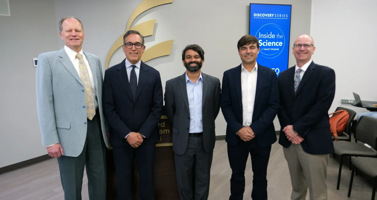
Sanford Burnham Prebys goes Inside the Science of Heart Disease
Apr 21, 2026Event recording now available for panel discussion with scientists held on April 15, 2026.
 Oct 22, 2025
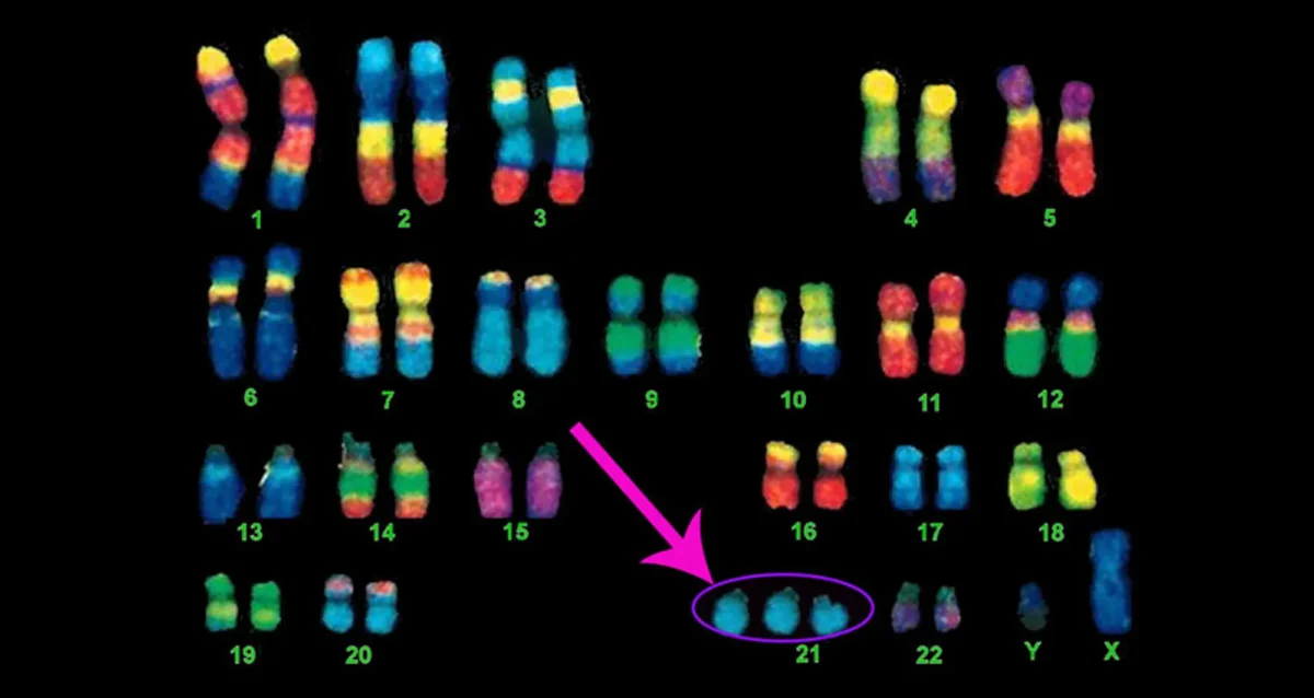
Hunting for the Chromosomal Genes that Break the Heart
Oct 22, 2025Using advanced CRISPR technology, researchers identify a protein that in overabundance is linked to Down syndrome and congenital heart disease.
 Sep 25, 2025
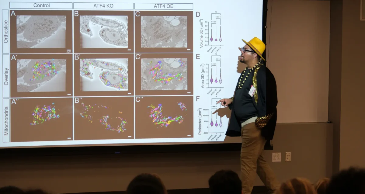
Celebrating research and mentorship during National Postdoc Appreciation Week
Sep 25, 2025The 24th Annual Biomedical Research Symposium brought together postdocs, graduate students, staff scientists, administrators, and faculty for a day that…
 Aug 7, 2025
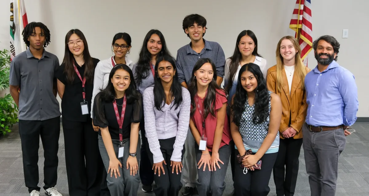
Summer interns SPARK interest in regenerative medicine
Aug 7, 2025Eleven participants presented posters at event marking the end of six weeks of hands-on biomedical research experience at Sanford Burnham…
 Jan 19, 2024
Sanjeev Ranade wants to get to the heart of congenital disease
Jan 19, 2024New Sanford Burnham Prebys researcher studies the underlying factors that cause in heart defects in 1 in 100 newborns.
 Nov 2, 2023
La Jolla’s Sanford Burnham Prebys adding six scientists in hopes of launching new era of collaboration
Nov 2, 2023The newcomers, brought in with help from a $70 million donation, are versed in different technologies that can be applied…
Pier Lorenzo Puri earned his MD at the University of Rome “la Sapienza” in 1991. Dr. Puri completed his internship in Internal Medicine at the hospital “Policlinico Umberto I” (Rome) from 1992 to 1997, and defended an experimental thesis on the vascular effects of angiotensin II to graduate as Specialist in Internal medicine at the University of Rome “la Sapienza” in 1997. During this time he was frequently working at the Freien University of Berlin, as visiting scientist at the Deprtment of Biochemistry and Molecular Biology, to perform experiments of protein and DNA microinjection in cultured cells. Dr. Puri trained as a post-doctoral fellow at the University of California San Diego (UCSD), in the department of Cell Biology, under the supervision of Dr. Wang, from 1997 to 2001. He was appointed as Staff Scientist at the Salk Institute (La Jolla) in 2001, and became an Assistant Telethon Scientist at the Dulbecco Telethon Institute in Rome in 2002. He was upgraded to Associate Telethon Scientist at the Dulbecco Telethon Institute in Rome since 2007 and became Senior Telethon Scientist, Dulbecco Telethon Institute, in 2012, but declined this position. Dr. Puri joined Sanford Burnham Prebys as an Assistant Professor in 2004. He has been promoted to Associate Professor in 2010 and full Professor in 2015. From 2008 to 2016 Dr. Puri served as Adjunct Professor of Pediatrics at the University of California, San Diego. From 2008 to 2013 Dr Puri was an Associate Member of Sanford Children’s Health Research Center. Dr Puri has been Director of the laboratory of Epigenetics and Regeneration at Fondazione S. Lucia, Roma, Italy, but stepped down this position since 2019.
Education
University of California San Diego, Postdoctoral, Department of Biology
University of Rome La Sapienza, PhD, Internal Medicine
University of Rome La Sapienza, MD, Internal Medicine
University of Rome La Sapienza, Undergraduate, Internal Medicine
Other Appointments
2020-2024: Member of the Science Advisory Board (SAB) European Commission-funded Consortium BIND (Brain Involvement In Dystrophinopathies)
2015-2019: Standing Member, NIH Study Section (SMEP)
2010-present: Member of Editorial Board of Skeletal Muscle
Related Disease
Aging-Related Diseases, Childhood Diseases, Molecular Biology, Muscular Dystrophy, Myopathy, Neurodegenerative and Neuromuscular Diseases, Rhabdomyosarcomas, Sarcopenia/Aging-Related Muscle Atrophy, Spinal Cord Injury, Transcription Factors
Phenomena or Processes
Adult/Multipotent Stem Cells, Aging, Cell Biology, Cell Cycle Progression, Cell Differentiation, Cell Signaling, Cellular Senescence, Development and Differentiation, Disease Therapies, DNA Damage Checkpoint Function, Epigenetics, Gene Regulation, Phosphorylation, Regenerative Biology, Signal Transduction, Transcriptional Regulation
Anatomical Systems and Sites
General Cell Biology, Musculoskeletal System
Research Models
Clinical and Transitional Research, Cultured Cell Lines, Human Adult/Somatic Stem Cells, Mouse Embryonic Stem Cells, Mouse Somatic Stem Cells, Primary Human Cells
Techniques and Technologies
Bioinformatics, Cellular and Molecular Imaging, Gene Expression, Genomics
Puri’s lab group investigates the molecular and epigenetic regulation of gene expression in skeletal muscle progenitors and other muscle-resident cell types (including fibro-adipogenic progenitors, cells from the inflammatory infiltrate, cellular components of neuro-muscular junctions) during physiological and pathological perturbations of skeletal muscle homeostasis.
- We use molecular, biochemical and epigenetic tools to understand structural and functional principles of the 3D genome organization that regulates gene expression during muscle regeneration and diseases.
- A topic of particular interest is the analysis of chromatin interactions that define the 3D genome organization and the identification of structural and functional interactions that regulate cell type-specific patterns of gene expression in response to cues released within the skeletal muscle regenerative environment in health and disease conditions, such as muscular dystrophies and other neuromuscular diseases.
- The knowledge derived from our studies is instrumental to elucidate the pathogenesis of muscular disorders and discover pharmacological interventions that promote muscle regeneration to repair diseased muscles.
- Current translational focus is devoted to:
- the study of the therapeutic potential of HDAC inhibitors for treatment of Duchenne Muscular Dystrophy (DMD)
- the identification of genome variants associated to DMD patient-specific patterns of expression of disease-modifier genes that can account for individual trends of disease progression beyond the common genetic deficiency of dystrophin
- the effect of dystrophin deficiency and restoration by gene therapy on 3D genome and transcriptional output of DMD myofibers; the therapeutic potential of extracellular vesicles released by fibro-adipogenic progenitors of DMD skeletal muscles exposed to HDACi.
Puri Lab
Pier Lorenzo Puri’s Research Report
Puri’s lab group investigates the molecular and epigenetic regulation of gene expression in skeletal muscle progenitors and other muscle-resident cell types (including fibro-adipogenic progenitors, cells from the inflammatory infiltrate, cellular components of neuro-muscular junctions) during physiological and pathological perturbations of skeletal muscle homeostasis.
We use molecular, biochemical and epigenetic tools to understand structural and functional principles of the 3D genome organization that regulates gene expression during muscle regeneration and diseases
A topic of particular interest is the analysis of chromatin interactions that define the 3D genome organization and the identification of structural and functional interactions that regulate cell type-specific patterns of gene expression in response to cues released within the skeletal muscle regenerative environment in health and disease conditions, such as muscular dystrophies and other neuromuscular diseases.
The knowledge derived from our studies is instrumental to elucidate the pathogenesis of muscular disorders and discover pharmacological interventions that promote muscle regeneration to repair diseased muscles
Current translational focus is devoted to:
- the study of the therapeutic potential of HDAC inhibitors for treatment of Duchenne Muscular Dystrophy (DMD)
- the identification of genome variants associated to DMD patient-specific patterns of expression of disease-modifier genes that can account for individual trends of disease progression beyond the common genetic deficiency of dystrophin
- the effect of dystrophin deficiency and restoration by gene therapy on 3D genome and transcriptional output of DMD myofibers; the therapeutic potential of extracellular vesicles released by fibro-adipogenic progenitors of DMD skeletal muscles exposed to HDACi.
1. Epigenetic regulation of skeletal myogenesis by histone acetyltransferases and deacetylases
Our earlier identification and characterization of acetyltransferases p300/CBP and PCAF, as transcriptional co-activators, and the histone deacetylases HDACs, as transcriptional co-repressors, of the myogenic determination factor MyoD1-3, respectively, inspired the experimental rationale toward exploiting pharmacological inhibition of HDAC to promote skeletal myogenesis.
- p300 is required for MyoD-dependent cell cycle arrest and muscle-specific gene transcription. Puri PL, Avantaggiati ML, Balsano C, Sang N, Graessmann A, Giordano A, Levrero M. EMBO J 1997 Jan 15 ;16(2):369-8
- Differential roles of p300 and PCAF acetyltransferases in muscle differentiation. Puri PL, Sartorelli V, Yang XJ, Hamamori Y, Ogryzko VV, Howard BH, Kedes L, Wang JY, Graessmann A, Nakatani Y, Levrero M. Mol Cell 1997 Dec ;1(1):35-45
- Class I histone deacetylases sequentially interact with MyoD and pRb during skeletal myogenesis. Puri PL, Iezzi S, Stiegler P, Chen TT, Schiltz RL, Muscat GE, Giordano A, Kedes L, Wang JY, Sartorelli V. Mol Cell 2001 Oct ;8(4):885-97
- Stage-specific modulation of skeletal myogenesis by inhibitors of nuclear deacetylases. Iezzi S, Cossu G, Nervi C, Sartorelli V, Puri PL. Proc Natl Acad Sci U S A 2002 May 28 ;99(11):7757-62
- Deacetylase inhibitors increase muscle cell size by promoting myoblast recruitment and fusion through induction of follistatin. Iezzi S, Di Padova M, Serra C, Caretti G, Simone C, Maklan E, Minetti G, Zhao P, Hoffman EP, Puri PL, Sartorelli V. Dev Cell 2004 May ;6(5):673-84
2. HDAC inhibitors as pharmacological intervention in DMD and other muscular dystrophies
Puri lab discovered that dystrophin-activated nNOS signalling controls HDAC2 activity, thereby revealing a previously unrecognized link between constitutive activation of HDAC2 and alteration of the epigenetic landscape of dystrophin-deficient muscles6,7. This discovery established the rationale for using HDAC inhibitors to counter the progression of Duchenne muscular dystrophy (DMD), by correcting aberrant HDAC activity in dystrophin-deficient muscles8-11.
- Functional and morphological recovery of dystrophic muscles in mice treated with deacetylase inhibitors. Minetti G. C., Colussi c., Adami R., Serra C., Mozzetta C., Parente V., Illi B., Fortuni S., Straino S., Gallinari P., Steinkhuler C., Capogrossi M., Sartorelli V., Bottinelli R., Gaetano C., Puri P.L. Nat. Med 2006 Oct ;12(10):1147-50.
- A Common Epigenetic Mechanism Underlies Nitric Oxide Donors and Histone Deacetylase Inhibitors Effect in Duchenne Muscular Dystrophy. Colussi C., Mozzetta C., Gurtner A. , Illi B., Straino S., Ragone G., Pescatori M., Zaccagnini G., Rosati G., Minetti G., Martelli F., Ricci E., Piaggio G., Gallinari P., Steinkulher C., Capogrossi M.C., Puri P.L*, Carlo Gaetano. Proc Natl Acad Sci U S A 2008 Dec 9 ;105(49):19183-7 *Corresponding author.
- Fibroadipogenic progenitors mediate the ability of HDAC inhibitors to promote regeneration in dystrophic muscles of young, but not old mdx mice. Mozzetta C., Consalvi S., Saccone V., Tierney M., Diamantini A., Mitchel K.J., Marazzi G., Borsellino G., Battistini L., Sassoon D., Sacco A., Puri P.L. EMBO Mol Med 2013 Apr ;5(4):626-39
- Histological effects of givinostat in boys with Duchenne muscular dystrophy. Neuromuscul Disord. Bettica P, Petrini S, D’Oria V, D’Amico A, Catteruccia M, Pane M, Sivo S, Magri F, Brajkovic S, Messina S, Vita GL, Gatti B, Moggio M, Puri PL, Rocchetti M, De Nicolao G, Vita G, Comi GP, Bertini E, Mercuri E. Neuromuscul Disord 2016 Oct ;26(10):643-649
- HDAC inhibitors tune miRNAs in extracellular vesicles of dystrophic muscle-resident mesenchymal cells. Sandonà M, Consalvi S, Tucciarone L, De Bardi M, Scimeca M, Angelini DF, Buffa V, D’Amico A, Bertini ES, Cazzaniga S, Bettica P, Bouché M, Bongiovanni A, Puri PL, Saccone V. EMBO Rep 2020 Sep 3 ;21(9):e50863
- Determinants of epigenetic resistance to HDAC inhibitors in dystrophic fibro-adipogenic progenitors. Consalvi S, Tucciarone L, Macrì E, De Bardi M, Picozza M, Salvatori I, Renzini A, Valente S, Mai A, Moresi V, Puri PL. EMBO Rep 2022 Jun 7 ;23(6):e54721
3. Control of chromatin structure in muscle cells by regeneration-induced signaling pathways
Upon the discovery and characterization of intracellular signaling pathways (i.e. p38, ERK and AKT cascades) that regulate muscle gene expression in myoblasts, in earlier studies during Puri’s postdoctoral training, Puri lab has revealed the mechanism by which muscle environmental cues are converted into epigenetic changes that regulate gene expression in healthy and diseased muscles, via extracellular signal-activated kinase targeting of chromatin-modifying enzymes. These studies provided the first evidence that regeneration activated p38 and AKT signaling cooperatively direct assembly and activation of histone acetyltransferases and chromatin remodeling SWI/SNF complex at myogenic loci in muscle progenitors12,13,15. Moreover, we discovered that regeneration-activated p38 targets Polycomb Repressory Complex (PCR2) at Pax7 locus to promote formation of repressive chromatin during satellite cells a ctivation14.
- p38 Pathway Targets SWI/SNF Chromatin Remodeling Complex to Muscle-Specific Loci. Simone C., Forcales S.V., Hill D., Imbalzano A.L., Latella L., and Puri P.L. Nat Genet 2004 Jul ;36(7):738-43
- Functional interdependence at the chromatin level between the MKK6/p38 and IGF1/Pi3K/AKT pathways during muscle differentiation. Serra C., Palacios D., Mozzetta C Forcales S., Ripani M., Morantte I., Jones D. Du K., Jahla U., Simone C., Puri P.L. Mol Cell 2007 Oct 26 ;28(2):200-13
- TNF/p38 alpha/Polycomb signalling to Pax7 locus in satellite cells links inflammation to the epigenetic control of muscle regeneration. Palacios D., Mozzetta C., Consalvi S., Caretti G., Saccone V., Proserpio V., Marquez V.E., Valente S., Mai A., Forcales S., Sartorelli V., Puri P.L. Cell Stem Cell 2010 Oct 8 ;7(4):455-69
- Signal dependent incorporation of MyoD-BAF60c into Brg1-based SWI/SNF chromatin-remodeling complex. Forcales S., Albini S., Giordani L., Malecova B., Cignolo L., Chernov A., Coutinho P., Saccone V., Consalvi S., Williams R., Wang K., Wu Z., Baranovskaya S., Miller A., Dilworth F., Puri P.L. EMBO J 2012 Jan 18 ;31(2):301-16
4. Epigenetic basis for activation of the myogenic program in ESCs and other pluripotent cell types
Puri lab studied the epigenetic determinants of human embryonic stem cells (hESCs) and induced pluripotent stem cells (hiPSCs) commitment to skeletal myogenesis, by investigating the hESC resistance to direct conversion into skeletal muscle upon ectopic expression of MyoD, which can otherwise reprogram somatic cells into the skeletal muscle lineage. These studies showed that hESC and hiPSC resistance to myogenic conversion is caused by the lack of expression of one structural component of the SWI/SNF chromatin remodelling complex – BAF60C – which is specifically induced in embryoid bodies13. Based on these studies, we have recently established a protocol of hESC-derived 3D contractile myospheres that offers the unprecedented opportunity to dissect and analyze the epigenetic dynamics that underlie the formation of skeletal muscles and to identify changes in the epigenome induced by contractile activity in healthy vs dystrophin-deficient myofibers16,20. We have also determined the identity of the general transcription factors implicated in the activation of skeletal myogenesis17, and we have discovered that replicative senescence is associated with acquisition of resistance to MYOD-mediated activation of muscle gene expression, caused by the constitutive activation of DNA damage repair (DDR) response that impairs cell cycle progression and MYOD activity18. Finally, our recent work has elucidated the mechanism by which MYOD regulates high-order chromatin interactions to define the tri-dimensional (3D) nuclear architecture for the activation of skeletal myogenesis during human somatic cell reprogramming into skeletal muscles19.
- Epigenetic reprogramming of human embryonic stem cells (hESCs) into skeletal muscle cells and generation of contractile myospheres. Albini S., Coutinho P., Malecova B., Giordani L., Savchenko A., Forcales S, Puri P.L. Cell Rep 2013 Mar 28;3(3):661-70
- TBP/TFIID-dependent activation of MyoD target genes in skeletal muscle cells. Malecova B., Dall’Agnese A., Madaro L., Gatto S., Coutinho Toto P., Albini S., Ryan T., Tora L., Puri PL. Elife 2016 Feb 25 ;5:e12534
- 18) DNA damage signaling mediates the functional antagonism between replicative senescence and terminal muscle differentiation. Latella L., Dall’Agnese A, Boscolo F., Nardoni C. Cosentino M., Lahm A, Sacco A., Puri P.L. Genes Dev 2017 Apr 1 ;31(7):648-659
- Transcription Factor-Directed Re-Wiring of Chromatin Architecture for Somatic Cell Nuclear Reprogramming Toward Trans-differentiation. Dall’Agnese A., Caputo L., Nicoletti C., di Iulio J., Schmitt A., Gatto S., Diao Y., Ye Z., Forcato M., Perera R., Bicciato S., Telenti A., Ren B., Puri P.L. Mol Cell 2019 Nov 7 ;76(3):453-472.e8
- Acute conversion of patient-derived Duchenne muscular dystrophy iPSC into myotubes reveals constitutive and inducible over-activation of TGFβ-dependent pro-fibrotic signaling. Caputo L, Granados A, Lenzi J, Rosa A, Ait-Si-Ali A, Puri PL, Albini S. Skelet Muscle 2020 May 2 ;10(1):13
5. Identification, functional, phenotypic and molecular characterization of muscle-interstitial cells – (the fibroadipogenic progenitors – FAPs) in healthy and diseased muscles.
Our work has elucidated the molecular determinants of the interplay between adult muscle stem cells and cellular components of their functional niche (i.e. FAPs), by identifying regulatory networks implicated in compensatory or pathogenic regeneration, and suggesting “disease stage-specific” responses to pharmacological treatment of neuromuscular disorders, such as DMD. Indeed, we have shown that HDACi promote compensatory regeneration and prevent fibro-adipogenic degeneration in mdx mice at early stages of diseases, by targeting a population of muscle interstitial cells – FAPs8 – and have identified a HDAC-regulated network that controls expression of myomiRs and alternative incorporation of BAF60 variants into SWI/SNF complexes to direct the pro-myogenic or fibro-adipogenic FAP activity21. Furthermore, we have recently identified specific subpopulations of FAPs (subFAPs) in physiological conditions and disease22 and we have discovered that specific subFAPs expand and adopt pathogenic phenotypes upon muscle denervation23 or in muscles of patients affected by type 2 diabetes24.
- HDAC-regulated myomiRs control BAF60 variant exchange and direct the functional phenotype of fibro-adipogenic progenitors in dystrophic muscles. Saccone V, Consalvi S, Giordani L, Mozzetta C, Barozzi I, Sandoná M, Ryan T, Rojas-Muñoz A, Madaro L, Fasanaro P, Borsellino G, De Bardi M, Frigè G, Termanini A, Sun X, Rossant J, Bruneau BG, Mercola M, Minucci S, Puri P.L. Genes Dev 2014 Apr 15 ;28(8):841-57
- Spectrum of cellular states within Fibro-Adipogenic Progenitors upon physiological and pathological perturbations of skeletal muscle. Malecova B., Gatto S., Etxaniz U, Passafaro M. Cortez A., Nicoletti C., Giordani L., Torcinaro A., De Bardi M., Bicciato S., De Santa F., Madaro L, Puri PL. Nat Commun 2018 Sep 10 ;9(1):3670
- Denervation-activated STAT3-IL6 signaling in fibro-adipogenic progenitors (FAPs) promotes myofibers atrophy and fibrosis. Madaro L, Passafaro M., Sala D., Etxaniz U., Lugarini F, Proietti D., Alfonsi MV, Nicoletti C., Gatto S., De Bardi M., Rojas-García R, Giordani L., Marinelli S., Pagliarini V., Sette C, Sacco A, Puri PL. Nat Cell Biol 2018 Aug ;20(8):917-927
- Human skeletal muscle CD90+ fibro-adipogenic progenitors are associated with muscle degeneration in type 2 diabetic patients. Farup J, Just J, de Paoli F, Lin L, Jensen JB, Billeskov T, Roman IS, Cömert C, Møller AB, Madaro L, Groppa E, Fred RG, Kampmann U, Gormsen LC, Pedersen SB, Bross P, Stevnsner T, Eldrup N, Pers TH, Rossi FMV, Puri PL, Jessen N. Cell Metab 2021 Nov 2 ;33(11):2201-2214.e11
 Aug 12, 2025
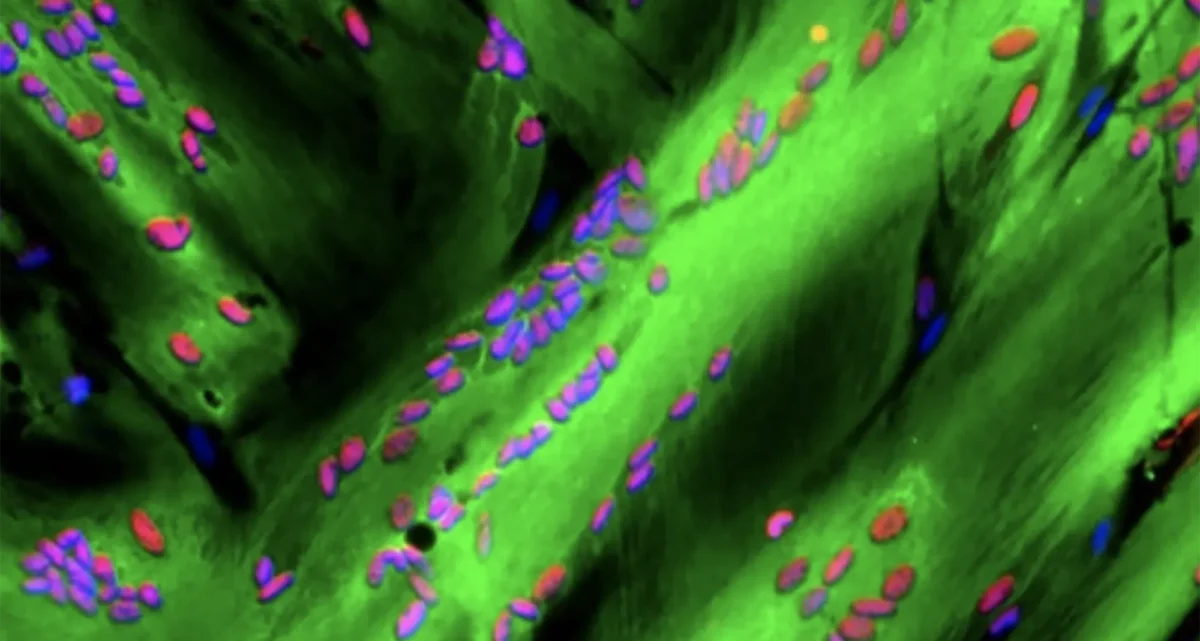
Muscle’s master regulator moonlights as gene silencer
Aug 12, 2025Study uncovers new repressor role for the transcription factor that triggers muscle stem cells to turn into muscle.
 Mar 12, 2025
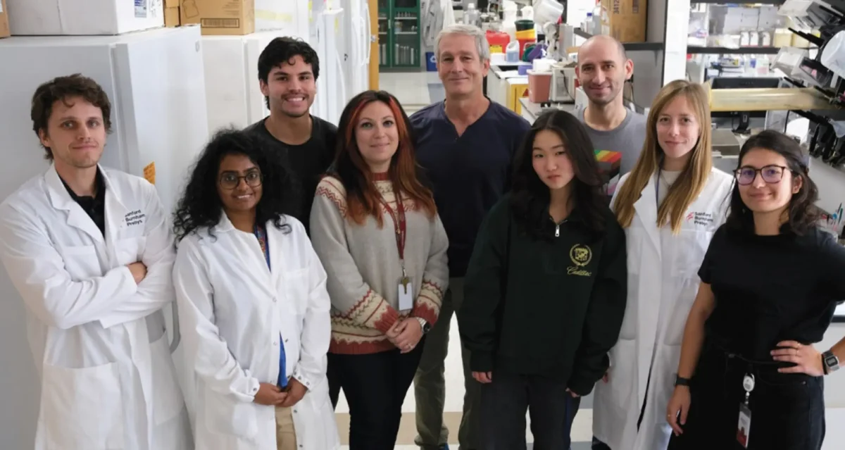
Meet the La Jolla researcher who helped discover new muscular dystrophy drug
Mar 12, 2025People in Your Neighborhood: Dr. Pier Lorenzo Puri of Sanford Burnham Prebys says he was ‘obsessed with doing something’ to…
 Nov 13, 2024
Decades of dedication led to FDA approval of a new treatment for Duchenne Muscular Dystrophy
Nov 13, 2024Nearly 30 years of discoveries by a Sanford Burnham Prebys scientist and collaborators lead to federal approval of the first…
 Dec 14, 2020
Our top 10 discoveries of 2020
Dec 14, 2020This year required dedication, patience and perseverance as we all adjusted to a new normal—and we’re proud that our scientists
 Sep 14, 2020
Scientists uncover a novel approach to treating Duchenne muscular dystrophy
Sep 14, 2020The study reveals a promising new therapeutic approach for the incurable muscle-wasting condition.
 Sep 10, 2019
How your DNA takes shape makes a big difference in your health
Sep 10, 2019The more we learn about our genome, the more mysteries arise. For example, how can people with the same disease-causing…
Elena Pasquale earned her PhD in biology from the University of Parma, Italy. She did postdoctoral work at Cornell University, after which she was appointed Research Assistant Professor at University of Parma. Following a second postdoctoral training period at the University of California in San Diego, Dr. Pasquale was appointed Assistant Research Biologist at that institution. Dr. Pasquale was recruited to Sanford Burnham Prebys in 1990.
Related Disease
Cancer, Neurodegenerative and Neuromuscular Diseases, Skin Cancer and Melanoma
Cancer, Neurodegenerative and Neuromuscular Diseases
Receptor tyrosine kinases of the Eph family and their ligands, the ephrins, represent an important cell communication system that controls a vast array physiological and disease processes. For example, Eph receptors and ephrins take part in the formation of blood vessels, including the blood vessels that feed tumors, and regulate the malignant properties of cancer cells and their interplay with the tumor microenvironment. They also regulate the formation, plasticity and regeneration of neuronal circuits as well as neurodegenerative processes such as those occurring in amyotrophic lateral sclerosis (ALS) and Alzheimer’s disease. The signal transduction mechanisms of Eph receptors are intriguing, and complex, because these receptors engage in multiple modes of signaling. Binding to ephrin ligands on the surface of neighboring cells induces canonical signaling involving receptor clustering, autophosphorylation on tyrosine residues, and kinase activity-dependent downstream signaling. Binding to the Eph receptors can also cause the ephrins, which have a cytoplasmic domain or a GPI-anchor, to transmit signals. This leads to bidirectional signals emanating from Eph receptor-ephrin complexes positioned at sites of cell-cell contact. In addition, at least some Eph receptors can also signal through non-canonical mechanisms that are independent of ligand binding and kinase activity, for example through interplay with other receptor tyrosine kinase families and with serine/threonine kinases.
Our research investigates Eph receptor signaling activities in order to understand their role in normal physiology and in pathological conditions such as cancer and neurodegenerative disorders. This knowledge is useful for the development of disease treatments based on modulating Eph receptor/ephrin activities. Ongoing efforts in our laboratory also focus on the development of agents targeting Eph receptors for research and translational applications.
Elena Pasquale’s Research Report
We discovered several Eph receptors and ephrins, and research in our laboratory is dedicated to the characterization of Eph receptor signal transduction mechanisms and biological functions using biochemical, mass spectrometry, molecular biology and cell biology approaches in conjunction with animal models. We have identified tyrosine and serine/threonine phosphorylation sites of Eph receptors and ephrins using mass spectrometry and investigated the signaling role of these phosphorylation sites. For example, our past work showed that two conserved tyrosine phosphorylation sites in the juxtamembrane segment of the Eph receptors not only mediate association with binding partners but also regulate receptor kinase activity. We also found that the SRC and ABL non-receptor tyrosine kinases and the SHEP1 scaffolding protein are binding partners of the Eph receptors, and we identified signaling connections between Eph receptors and integrins. We also found that EphA4 is highly expressed in the adult brain, where it regulates synaptic connections. More recent work in our laboratory focuses on elucidating signaling pathways that mediate the activities of Eph receptors in cancer cells.
Tumor Suppression and Tumor Promotion by Eph Receptors
Many Eph receptors are highly expressed in tumors, but their role in cancer is incompletely understood and likely depends on the cellular context. Certain Eph receptors and ephrins promote tumor angiogenesis. We showed that the EphA2 receptor is upregulated in the tumor vasculature together with the ephrin-A1 ligand, which suggested a role in tumor angiogenesis that is now well established. We also found that the EphB4 receptor expressed on the surface of breast cancer cells can promote tumor xenograft growth by enhancing blood vessel formation through interactions with its preferred ligand, ephrin-B2, present in tumor endothelial cells. Additional intriguing roles for the Eph receptors in cancer progression have also emerged. We found that canonical signaling by the EphB4 receptor is low in breast cancer cells and that ephrin-induced stimulation of EphB4 kinase activity inhibits breast cancer cell malignancy in culture and tumor growth in vivo (Figure 1A) through inhibition of the CRK proto-oncogene. More recently, we elucidated an additional mechanism of tumor suppression mediated by canonical ephrin-induced EphA2 signaling (Figure 1A), which leads to inhibition of the AKT-mTORC1 oncogenic pathway through interplay of EphA2 with a phosphatase that dephosphorylates the AKT serine/threonine kinase.

Figure 1. Dual activities of Eph Receptors in Cancer Cells. (A) Eph receptor-ephrin binding at cell-cell contact sites results in the dimerization/clustering of Eph receptor-ephrin complexes, and initiation of canonical signals through the receptor cytoplasmic domain. Signals through the ephrins can also be generated. Tyrosine phosphorylation sites (yellow circles) promote Eph kinase activity and also provide binding sites for signaling proteins containing SH2 domains. Other effectors also mediate Eph signals, including PDZ domain-containing proteins. The Eph receptor domains are indicated on the left; LBD, ligand-binding domain. (B) Eph receptors can potentiate the oncogenic effects of other receptors. These activities are independent of ephrin binding and/or kinase activity and their mechanism is not well understood but in some cases depends on Eph receptor phosphorylation on serine/threonine residues (red circle).
There is also evidence that some Eph receptors can increase cancer cell malignancy through non-canonical ephrin-independent and/or kinase-independent signaling activities (Figure 1B), which is the subject of ongoing work. These tumor promoting activities include inducing invasiveness and metastasis, epithelial-to-mesenchymal transition, stem cell-like features and drug resistance.
Eph Receptor Mutations in Cancer
The Eph receptors are frequently mutated in many types of cancer. In particular, genome-wide sequencing studies have detected somatic mutations in one or more Eph receptors in 25%-45% of melanomas, 15%-45% of lung cancers, 25-40% of colorectal cancers and 12%-25% of head and neck and uterine cancers (Figure 2), but very limited information is available on the effects of the mutations. Studies by ours and other groups have shown that a number of EphA2 and EphA3 mutations inactivate Eph receptor canonical signaling by disrupting ephrin binding or kinase activity, consistent with a role of canonical signaling in tumor suppression. Ongoing work in our laboratory focuses on characterizing the functional effects of Eph receptor mutations in cancers such as melanoma, and investigating whether the mutations shift the balance of the Eph receptor signaling activities from tumor suppression to tumor promotion. We are also interested in the interplay of Eph receptor mutations with mutations affecting well-established oncogenes and tumor suppressor genes. Understanding the effects of Eph receptor mutations in cancer cells will help shed light on the role of the Eph receptor/ephrin system in cancer cell transformation, malignant progression and drug resistance.
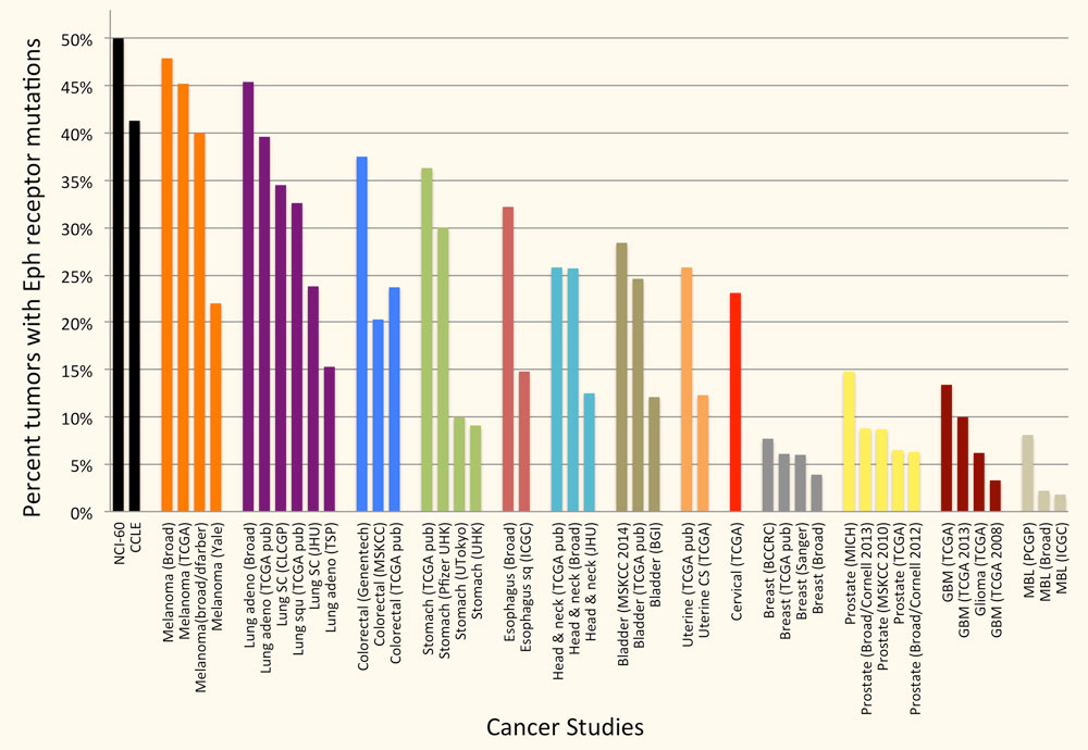
Figure 2. A large percentage of tumor specimens and cell lines harbor one or more Eph receptor mutations. Groups of bars of the same color represent studies of the same cancer type. The cancers with most Eph receptor mutations are shown; other tumor types have fewer or no Eph receptor mutations. The graph is based on data from cBioPortal for Cancer Genomics (www.cbioportal.org).
Peptides Targeting Eph Receptors
We have identified a number of peptides that bind to Eph receptors and inhibit ephrin binding by using phage display approaches. Collaborating groups have elucidated the structural features of several of these peptides in complex with the ligand-binding domain of Eph receptors, demonstrating that the peptides bind to the ephrin-binding pocket in the ligand-binding domain (Figure 3A). Most of the peptides are antagonists, but the peptides targeting EphA2 are agonists that activate receptor signaling and endocytosis similarly to the natural ephrin ligands. Interestingly, some of the identified peptides are highly specific and bind to only one Eph receptor family member. This is unlike the natural ephrin ligands, each of which promiscuously binds to multiple Eph receptors. Thus, Eph receptor-targeting peptides represent valuable pharmacological tools to study the functional importance of specific Eph receptors in tumors and the nervous system. Furthermore, they could be used as leads to develop therapies against cancer and neurological disorders, and to promote neural repair after nervous system injury (Figure 3B). Finally, our peptides have been used by other groups to deliver conjugated imaging agents, drugs and nanoparticles to Eph receptor-positive tumors. Current work focuses on identifying novel Eph receptor-targeting agents (such as peptides and small molecules) as well as improving the existing ones in collaboration with medicinal chemists and structural biologists, and evaluating them in cell culture and in vivo animal models.
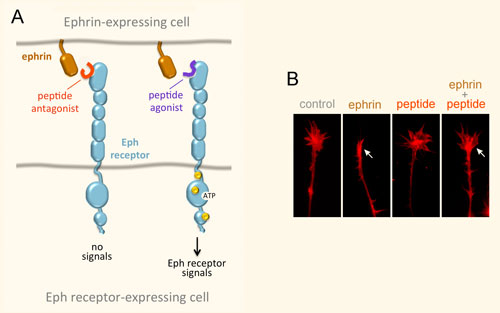
Figure 3. Peptides can target the ephrin-binding pocket of Eph receptors with high affinity and specificity, affecting receptor function. (A) Peptides targeting the Eph receptors can function as antagonists that inhibit ephrin binding and receptor signaling, or in some cases as agonists that mimic the ephrins by activating Eph receptor signaling. Yellow circles indicate tyrosine phosphorylation sites in the activated Eph receptor. (B) An EphA4 peptide antagonist blocks ephrin-induced growth cone collapse in EphA4-expressing axons, suggesting its usefulness for promoting neural repair. The arrow in the second panel marks a growth cone collapsed due to ephrin treatment; the arrow in the fourth panel marks a growth cone that did not collapse following ephrin treatment in the presence of a peptide antagonist.
 Jan 7, 2025
Mutations in protein receptor gene linked to Alzheimer’s disease
Jan 7, 2025New research on four variants in the EPHA1 gene reveals how its genetic typos may contribute to risk of dementia.
 Nov 27, 2023
The “Eph” system may pave the way for novel cancer therapies
Nov 27, 2023Over the past three decades, researchers have been investigating an important cell communication system called the “Eph system,” and the…
- May 13, 2019
Targeting long-sought EphA2 receptor becomes crystal clear
May 13, 2019Scientists have long sought to target a cellular receptor called EphA2 because of its known role in many disorders, including…
 Jul 31, 2017
SBP researchers awarded Padres Pedal the Cause collaborative grants
Jul 31, 2017Sanford Burnham Prebys Medical Research (SBP) is pleased to announce that it has been awarded five collaborative grants with the…
 May 16, 2017
What SBP Scientists are Researching to Battle Skin Cancer
May 16, 2017Skin cancer is one of the most common of all cancers, and melanoma accounts for about 1 percent of skin…
 Oct 29, 2015
The Collaboration 4 Cure Alzheimer’s research awards announced at SBP
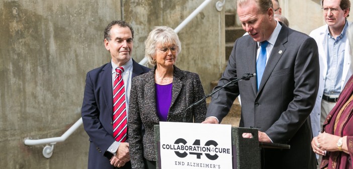
Oct 29, 2015On October 28, Mayor Kevin Faulconer, County Supervisor Diane Jacob, San Diego philanthropist Darlene Shiley, and Mary Ball, president and…
Andrei Osterman is a Professor in the Immunity and Pathogenesis Program Program at the Infectious and Inflammatory Disease Center of Sanford Burnham Prebys (since August 2003). He received his doctorate from Moscow State University in 1983, did postdoctoral work UT Southwestern Medical Center, and held the position of the Director and then Vice President of Research at Integrated Genomics in 1999-2003. Dr. Osterman is one of the founders of the Fellowship for Interpretation of Genomes (FIG), a nonprofit research organization that launched the Project to Annotate 1,000 Genomes in 2003. FIG provides the open-source integration of all publicly available genomes and tools for their comparative analysis, annotation, and metabolic reconstruction.
Related Disease
Breast Cancer, Cancer, Infectious Diseases, Radiation Damage, Skin Cancer and Melanoma
The main focus of Dr. Osterman’s research team is on fundamental and applied aspects of the key metabolic subsystems in a variety of species, from bacteria to human. This group uses a systems biology approach to reconstruct and explore metabolic and transcriptional regulatory networks. This approach combines comparative genomics and other bioinformatic techniques with biochemical and genetic experiments for pathway, gene and target discovery. Using this approach this group predicted and experimentally verified numerous enzyme families in the metabolism of cofactors, carbohydrates, and amino acids. Recent breakthroughs included prediction and characterization of novel transporters, transcriptional regulators and carbohydrate utilization pathways in a number of model bacterial systems. Applications in the field of infectious disease include identification of novel drug targets and structure-based development of novel anti-infective agents. New directions in cancer research are based on application of metabolic profiling technology for identification of novel diagnostic and therapeutic targets. Other directions of the on-going research include bioinformatics of regulatory proteolysis and applications of structural modeling for exploration of metabolic networks and gene discovery.
 Jun 3, 2026
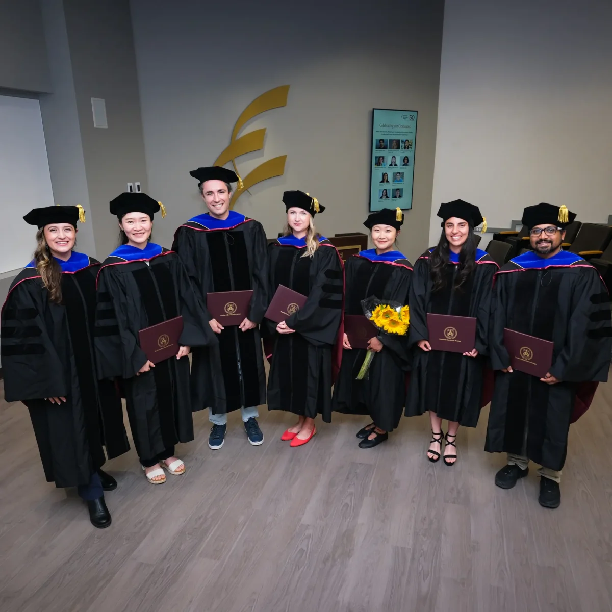
Sanford Burnham Prebys recognizes 10 doctoral degree recipients
Jun 3, 2026The Institute’s Graduate School of Biomedical Sciences held its third Commencement ceremony to celebrate new alumni.
 Nov 7, 2025
Evolving antibiotic resistance under pressure
Nov 7, 2025Study shows how a bacterial pathogen avoids biting the dust through genomic mutations, may lead to personalized treatment.
 Jul 16, 2025
Bacterial genomes hold clues for creating personalized probiotics
Jul 16, 2025Studying the genomes of beneficial bacteria may lead to new targeted probiotic treatments.
 Oct 2, 2024
Gut microbiome repair in children with severe acute malnutrition
Oct 2, 2024Researchers around the world, including Andrei Osterman, have been investigating potential remedies for child malnutrition.
 Aug 13, 2024
Dodging AI and other computational biology dangers
Aug 13, 2024Sanford Burnham Prebys scientists say that understanding the potential pitfalls of using artificial intelligence and computational biology techniques in biomedical…
- Aug 8, 2024
Scripting their own futures
Aug 8, 2024At Sanford Burnham Prebys Graduate School of Biomedical Sciences, students embrace computational methods to enhance their research careers
After a successful teaching career at the University of Michigan I have had the privilege to “reboot” my research career at Sanford Burnham Prebys where I have had the opportunity to develop novel methodologies to understand cardiomyopathy. I have also had the opportunity to work with NASA scientists to do experiments on the International Space Station.
Education
Postdoctoral Fellow, Stanford University, Palo Alto, CA, Neurochemistry
Postdoctoral Fellow, University of Texas Medical School, Houston, TX, Neuroscience NIMH
PhD, Wesleyan University, Middletown, CT, Neuroscience NIMH
B.A., Lehigh University, Bethlehem, PA, Biology
Prestigious Runding Awards or Major Collaborative Grants
2015-2020: NIH R01 HL132241-01A1 – Using Drosophila genetics to identify molecular links between ion channel dysfunction and pathological cardiac remodeling. (PI) 2013-2018 NASA NRA #NNH12ZTT001N – The effects of microgravity on cardiac function, structure and gene expression using the Drosophila model. (Co-I)
Honor and Awards
2014: Space Florida International Space Station Research Competition Winner – Co-investigator – One of three Basic Research proposals selected for launch aboard SpaceX3 – Mission completed, live flies returned on May 18,2014
2001: Excellence in Teaching Award, University of Michigan
1997: Excellence in Teaching Award, University of Michigan
1986-1988: National Institute of Mental Health Fellowship
1983-1985: National Institute of Mental Health Fellowship
1981: Sigma Xi Research Award 1980 MBL Scholarship, Neural Systems and Behavior Course
1971-1975: National Merit Scholarship, Lehigh University
Board Appointments
2018-present: Board member American Society for Gravitational and Space Research
Related Disease
Aging, Aging-Related Diseases, Atrial Fibrillation, Cardiac Fibrosis, Cardiomyopathies, Heart Disease, Metabolic Syndrome, Molecular Biology
Phenomena or Processes
Actin Cytoskeleton, Calcium Signaling, Cardiovascular Biology, Cell Signaling, Extracellular Matrix, Fuel Metabolism, Integrins, Metabolic Networks, Phosphorylation, Stress Response Pathways
Anatomical Systems and Sites
Cardiovascular System, Heart
Research Models
Drosophila, Larval Zebrafish Heart, Zebrafish
Techniques and Technologies
Biophysiology, Cellular and Molecular Imaging, Fluorescence Microscopy, Gene Silencing, Genetics, In vivo Modeling, Ion Channels, Live Imaging, Microarrays, Microscopy and Imaging, Molecular Genetics, RNA Interference (RNAi), Semi-automated Optical Heartbeat Analysis (SOHA), Systems Biology, Transgenic Organisms
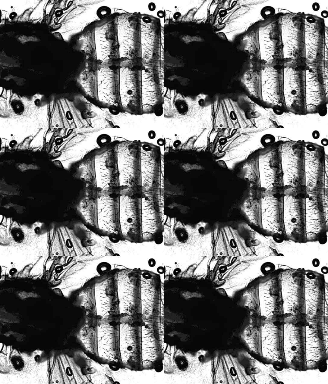
The Ocorr Lab is investigating the cellular and molecular basis of adult heart function and cardiomyopathies using the genetic model system Drosophila.
We use functional, electrophysiological, biochemical and immunohistochemical techniques that allow us to examine the roles of genes and gene products in cardiac channelopathies and stress-related cardiomyopathies.
Our lab pioneered the development of a novel methodology (Semi-automatic Optical Heartbeat Analysis, SOHA) that permits the quantification of heartbeat parameters in model systems with small hearts.
Using this system we have identified several ion channels in the fly heart that play prominent roles in repolarization of the human heart and cause arrhythmia in both the fly and in humans when mutated. We also have developed a number of other disease models including a diabetic-like cardiomyopathy induced by high sugar diet and hypoxia-induced cardiomyopathy.
Recently we have begun collaborations with NASA (by winning a Space Florida International Space Station Research Competition). We are using the fly to uncover the molecular/cellular basis for cardiac and muscle atrophy in astronauts exposed to extended periods of microgravity despite extensive exercise regimes aboard the ISS. Our flies were launched aboard SpaceX 3 for a month-long exposure to micro-gravity.
Karen Ocorr’s Research Report
My lab is working to understand the cellular and molecular basis of heart disease. One project is focused on the genetic basis of Atrial Fibrillation. This project is a collaborative one with the lab of Alexandre Colas. We are combining two model systems, the fly in my lab and human induced cardiomyocytes in his lab, to identify AFib genes that have been implicated from patient studies. Another project focuses on the role of metabolism in cardiomyopathies. This is because obesity and metabolic syndrome are linked to an increased risk of heart disease. We are studying the role of a key metabolic signaling molecule in hypertrophic cardiomyopathy. A separate effort is focused on the role of gravity in heart function. These studies will provide important information for future habitants of colonies on the moon and Mars. But they are also relevant to patients who are bedridden and to patients with muscle wasting (sarcopenia).
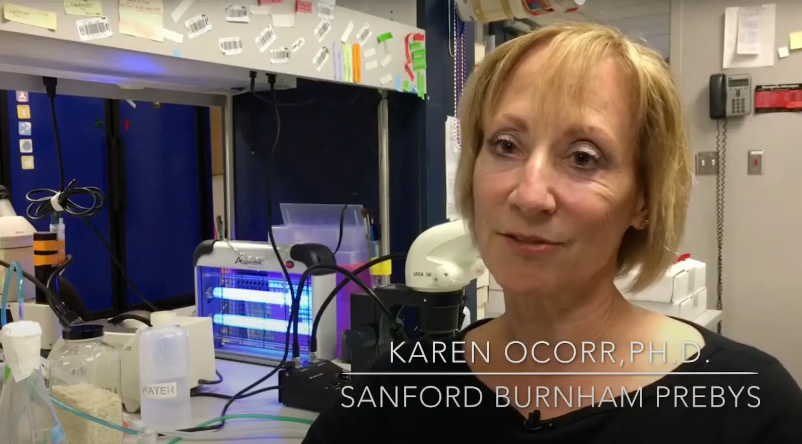
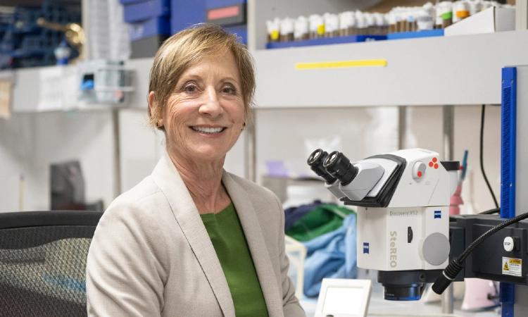 Aug 2, 2024
Controlling thickness in fruit fly hearts reveals new pathway for heart disease
Aug 2, 2024A new study in Cell Reports details how a protein previously associated with regulating metabolism in the liver also plays…
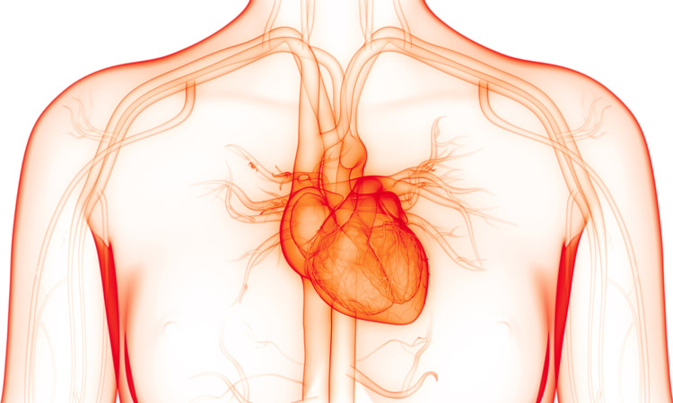 Aug 15, 2023
Scientists unite to get to the heart of AFib
Aug 15, 2023A collaborative study led by Institute researchers is paving the way to identifying gene networks that cause atrial fibrillation (AFib),…
 Sep 28, 2021
NASA awards $2 million to study effects of low gravity on muscle and neuron function
Sep 28, 2021As part of their larger initiative to understand the effects of space conditions on different organisms, NASA has awarded $2…
- Dec 14, 2020
Our top 10 discoveries of 2020
Dec 14, 2020This year required dedication, patience and perseverance as we all adjusted to a new normal—and we’re proud that our scientists
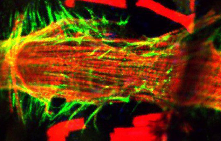 Nov 23, 2020
Fruit flies reveal new insights into space travel’s effect on the heart
Nov 23, 2020Scientists identify protein production changes in the space flies that weren’t on the radar of heart researchers—opening new avenues for…
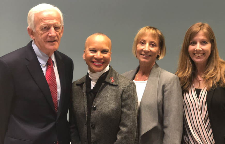 Feb 15, 2019
5 takeaways from Insights: Heart Disease
Feb 15, 2019It’s easy to forget about the fist-sized organ in our chest. But the heart is arguably the most important muscle
After receiving his early training in clinical chemistry/biochemistry at the University of Buenos Aires, Argentina, Dr. Millán first joined the La Jolla Cancer Research Foundation (LJCRF) in 1977, the predecessor of Sanford Burnham Prebys, as a trainee in clinical enzymology. He completed his PhD studies in Medical Biochemistry at the University of Umeå, Sweden and after post-doctoral stints in Copenhagen and LJCRF he was appointed to the faculty at SBP in 1986. He served as Professor of Medical Genetics in the Department of Medical Biosciences at his alma mater, Umeå University, Sweden, from 1995-2000. He was appointed Sanford Investigator at the Sanford Children’s Health Research Center at Sanford Burnham Prebys in 2008.
Honors and Recognition
2018: ASBMR Lawrence G. Raisz Award for Pre-clinical Research.
2001: Gold Medal of the Royal Academy of Medicine and Surgery, Murcia, Spain
1992: Honorary title of AcadémicoCorresponsal at the Royal Academy of Medicine and Surgery, Murcia, Spain.
Other Affiliations
Member, Scientific Advisory Board, Soft Bones
Related Disease
Arthritis, Bone Mineralization Disorders, Cardiovascular Diseases, Colorectal Cancer, Crohn’s Disease (Colitis), Heart Disease, Inherited Disorders, Metabolic Syndrome, Peripheral Vascular Disease, Testicular Cancer
Phenomena or Processes
Cardiovascular Biology, Disease Therapies, Extracellular Matrix, Protein Structure-Function Relationships
Anatomical Systems and Sites
Cardiovascular System, Musculoskeletal System, Vasculature
Research Models
Mouse
The Millán laboratory works on understanding the mechanisms that control normal skeletal and dental mineralization and elucidating the pathophysiological abnormalities that lead to heritable soft bones conditions such as Hypophosphatasia (HPP) and to soft-tissue calcification, including vascular calcification, that is a hallmark in patients affected by a variety of rare genetic diseases as well as in chronic kidney disease. Dr. Millán’s research has already contributed to the implementation of a novel therapy for HPP, a genetic disease caused by deficiency in tissue-nonspecific alkaline phosphatase (TNAP) function, that leads to accumulation in the extracellular space of inorganic pyrophosphate (PPi), a potent inhibitor of mineralization. HPP is characterized by defective mineralization of bones (rickets or osteomalacia), and teeth that display a lack of acellular cementum, hypomineralized dentin and enamel, and periodontal defects. Dr. Millán’s team has demonstrated the effectiveness of enzyme replacement therapy using mineral-targeted recombinant TNAP (asfotase alfa) to prevent the skeletal and dental defects in the TNAP knockout mouse model of infantile HPP. This therapy was approved in 2015 for the treatment of patients with pediatric-onset HPP.
Current efforts, in collaboration with Professor Miyake’s group in Japan (https://www.nms-gt.org/en/members), focus on developing gene therapy as an alternative approach to treat HPP. Dr. Millán’s group has also identified key pathophysiological changes that lead to calcification of the arteries in animal models of generalized arterial calcification of infancy, pseudoxanthoma elasticum and related genetic diseases as well as in animal models of chronic kidney disease. His group, in collaboration with scientists at the Conrad Prebys Center for Chemical Genomics at Sanford Burnham Prebys, has developed proprietary compounds able to ameliorate the soft-tissue calcification in these conditions and clinical trials are now underway using these first-in-class small molecule inhibitors.
 Feb 3, 2025
Gene therapy may be “one shot stop” for rare bone disease
Feb 3, 2025New study shows a single shot may be able to treat a skeletal disease currently requiring nearly daily injections.
 Jul 21, 2023
José Luis Millán joins international initiative to study calcification in aging
Jul 21, 2023José Luis Millán, PhD, has joined a five-year, $13 million program that will study misplaced calcification in the eyes and…
 May 10, 2023
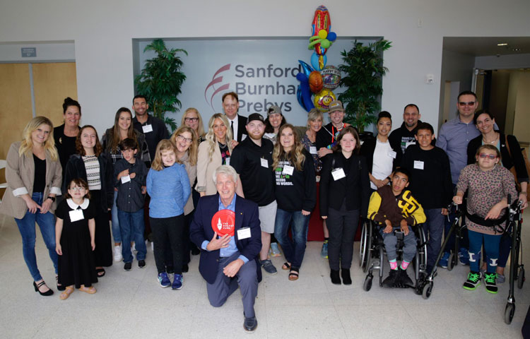
Where science meets patients: Sanford Children’s Research Center hosts inaugural symposium
May 10, 2023The event celebrated 16 years of progress at the Center and connected scientists with the people most impacted by their work.
 Dec 19, 2022
Sanford Burnham Prebys announces start of Phase 2 clinical trial of DS-1211 in individuals with PseudoXanthoma Elasticum
Dec 19, 2022San Diego Biotech Networks News
 Nov 30, 2022
Sanford Burnham Prebys announces start of Phase 2 clinical trial of DS-1211 in individuals with pseudoxanthoma elasticum
Nov 30, 2022A Phase 2 clinical trial has started of DS-1211 in individuals with pseudoxanthoma elasticum (PXE), a rare multisystem genetic disease…
 Jun 7, 2021
Study supports gene therapy as a promising treatment for soft bone disease
Jun 7, 2021A single dose of AAV8-TNAP-D10 may be safe and effective for hypophosphatasia A preclinical study led by scientists at Sanford
Jamey Marth is a Professor at Sanford Burnham Prebys. Dr. Marth’s previous positions included Professor of Medical Genetics at the Biomedical Research Centre, University of British Columbia; Investigator of the Howard Hughes Medical Institute and Professor of Cellular and Molecular Medicine at the University of California San Diego; and Professor and Director of the Center for Nanomedicine at the University of California Santa Barbara. Dr. Marth received a PhD degree in Pharmacology from the University of Washington where he trained in the laboratories of Roger M. Perlmutter, MD, PhD, and Nobel-laureate Edwin G. Krebs, MD.
Education
1987: PhD, University of Washington, Pharmacology
1984: BSc, University of Oregon, Genetics and Chemistry
Honors and Recognition
2024: Distinguished Service Award, Society for Glycobiology
2017: Karl Meyer Award, Society for Glycobiology
2009-2020: John Carbon Chair in Biochemistry and Molecular Biology
2009-2019: Duncan and Suzanne Mellichamp Chair in Systems Biology
2009: Julius Stone Lectureship Award: Society for Investigative Dermatology
1995-2009: Investigator Award, Howard Hughes Medical Institute
1991-1995: Faculty Scholarship, The Medical Research Council of Canada
Related Disease
Cancer, Colitis, Diabetes – General, Inflammatory/Autoimmune Disease, Sepsis
Dr. Marth is a molecular and cellular biologist specializing in diseases attributable to protein glycosylation. His education and training span molecular genetics, biochemistry, pharmacology, cell biology, immunology, hematology, developmental biology, microbiology, and glycobiology.
As an enzymatic process essential to cells, glycosylation produces saccharides linked by glycosidic bonds to proteins, lipids, and themselves, termed glycans. The vast majority of secreted and cell surface proteins are post-translationally modified by glycosylation during transit through the secretory pathway, termed glycoproteins. A widely used college level cell biology textbook authored by others denotes glycans as one of the four main families of the organic molecules of all cells, with lipids, proteins, and nucleic acids. Together they compose the macromolecules and other assemblies of the cell. The structures of glycans (and lipids) are, however, synthesized by template-independent processes, rendering them hard to predict and study. However, cells produce and regulate an abundant and diverse glycome of glycosidic linkages in which some of the biological information is decoded by one or more glycan-binding receptors, termed lectins.
Glycans and lectins represent a significant percentage of genes in the genomes of organisms, with several hundred present in mammals. Because glycan biosynthesis, diversification, and degradation rely upon corresponding gene and enzyme function, glycan function can be investigated similarly to other enzymatic and metabolic pathways, such as protein phosphorylation. In contrast however, studies of intact organisms are typically required to uncover the functions of protein glycosylation in mammals. Dr. Marth’s laboratory identified this model system requirement and has further focused on discovering how glycosidic linkages regulate proteins modified by N- and O-glycans and contribute to the molecular origins of common diseases and syndromes including colitis, diabetes, autoimmune disease, and sepsis.
To understand the nature and extent of the information within glycosidic linkages, the Marth Laboratory has applied multiple molecular approaches to investigate protein glycosylation in mice and humans. This includes the development of enabling technologies with broad applicability, such as conditional mutagenesis by Cre-lox recombination in living animals to determine gene function with temporal and spatial selectivity. His laboratory also develops and studies disease models that better represent real-world models of environmental factors that trigger common acquired human diseases. The laboratory is based on interdisciplinary research focused upon physiology and disease process regulated by protein glycosylation.
The physiological systems regulated by protein glycosylation are broad even when comparing among sequential biosynthetic steps, and our findings continue to indicate the presence of undiscovered information of medical relevance residing in the glycan linkages of glycoproteins.
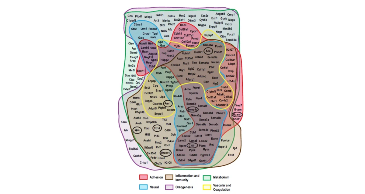 Jul 31, 2025
Signal boost uncovers hundreds of hidden binding partners for blood protein receptor
Jul 31, 2025Study identifies receptor-ligand interactions, links receptor dysfunction to age-associated defects in multiple organs.
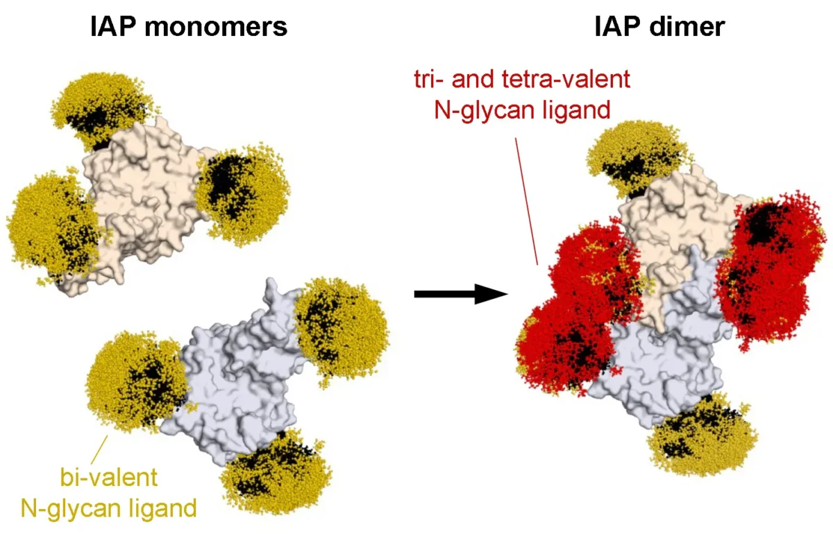 May 14, 2025
Rediscovering the first known cellular receptor
May 14, 2025Scientists from the Marth lab apply new techniques to reexamine a receptor linked to sepsis.
 Oct 31, 2024
Jamey Marth interviewed by The Scientist
Oct 31, 2024The Sanford Burnham Prebys scientist discussed the Cre-loxP recombination system, a mainstay genetic engineering technology.
 Sep 28, 2021
Jamey Marth awarded $13.5 million by NIH to investigate the pathogenesis and treatment of sepsis
Sep 28, 2021Sanford Burnham Prebys professor Jamey Marth, PhD, has been awarded $13.5 million from the National Heart, Lung, and Blood Institute to
 Jul 13, 2021
Study finds promising therapeutic target for colitis
Jul 13, 2021Neu3 controlled the emergence of disease in a model of human colitis An international research group, led by Jamey Marth, PhD,
 Oct 10, 2017
Jamey Marth honored for research linking glycans to diabetes, lupus, sepsis
Oct 10, 2017Jamey Marth, Ph.D., is the 2017 recipient of the Society for Glycobiology’s Karl Meyer Award. The international award is given…
Dr. Kumsta earned her degree as a Diplom Biologist/M.Sc. and PhD from the Technical University of Munich, Germany and performed her thesis research in the laboratory of Dr. Ursula Jakob at the University of Michigan. Dr. Kumsta joined Sanford Burnham Prebys and the lab of Malene Hansen as a postdoctoral fellow in 2009. In 2018 Caroline was promoted to Research Assistant Professor and then to Assistant Professor in 2021. Dr. Kumsta’s research focuses on the role of autophagy in hormetic stress responses, aging, and neurodegeneration in C. elegans and human tissues.
Education and Training
2018-2021: Research Assistant Professor, Sanford Burnham Prebys
2016-2018: Staff Scientist, , Sanford Burnham Prebys
2009-2016: Postdoctoral Fellow with Dr. Malene Hansen, Sanford Burnham Prebys
2009: Postdoctoral Associate with Dr. Ursula Jakob, Department of Molecular, Cellular and Developmental Biology, University of Michigan, Ann Arbor, USA
2005-2008: Doctor rerum naturalium (PhD) with Dr. Ursula Jakob, University of Michigan and Technical University of Munich
1999-2005: Diplom-Biologin Univ. (MS), Technical University of Munich, Germany
Honors and Awards
2024: R01 NIA: Hormetic regulation of autophagy in aging
2024: Selected as Member of Council of International Rising Stars (COIRS) of The Autophagy, Inflammation and Metabolism Center of Biomedical Research (NIH-funded AIM Center, University of New Mexico)
2023: P30 NIA: Pilot Grant, San Diego – Nathan Shock Center: Heterogeneity of autophagy during aging
2022: W.O.W. Award – For Wonderful Original Work, presented at the Annual Retreat of the Sanford Burnham Prebys Faculty
2022: NIA Fellow Award at 2022 Autophagy Gordon Research Conference
2015: Best Oral Presentation Prize, EMBO Workshop: The Regulation of Aging and Proteostasis
2013: AFAR Postdoctoral Fellowship: Transcriptional regulation of autophagy in promoting proteostasis upon hormetic stress
2013: Best Oral Presentation at the 12th Annual Poster Symposium at Sanford Burnham Prebys Medical Discovery Institute
2011: Fishman Fund Career Development Award
2011: AACR Postdoctoral Fellowship: Translational Control of Tumor Formation in C. elegans
Related Disease
Aging-Related Diseases
Phenomena or Processes
Autophagy
Research Models
C. elegans
“We are aging—not just as individuals but as a world. In 2020, about 727 million people worldwide were 65 and older. By 2050, that total is projected to increase to 1.5 billion — 1 in every 6 of the earth’s inhabitants.” – Global Aging, National Institute on Aging
As we age, we face an increased risk of developing age-related disorders, such as neurodegenerative diseases, because of the increased cellular accumulation of damaged biomolecules, including protein aggregates, which contributes to cellular decline. The activation of cyto-protective mechanisms, such as the cellular recycling process of autophagy and stress responses, contributes to improving cellular function and preventing aging-induced dysfunction. Discovering mechanisms that help maintain cellular integrity during aging, is therefore an important step towards developing new strategies for maintaining cellular homeostasis and organismal health, which could have great impact by increasing healthspan and eliminating age-related diseases.
 Aug 19, 2024
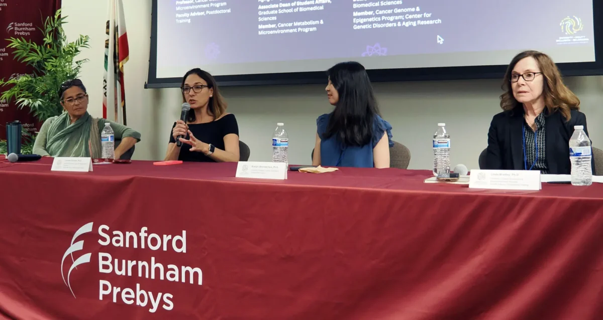
Women in Science event at Sanford Burnham Prebys examines how female faculty members navigate research careers
Aug 19, 2024Topics at the event included work/life balance, caregiving and family obligations, and gender disparities in academic rank at research and…
 Jul 11, 2024
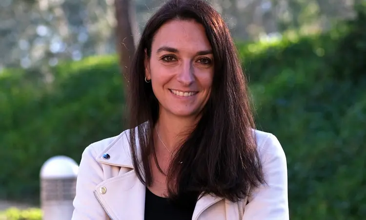
Caroline Kumsta awarded $2.9M to study how short-term stress improves health and life expectancy
Jul 11, 2024By learning how small amounts of stress activate autophagy, researchers may create new approaches to combat age-related disease
 Feb 29, 2024
Time to talk about aging research
Feb 29, 2024Hundreds of scientists gather in San Diego and virtually to share knowledge on the science of aging
 Feb 9, 2024
The heterogeneity of aging, a symposium
Feb 9, 2024Aging research is always timely, and here’s a date: On March 6, the San Diego Nathan Shock Center, a consortium…
 Feb 7, 2024
Speaking of International Day of Women and Girls in Science
Feb 7, 2024Designated by the United Nations, the 9th International Day of Women and Girls in Science is Sunday, February 11, preceded…
 Jan 3, 2024
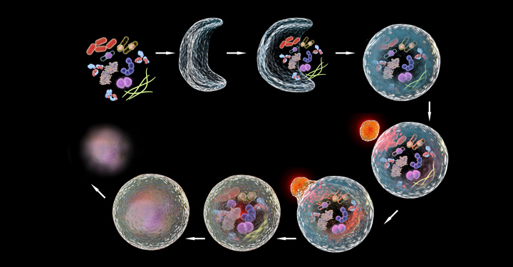
New roles for autophagy genes in cellular waste management and aging
Jan 3, 2024Autophagy genes help extrude protein aggregates from neurons in the nematode C. elegans.
“Cancer is one the main causes of death in the US. Our research is focused on understanding how we can harness the power of our immune system to attack and kill cancer cells and cure patients. I chose to join Sanford Burnham Prebys because of their collaborative research culture and state-of-the- art core facilities. I believe that teamwork is the foundation of scientific breakthroughs, and the Institute provides a perfect supportive and friendly environment to achieve this.”
Originally from the Netherlands, Kelly received her BS and MS from Utrecht University, and performed her PhD studies at the Netherlands Cancer Institute in Amsterdam working with Prof. Karin de Visser studying the role of the immune system in the metastatic spread of breast cancer. For her postdoctoral training, Kelly joined the lab of Prof. Max Krummel at the University of California San Francisco to study how tumor-associated myeloid cells affect anti-tumor T cell responses. Kelly is currently setting up her independent research team at Sanford Burnham Prebys.
Education
2016-2023: Postdoctoral Training, University of California San Francisco (Mentor: Prof. Max Krummel, focus on immune evasive cancer)
2017: PhD, Netherlands Cancer Institute Amsterdam/ Leiden University (Mentor: Prof. Karin de Visser, focus on role immune system in breast cancer metastasis, 2011-2016)
2011: MS, Biomedical Sciences, Utrecht University, Netherlands
2008: BS, Biology, Utrecht University, Netherlands
Honors and Recognition
2022: Selected Attendee for SITC Women in Cancer Immunotherapy Network (WIN) Leadership Institute
2022: Selected Attendee for Arthur and Sandra Irving Cancer Immunology Symposium
2022: Ray Owen Poster Award for Outstanding Poster Presentation at 60th Midwinter Conference for Immunologists at Asilomar (sponsored by AAI)
2020-2022: Parker Scholar Award awarded by the Parker Institute for Cancer Immunotherapy (PICI)
2018: Honorable Mention of poster presentation at UCSF/UCB/UCM Immunology Retreat
2018: Poster presentation ‘Excellence in Research Award’ awarded by the National Philanthropic Trust
2017-2019: NWO Rubicon postdoctoral fellowship awarded by the Netherlands Organization for Scientific Research (NWO)
2015: Best presentation award at the annual Tumor Cell Biology meeting of the Dutch Cancer Society (KWF)
2014: Travel scholarship awarded by the Dutch Foundation for Pharmacological Sciences (NSFW)
2010: Master scholarship awarded by the Dutch Cancer Society (KWF)
Memberships
2022-present: Society for Immunotherapy of Cancer (SITC)
2020-present: Parker Institute for Cancer Immunotherapy (PICI)
2012-present: American Association for Cancer Research (AACR)
Related Disease
Breast Cancer, Cancer Biology, Immune Disorders, Inflammation, Innate Immunity, Metastasis, Skin Cancer and Melanoma, Tumor Microenvironment, Tumorigenesis
Phenomena or Processes
Adaptive Immunity, Innate Immunity
Research Models
Mouse, Primary Cells
Techniques and Technologies
Cellular and Molecular Imaging
The Kersten lab studies the interactions between the immune system and cancer.
We are fascinated by the crosstalk between the immune system and cancer. Our goal is to understand how cancer cells hijack the normal physiology and function of immune cells to support tumor growth and evade destruction by the immune system.
Kelly Kersten’s Research Report
Cancer immunotherapy, harnessing a patient’s immune system to fight cancer, has revolutionized the way we treat cancer. However, a large proportion of patients do not respond to this type of therapy, and we do not fully understand why. In the Kersten lab, we study how different immune cells affect anti-tumor immunity with the ultimate goal to improve therapies to fight cancer. Research in our lab is focused on understanding how interactions between different immune cells in the tumor microenvironment, specifically macrophages and T cells, affect anti-tumor immunity and responsiveness to immunotherapy. Why do T cells become dysfunctional and exhausted? How do exhausted T cells modulate the composition of immune cells in tumors? And how do macrophages shut down anti-tumor T cells? Our research aims to define the molecular mechanisms that regulate these reciprocal signals to design novel anti-cancer therapies. How immune cells function is highly context-dependent. Upon infiltration in the tumor microenvironment, immune cells face extremely harsh conditions characterized by nutrient deprivation, hypoxia and metabolic challenges resulting in their failure to function properly. We study how different environmental factors impact immune cell phenotype and function, with the goal to optimize their cancer-killing properties.
 May 21, 2026
Women in Science Lecture series spotlights structural biology and immunology leader
May 21, 2026The series highlights the groundbreaking work and unique perspectives of women leaders in the biomedical sciences.
 Dec 12, 2025
Sanford Burnham Prebys scientists garner eight cancer research grants from Curebound to advance therapeutic treatments and cures
Dec 12, 2025Ten scientists at Sanford Burnham Prebys Medical Discovery Institute were awarded eight grants yesterday from Curebound, a San Diego-based philanthropic organization
 Sep 2, 2025
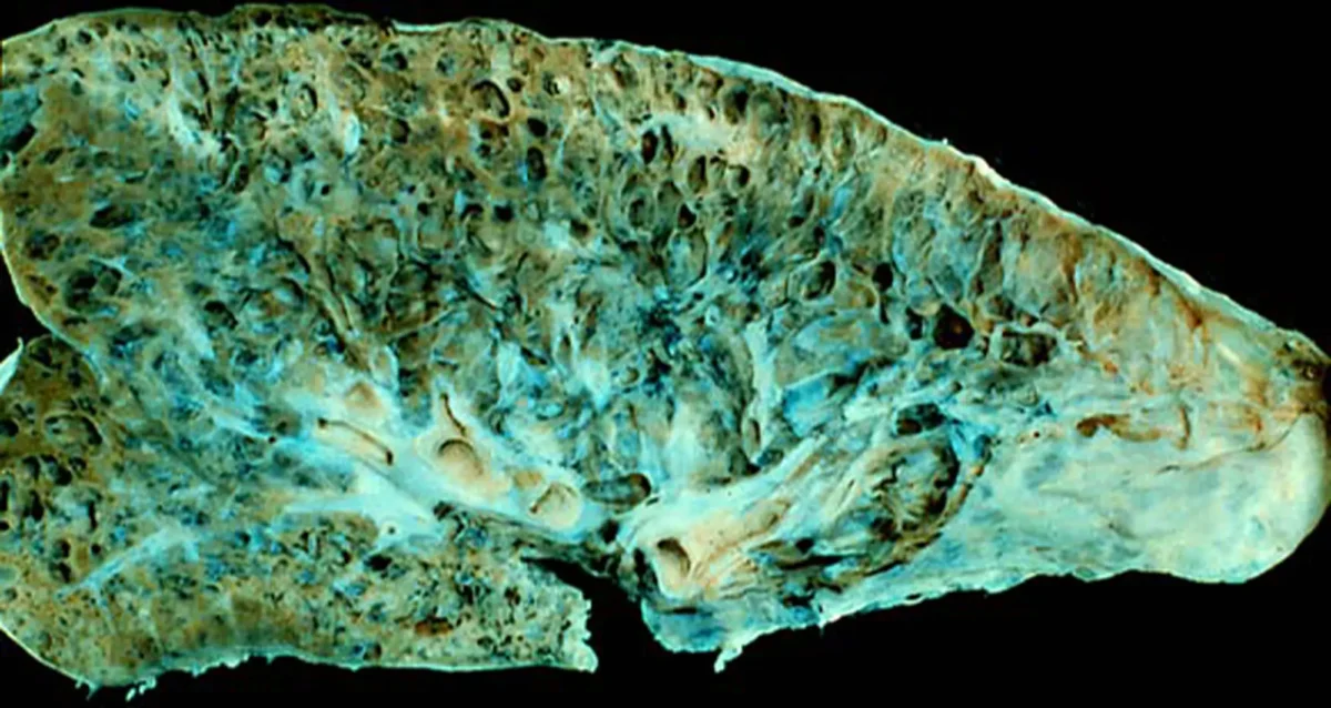
How Cellular Crosstalk Translates into Idiopathic Pulmonary Fibrosis
Sep 2, 2025Rare but deadly condition begins with metabolic changes to make a pro-fibrotic metabolite.
 May 2, 2025
Kelly Kersten awarded Melanoma Research Alliance grant to support research on melanoma immunotherapy
May 2, 2025The newly created award is part of the alliance’s $9.3 million commitment to melanoma research funding in 2025.
 Oct 31, 2024
Raising awareness of breast cancer research at Sanford Burnham Prebys
Oct 31, 2024The October Science Connect Series event was themed around Breast Cancer Awareness Month.
 Jun 4, 2024
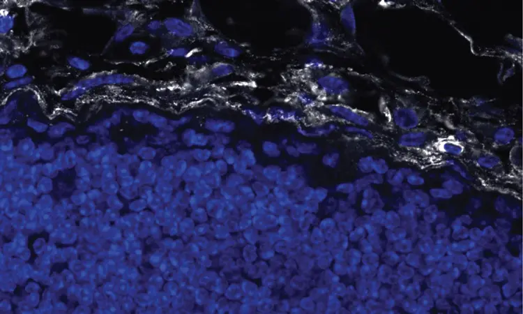
How tumor stiffness alters immune cell behavior to escape destruction
Jun 4, 2024Immunotherapy is based on harnessing a person’s own immune system to attack cancer cells. However, patients with certain tumors do…