Shared Resource Archives - Sanford Burnham Prebys

Exploratory Pharmacology

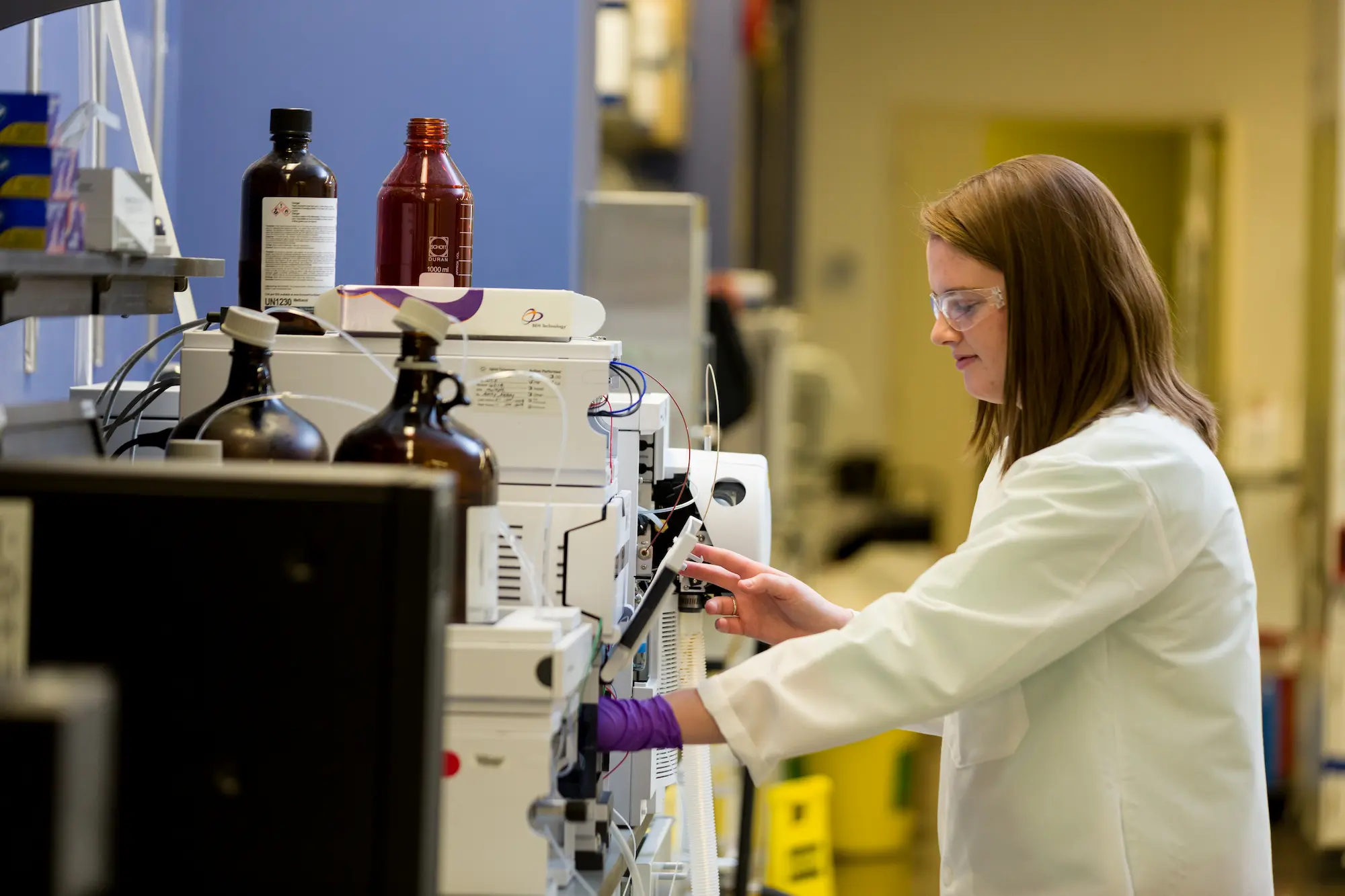
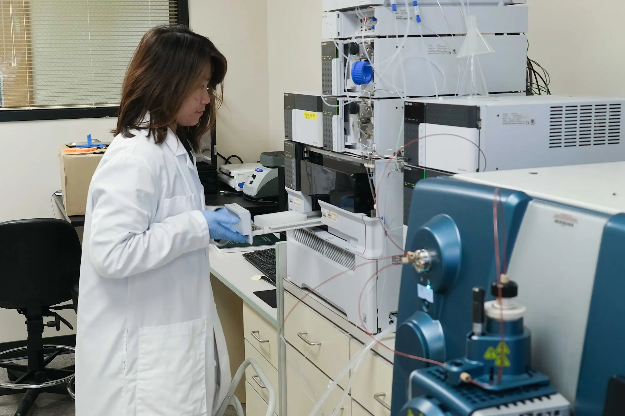
scientist working in a lab

Overview

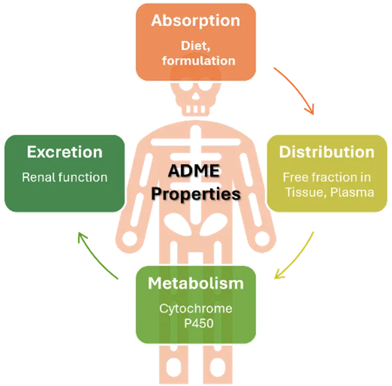
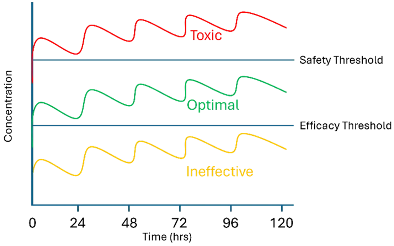
ADME/PK assays enable informed decision making in drug discovery and development. At Sanford Burnham Prebys, the ADME/PK core is focused on developing and running in vitro and in vivo assays to investigate various absorption, digestion, metabolism and excretion parameters of molecules. Our aim is to deliver quality data in a short time to enable medicinal chemists to effectively develop structure-activity relationships and improve drug-like properties in drug discovery and development programs. This facility is integral part of Conrad Prebys Center for Chemical Genomics and works in collaboration with other core facilities across the organization.

ADME/PK core also provides support for grants in terms of planning and budgeting ADME/PK activities, scientific input on drug discovery projects, performing DMPK experiments, and fulfilling reporting requirements. We also provide consultations on standalone service in experimental design, experimentation and reporting DMPK experiments.


Services

Assays

  • Stability
    Liver microsome, hepatocyte, plasma and formulation stability
  • Solubility
    Kinetic and thermodynamic solubility
  • Free fraction determination
    Plasma protein binding and brain fractionation
  • PK and bioavailability studies
    IV, IP, PO, SC, topical etc. 
  • Dose predictions
    Clearance, Brain weight and Maximum Lifespan Potential based First-in-Human predictions
  • PD and efficacy studies
    In vivo models for assessing target engagement, functional activity and assessment of drug like properties

Equipment & Resources

Shimadzu UPLCs

Sciex QTRAP 6500+

Brings about significant enhancement in our bioanalysis capabilities

  • Linearity: Triple quadrupole has one of the best range of linearity with MRM3 mode (up to 6 orders of magnitude)
  • Sensitivity: LLOQ in pg/mL to fg/ml ranges (results vary for analytes; Attograms sensitivity reported for some analytes)
  • Agility: Polarity switching (1millisecond)
  • Versatility: Mass range of m/z 5 – 2,000
  • Robustness: Multi-day consecutive injection feasible
  • Efficiency: Increased ionization efficiency and heat transfer with the new IonDrive Turbo V source
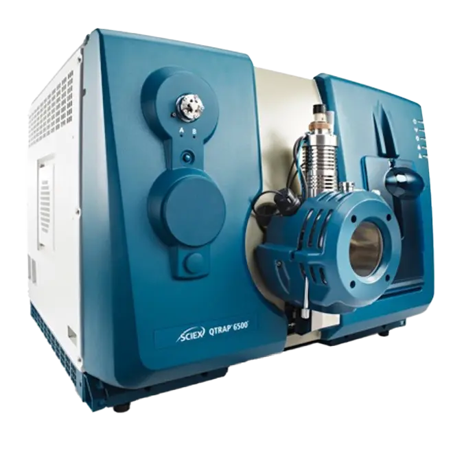
Product photo of Sciex-QTRAP-6500

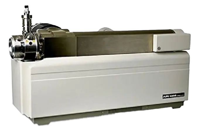
Sciex API 4000

For ADME and bioanalysis

  • Linearity: Triple quadrupole has one of the best range of linearity with MRM3 mode (up to 6 orders of magnitude)
  • Sensitivity: LLOQ in ng/mL ranges (results vary for analytes; picograms sensitivity reported for some analytes)
  • Agility: Polarity switching
  • Versatility: Mass range of m/z 5 – 1,000
  • Robustness: Multi-day consecutive injection feasible
  • Efficiency: Increased ionization efficiency and heat transfer with the new IonDrive Turbo V source

Price List

For a Price List, please email us.


Leadership

Raghuveer Ramachandra, PhD
Associate Director
Medicinal Chemistry

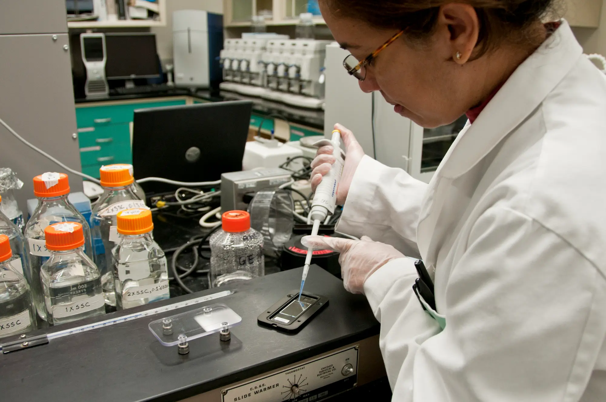
scientist working in a lab

Overview

The Medicinal Chemistry Facility at Sanford Burnham Prebys Medical Discovery Institute is tasked with providing general synthetic and medicinal chemistry resources and expertise to the biomedical research community at the Institute and with external academic partners. Our team is staffed with industry-trained chemists and has a solid track record in all aspects of synthetic problem solving.


Services

Chemistry Consulting for Projects and Grant Applications

  • Analysis of prior art and chemical landscape

Synthetic and Medicinal Chemistry

  • Evaluation of structure-activity relationships (SAR), based on biological assay results, and next step decisions for progressing a project forward.
  • Synthesis of small molecule reference compounds from milligram to multi-gram scale.
  • Design and synthesis of chemical libraries for hit and lead identification, as well as diversity libraries based on a structural theme.
  • Synthesis of custom organic reagents and synthetic intermediates.
  • Analog synthesis and lead optimization targeting the improvement of potency, physiochemical, and pharmacokinetic properties.

Analytical Chemistry

  • 1H and 13C NMR with 1D and 2D spectra acquisition and multinuclear capability.
  • Verification of the structural identity and purity of samples.
  • HPLC and LC/MS (single quadrupole, triple quadrupole and TOF) analysis.
  • High resolution mass spectrometry for accurate mass determination.
  • Normal, chiral and reverse phase chromatography separations on analytical and/or preparative scales.

Equipment & Resources

The Medicinal Chemistry core occupies approximately 2000 sq. ft. of laboratory space and is equipped with cutting edge synthetic and analytical equipment.

Synthesis Equipment

  • Six 8 ft chemical fume hoods
  • Syrris AFRICA 4 channel microfluidics synthesis system
  • CEM 48 vial microwave system with an Explorer system for automated reaction processing
  • Parr shaker hydrogenation apparatus

Analysis and Purification Equipment

  • Waters automated preparative HPLC AutoPurification system with UV and MS based fraction collection
  • Two Biotage SP4 Flashmaster chromatography units with multi-column functionality and a variable dual wavelength detector
  • Two Shimadzu Discovery preparative HPLC systems with UV detection
  • sco Combiflash automated HPLC with UV detector
  • Waters Acquity LC/MS with ESI-MS and PDA detector for UV and MS interpretation of analytical samples
  • Shimadzu LC/MS 2010EV with ESI-MS and PDA detector
  • Applied Biosystems API3000 for LC/MS-MS studies
  • 400 MHz and 500 MHz Bruker NMR spectrometers with autosamplers

Price List

For a Price List, please call (858) 646-3100 ext. 5320 or email us.


Leadership

Steven Olson, PhD
Facility Director

Michael Jackson, PhD
Vice President, Drug Discovery & Development

Contact

Please call (858) 646-3100 ext. 5320 or use the button below to send us an email.

Contact the Facility

High-Throughput Screening

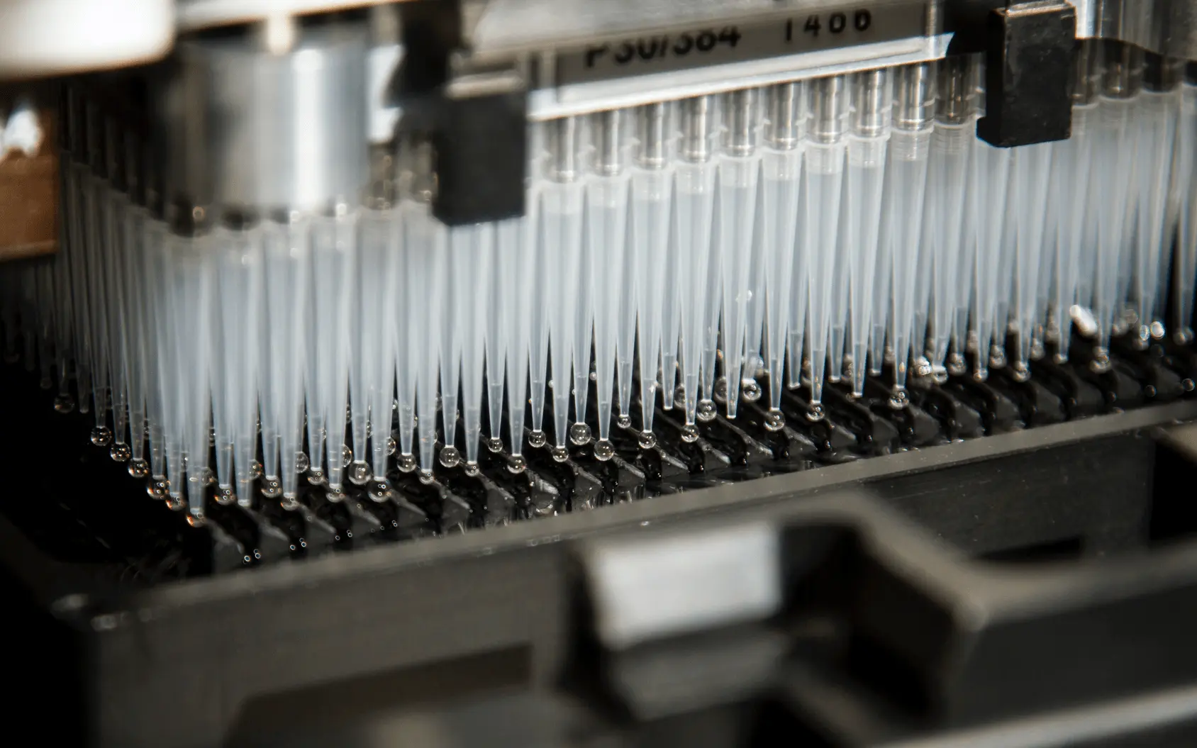
close-up photo of lab equipment

Overview

The High-Throughput Screening (HTS) facility is a part of Conrad Prebys Center for Chemical Genomics and has the necessary infrastructure to conduct chemical libraries screenings of biological targets. It provides diverse screening instrumentation, compound libraries and expertise on high throughput screens and automation in support of collaborative, and user- or facility-driven screening projects. HTS works closely with Assay Development and High-Content Screening teams to help select and implement the most suitable biochemical or cellular assays for each project, including diverse plate-reader and imaging assays.

The Prebys Center has existing collaborations with a limited number of pharma and biotech organizations, along with several clinical hospitals, to develop small molecule therapeutics. We continue to seek potential additional partnerships of mutual benefit. These partnerships typically involve a portfolio of projects the Prebys Center drives (in collaboration with the partner) through the early stages of the drug discovery process (assay development through lead optimization to in vivo POC). Often we engage in a pilot project with the partner, which then evolves into a broader collaboration/”shared pipeline.” We are not currently engaging in fee-for-service activities.


Services

  • Screening small-molecule compound libraries in 384- or 1536-well formats with various detection technologies (absorbance, fluorescence, luminescence, fluorescence polarization (FP), TR-FRET, AlphaScreen)
  • Help with the use of automated instrumentation
  • Preparation and handling of compound libraries for high throughput screening
  • Consultation on high throughput screening approaches
  • Conversion of cell-based and biochemical assays to high-throughput/automation compatible formats
  • Screening data analysis and support for HTS hit follow-up studies

Equipment & Resources

  • The High-throughput screening (HTS) system (HighRes Biosolutions)
    A fully integrated automated screening system equipped with a Staubli TX90XL robotic arm, a 420-plate random-access carousel for storage of compound and assay plates, a 210-plate STX240 Liconic incubator for cell-based assays, two Velocity 11 VSpin centrifuges, a Perkin Elmer ViewLux multimode CCD based uHTS plate reader, a Pherastar FS plate reader, a Labcyte Echo 555 Acoustic Liquid Handler, a Nexus XPeel and Agilent PlateLoc for re-sealing plates, a BioTek EL406 plate washer, a high-speed 10-position HighRes pintool device with ultrasonic baths and four multidrop Combi dispensers. It enables fully automated large-scale screening of biological assays with various formats.
  • Biomek FX liquid handling systems with 96- and 384-well plate heads and Span8 arms
  • Access™ Laboratory Workstation (Labcyte)
    An automated compound-dispensing system including an Echo 555 Acoustic Liquid Handler, a precise automation robotic arm, four random-access 20-plate racks, a de-lid station, a Deerak GX Series low-volume bulk dispenser, a Agilent V-Spin centrifuge, a Nexus XPeel and a Agilent PlateLoc and controlled by Labcyte TEMPO automation control software. This system allows fully-automated multiplate small-volume dispense operations especially useful for dose-response tests of large number of compounds, e.g. cherry-pick confirmation of primary HTS hits.
  • Plate processing workstation (Agilent)
    The workstation includes a PlateLoc sealer, Vcode barcode labeler, BenchCell 4x with four stackers and a Bravo liquid handler.
  • PerkinElmer Envision plate readers with 2 reagent injectors
  • Kalypsys washer/dispenser system for rapid and precise dispensing and plate washing in 96, 384, 1536 formats
  • Hamamatsu FDSS7000 rapid-kinetics fluorescence and luminescence plate reader with integrated 384-well liquid handling
  • Stand-alone Instruments
  • Labcyte Echo 555 Acoustic Liquid Handler
  • BMG PherastarPlus plate reader
  • Titertek Map-C plate washer
  • BioTek plate washers (EL405, EL406)
  • Molecular Devices M5 plate readers
  • Multiple liquid bulk dispensers (Wellmate, Multidrop Combi)

Price List

For a Price List, please call (858) 646-3100 ext. 3465 or email us.


Leadership

Ian Pass, PhD
Facility Director

Contact

Please call (858) 646-3100 ext. 3465 or use the button below to send us an email.

Contact the Facility

High Content Screening

Close-up shot of a scientists hands working with lab equipment

Overview

The High-Content Screening (HCS) core facility of the Conrad Prebys Center for Chemical Genomics is part of the Sanford Burnham Prebys screening center. As part of the Prebys Center, the HCS resource has access to the HTS plate and liquid handling infrastructure of the screening center as well as the screening center’s cell culture facility. For details on the other core facilities of the Prebys Center, please refer to the “Related Resources” below.

The HCS core facility provides assay development, screening, and data analysis/mining expertise and services for high content screens, where the readout is based on images obtained using high-throughput microscopy systems. For assay development and screening of non-imaging assays refer to Assay Development and High-Throughput Screening. The HCS team has expertise in all areas of high content image-based screens including sample preparation, image acquisition, image analysis, image data management, and algorithm development.

The HCS core has experience conducting phenotypic assays, ranging from functional assays, such as inhibition of phagocytosis and quantification of cytoskeletal changes, to transcriptional reporter modulation, such as activation of insulin promoter activity. HCS projects range from screening of several hundred chemical compounds to libraries of over 300,000 compounds. Additionally, the Prebys Center screening center is co-located with the Sanford Burnham Prebys Functional Genomics (FGC) core, fostering collaboration between the HCS and FGC resources for RNAi screening projects.


Services

The HCS core facility’s experienced staff is available to help investigators design and execute image-based assays by providing the following services:

  • Conceptualization and design of image-based assays for medium- and high-throughput screening.
    • For small scale screens:
      • Training/use of HCS instruments
      • Training/use of analysis software
      • Performing selection, development, and/or validation of image analysis methods and/or assay read-outs
    • For large scale screens:
      • Optimizing assay biology
      • Developing algorithms and read-outs
      • Performing assay miniaturization/validation
      • Screening in collaboration with HTS or Functional Genomics facilities
  • Aid in image data management, HC data analysis and visualization
     

HCS Project Examples

Image-based high content assays are usually employed when an assay utilizes multiplexed fluorescent labels, investigates sub cellular localization or spatial distribution of fluorescence. The technique can also be used when evaluating morphological changes of cells or cellular structures such as shape or size, quantifying co-localization of multiple labels, or monitoring the response of a sub-population of cells. Because of the high content nature of image-based assays, the multi-parametric assay read-outs can provide additional biological or disease relevant information. Some examples of medium and high throughput screening campaigns utilizing image-based high content assays are provided in the table below.


Equipment & Resources

Liquid Handling

Robotic liquid and plate handling equipment and resources are used in common by all groups within the Prebys Center and are described in detail under High Throughput Screening. For information on available screening libraries, refer to Compound Management for chemical libraries and Functional Genomics for RNAi libraries.

HCS imaging systems

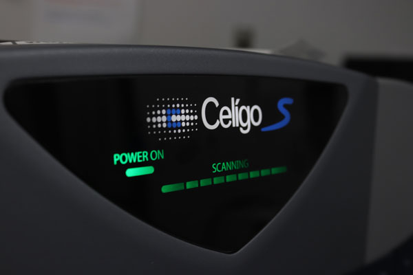
The HCS core facility contains state-of-the-art automated high-throughput microscopy instruments. Depending on the instrument, autofocusing is either image-based (IC200, Eidaq100) or laser-based (IN Cell 1000, Opera QEHS, Celigo). These instruments can be used with different objectives ranging from 4x to 60x magnification and have the ability to image standard clear-bottom multi-well plates (96, 384, 1536), preferably black plates with optical quality glass or plastic bottoms with a bottom thickness of ~200um. The INCell1000 and Opera QEHS in La Jolla have automatic plate loading capabilities (>40 plates). For a summary of each HCS instrument’s capabilities refer to the table below.

HCS image analysis and image data management software

A variety of image processing, data analysis, and visualization tools are available, including InCell Investigator (GE), CytoShop (Q3DM/Beckman Coulter), CyteSeer (Vala Sciences), Acapella (PE), Matlab (The Mathworks), Photoshop (Adobe), ImageJ (NIH), CellProfiler/CellVisualizer (nonprofit open source), and Volocity 3D/time course analysis software (PE). For screen data analysis and mining, CBIS (ChemInnovation) screening database software, as well as Spotfire (Tibco), Genedata Screener, and Prism (Graphpad) data analysis software are available. With these software packages the HCS team can rapidly apply available HCS solutions and algorithms, as well as develop custom HCS solutions, algorithms and tools for the wide variety of HCS projects.

To streamline HCS processing, visualization, and data management, the HCS team implemented a local storage system (LSS) dedicated to HCS based on the Columbus image-data management system (PE). The Columbus system provides ~12TB of fast access storage at the La Jolla site and handles image and data file management for currently ongoing HCS projects. The LSSs is connected via high-speed intranet and provide scientists with web-based access to image visualization, analysis, and data visualization tools. The Columbus image-data management system provides web-based upload capabilities for Opera, INCell 1000, IC200, Eidaq100, and Celigo instruments. It is integrated with the Volocity 3D/time course visualization and analysis package, and is also directly linked to Genedata Screener software for a complete HCS image-data analysis and visualization solution.


Price List

For a Price List, please call (858) 646-3100 ext. 3329 or email us.


Leadership

Susanne Heynen-Genel, PhD
Facility Director

Michael Jackson, PhD
Vice President, Drug Discovery & Development

Contact

Please call (858) 646-3100 ext. 3329 or use the button below to send us an email.

Contact the Facility

Assay Development

chemistry assay-pub domain

Overview

The Assay Development core facility performs design, development, validation and pilot screening of biological high-throughput assays. Our dedicated staff is skilled in various screening modalities to help design and develop de-novo assays or convert low-throughput lab protocols (e.g., cuvette- or gel-based) into plate-based HTS assays. To date, the facility has developed more than 250 cell-based and biochemical assays. Approximately half of these assays advanced to HTS, and the other half are employed in assay panels supporting HTS hit validation and Structure-Activity Relationship (SAR) studies. The facility is an integral part of Conrad Prebys Center for Chemical Genomics and works closely in collaboration with other cores and teams.

The HTS Assay Development core also provides direct assistance with preparing screening-related grants, guiding through assay development stages, helping design testing funnels, and generating preliminary data.


Services

  • Consultations on suitable screening alternatives and selection of the approaches consistent with project goals
  • Implementation and optimization of previously established screening protocols
  • Design and development of HTS assays
    • Cell-based and biochemical assays in 96-, 384- and 1536-well plate formats
    • Assays based on variety of HTS detection approaches (absorbance, fluorescence, fluorescence polarization, luminescence, AlphaScreen, BRET, FRET, TR-FRET)
    • Multiplexed cell-based assays for simultaneous assessment of multiple cellular functions, e.g. multiplexed qPCR assays, or reporter gene expression and cell viability
    • End-point and kinetic assays using common and special screening equipment
    • Functional and binding assays, including Differential Scanning Fluorimetry (also called protein thermal shift) assays detecting direct small-molecule binding through stabilization of target proteins in thermal denaturation
  • Assay optimization, miniaturization and implementation
    • Optimization of common assay parameters, such as cell density, ligand and enzyme concentrations, assay media or buffer
    • Evaluation of DMSO concentration effects
    • Optimization of assay sensitivity for identification of compounds with desired MOA
    • Evaluation and optimization of assay statistics and reproducibility
  • Focused library pilot screening
    • Assessment of assay performance measures (signal-to-noise, signal-to-background, assay dynamic range, Z’-factor) and their reproducibility
    • Evaluation and optimization of HTS hit rate
    • Access to a wide selection of focused libraries, e.g. collections of known drugs, kinase inhibitors, NCI compound sets (please inquire about the full list of focused libraries)
  • Support for Structure-Activity Relationship (SAR) studies:
    • Design and implementation of SAR panels relevant to the project goals
    • Development of secondary (e.g. orthogonal, counter-screen) assays
    • SAR data analysis and interpretation
    • Mechanism of action studies: determination of compound’s Ki, competition profile and time-dependent effect
  • Assistance with the preparation of screening grants
    • Consultation on available funding mechanisms (e.g. NIH PAR-12-058 and PAR-12-059)
    • Help in development of specific aims and project plan
    • Hands-on support in generation of preliminary data
    • Expertise with diverse target classes and multiple formats utilized at the facility and its application for selecting secondary assays and designing project workflow

Equipment & Resources

Major Robotic Equipment and Resources used by Assay Development at the Conrad Prebys Center for Chemical Genomics include:

  • Hamamatsu FDSS7000 rapid-kinetics (e.g. GPCR, glow luminescence, enzyme kinetics) fluorescence and luminescence plate reader with integrated 384-well liquid handling
  • LabCyte Access Laboratory Workstation for cherry picking of screening hits and preparation of SAR compound plates
  • NEPHELOstar plate reader for detecting light scattering in 96- and 384-well plate based solutions using nephelometry approach
  • Monochromator based multimode plate readers
    • Two Molecular Devices M5 instruments
    • Molecular Devices FlexStation 3
  • Two VIIA7, 7th generation real-time PCR instruments
    • Specifically designated for development and screening of qPCR and Differential Scanning Fluorimetry (protein melt) assays
    • 384-, 96-, fast 96-well plate and TaqMan array card assays
  • Filter based multimode plate readers
    • Molecular Devices Analyst HT equipped with stacker unit
    • Perkin Elmer EnVision equipped with liquid injector unit and 50-plate stackers
  • Microplate pipetting systems for handling serial dilutions and replication of plates for MOA studies
    • Agilent plate processing workstation – includes a Bravo liquid handler, BenchCell 4x with four stackers, a PlateLoc sealer, and Vcode barcode labeler; allows liquid dispensing using full-head and single-row or -column modes applicable for serial diluations
    • BioTek Precision – allows large-volume serial dilutions in 96- and 384-well plate formats and bulk dispense

Price List

For a Price List, please call (858) 646-3100 ext. 3462 or email us.


Leadership

Eduard Sergienko, PhD
Facility Director

Michael Jackson, PhD
Vice President, Drug Discovery & Development

Contact

Please call (858) 646-3100 ext. 3462 or use the button below to send us an email.

Contact the Facility

Proteomics

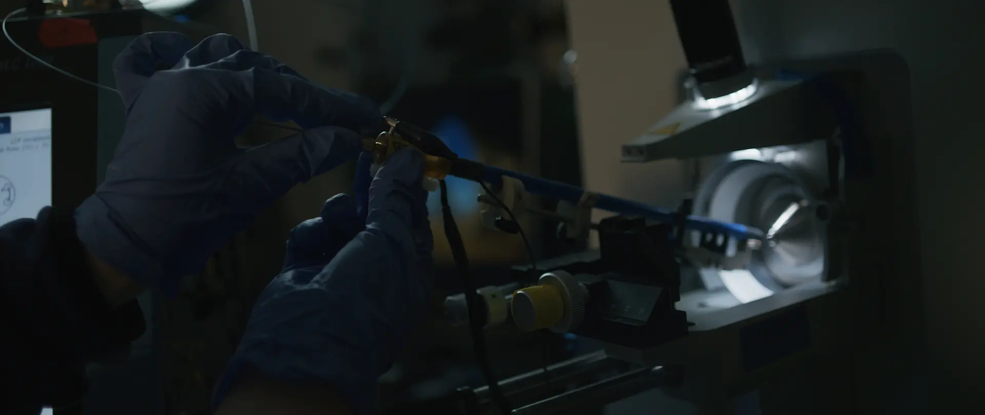
close-up of scientist doing work in a lab

Overview

The Sanford Burnham Prebys Proteomics Shared Resource provides state-of-the-art mass spectrometry-based proteomics services including identification, quantification and characterization of proteins and their post-translational modifications. We provide proteomics services to Institute scientists as well as outside non-profit and for-profits organization on a fee-for-service basis.

The Proteomics Shared Resources is located on the main campus and currently houses three mass spectrometry platforms. The facility is run by four highly committed proteomics experts, including one faculty member as scientific advisor.

Equipment Highlight

Seer Bio Proteograph SP100
The platform combined with TMT multiplexing has the capability to detect ~2500 protein groups in plasma samples, including low abundant proteins like cytokines.

Seer Fact Sheet


Services

The Proteomics Shared Resource Offers a Diverse Array of Mass Spectrometry Services Including:

  • Identification and Localization of Post-Translational Modifications (e.g., phosphorylation, ubiquitinylation, acetylation, methylation, nitrosylation)
  • Quantitative comparison of protein abundances in complex mixtures using label-free or label-based techniques (e.g., SILAC, TMT)
  • Targeted protein identification and quantification  
  • Protein/Peptide Identification
  • Identification of protein-protein interactions by Affinity Purification followed by Mass Spectrometry (APMS) analysis
  • Identification of biotinylated proteins
  • Comprehensive serum proteomic profiling 

We urge you to consult with us prior to starting your proteomics experiment. We will help you to design the best experiment that fits your purposes.


Equipment & Resources

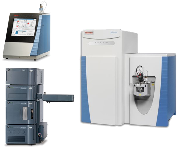
The Proteomics Shared Resource is equipped with 5 mass-spectrometry platforms, one automated liquid handling system for sample preparation and a robust computational infrastructure and software. Our mass spectrometers are coupled to either one- or two-dimension ultra-high performance nanoflow HPLC system (EASY nLC 1200, and 2D NanoAcquity).

Seer Bio Proteograph SP100

The platform combined with TMT multiplexing has the capability to detect ~2500 protein groups in plasma samples, including low abundant proteins like cytokines.

Seer Fact Sheet

Orbitrap Fusion Lumos

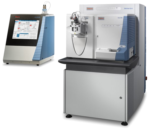
The Thermo Scientific Orbitrap Fusion Lumos Tribrid is the newest state-of-the-art mass spectrometer that allows system biologists to test the limits of proteome analysis. It incorporates the brightest ion source, a segmented quadrupole mass filter with improved selectivity and ion transmission, Advanced Vacuum Technology for improved ion transmission to the Orbitrap mass analyzer. These hardware improvements enable the new Tribrid instrument to excel in the most challenging applications, including analysis of low level proteins and PTMs, multiplexed relative quantitation using isobaric tags such as iTRAQ or TMT, and intact protein characterization. The Lumos is the most sensitive and versatile mass spectrometer currently available for proteomics work, and can do CID, HCD, and ETD fragmentation. Our two Lumos units are coupled to EASY 1200 UPLC system.

QExactive Plus

The QExactive Plus (QE+) is a highly sensitive, accurate mass instrument that uses an Orbitrap detector and HCD fragmentation. Our QE+ can be coupled to either a 2D M-Class NanoAcquity or an EASY 1200 UPLC system. With a fast scan speed and multiplexing capabilities, the QE+ is an outstanding system for mid-to-high complex proteomes. In addition, the QE+ also excels at targeted quantitation experiments, using the Product Reaction Monitoring (PRM) acquisition. In particular, we have developed an intelligent PRM approach which improves detection sensitivity at least 10x compared to conventional PRM method.

Orbitrap Elite

The Orbitrap Elite is a hybrid instrument consisting of an LTQ Velos Pro linear ion-trap coupled to a high-field Orbitrap detector. This allows high-mass accuracy scans to be acquired in the Orbitrap, and rapid sensitive scans to be acquired in the LTQ. The instrument can perform CID, HCD and ETD fragmentation. The high-field Orbitrap mass analyzer geometry and advanced signal processing technologies enable resolution of >240,000, superior spectral quality, and higher scan speed. The outstanding resolution increases analytical certainty by improving molecular weight determination for intact proteins and clearly resolving smaller, isobaric species. Our Orbitrap Elite is coupled to a EASY 1200 UPLC, and is generally used for low-to-mid complex proteomes.

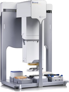
Agilent AssayMap BRAVO Platform

For automated sample preparation


Price List

For a complete list of services, please call (858) 646-1000 ext. 4180 or email us.

Prod IDProteomics ServiceInternal SubsidizedInternalExternal Non-ProfitExternal For-Profit
K800Total proteome profilingTMT 6plex$2,000$2,500$2,700$5,260
K801TMT 10plex$2,600$3,250$3,510$6,838
K802TMT 16plex$3,700$4,625$4,995$9,731
K015TMT 18plex$4,234$5,292.50$5,715.90$11,135.42
K803Phospho+Total proteome profilingTMT 6plex$3,100$3,875$4,185$8,153
K804TMT 10plex$3,600$4,500$4,860$9,468
K805TMT 16plex$4,943$6,178.75$6,673.05$13,000.09
K016TMT 18plex$5,560.88$6,951.09$7,507.18$14,625.10
K807Total proteome profilingLFQ$300$375$405$789
K808Total GoBig proteome profilingLFQ 6+$267$333.75$360.45$702.21
K809Phospho+Total proteome profilingLFQ 1x$500$625$675$1,315
K704Biotin AP-MS packageBioID$330$412.50$445.50$867.90
K705Biotin GoBig AP-MS package16+ BioID$300$375$405$789
K810On-bead’ AP-MS package1-5 samples$250$312$337.50$657.50
K8116+ samples$220$275$297$578.60
K812LC-MS/MS QE1-7 samples$170$212.50$229.50$447.10
K8138+ samples$150$187.50$202.50$394.50
K814LC-MS/MS Lumos1-7 samples$250$312.50$337.50$657.50
K8158+ samples$220$275$297$578.60

Leadership

Svetlana Maurya, PhD
Facility Associate Director

Elena Pasquale, PhD
Scientific Director

Contact

Svetlana Maurya, PhD

Main Responsibilities: Global proteomics analysis using label-free or isobaric labeling techniques, post-translational modification (PTM) analysis including phosphoproteomic analysis, biotinylated protein enrichment and identification, interactomics. Quantitative proteomics analysis and computational analysis of large-scale proteomics data using MaxQuant, Spectronaut, Spectromine, Proteome Discoverer. Facility billing/budgeting, Sample queue and general proteomics inquiries.

Please call (858) 646-3100 ext. 4180 or use the button below to send us an email.

Contact the Facility

Cancer Metabolism

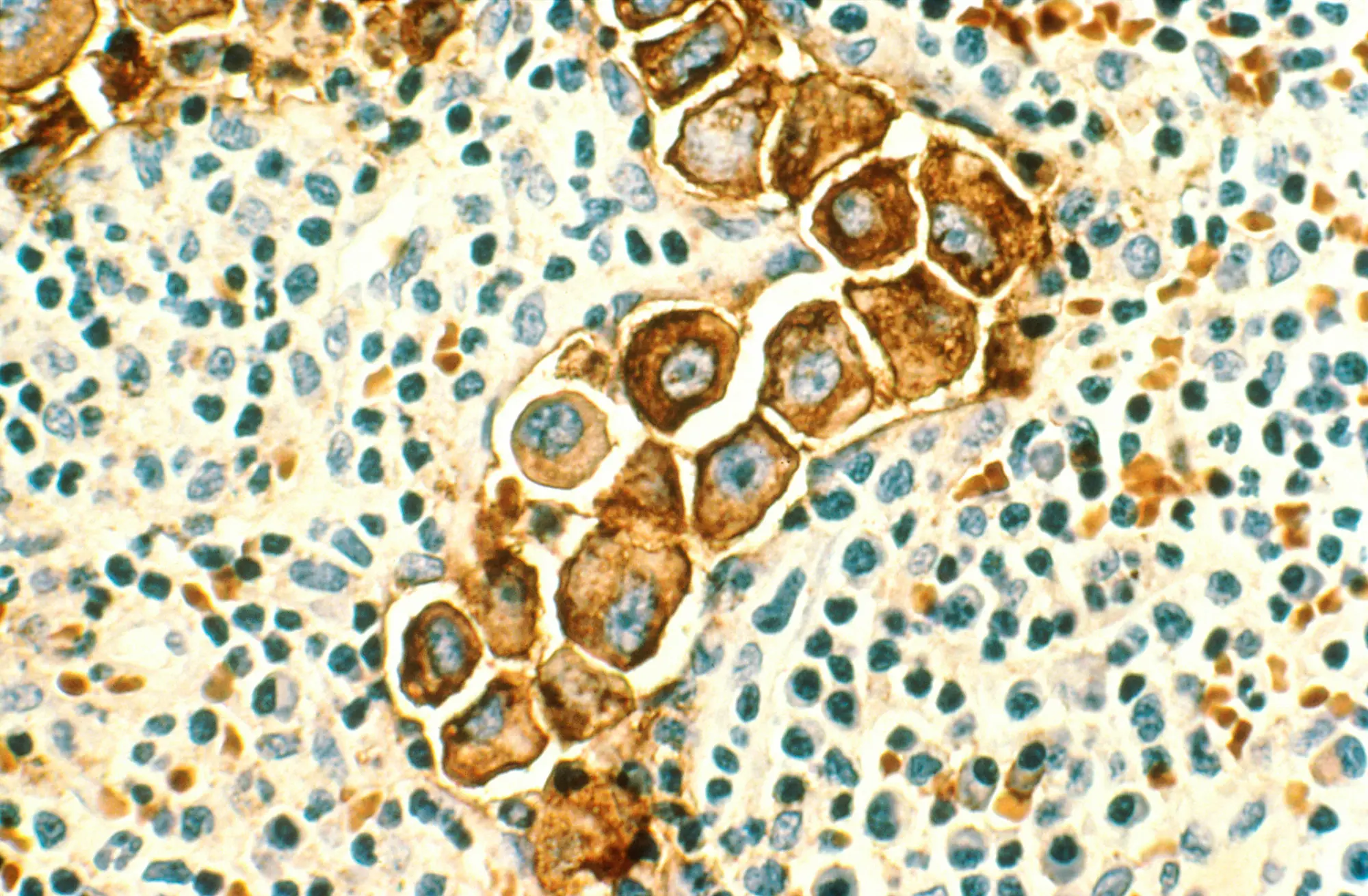
breast cancer in lymph nodes-nci

Overview

The Sanford Burnham Prebys Cancer Metabolism Core provides a hub within the Institute for analysis of metabolism using specialist instruments and methodologies, together with advice on planning experiments and analyzing data. All of the instruments and approaches are available on a fee-for-service basis to Cancer Center scientists, but also (as capacity permits) to outside users. Samples for most services can be shipped, but Seahorse analyzer users need to be local so they can bring live cells for analysis.


Services

The Primary Services Offered by the Core are:

  • GC/MS or GC/MS/MS-based analysis of metabolites, including:
    1. Quantification of polar metabolites (such as amino acids, components of glycolysis and TCA pathways) in panels designed for different sample types (cells, medium, tumor/ tissue, serum/ plasma, or “total”) and including 39-89 metabolites.
    2. Stable isotope (13C, 15N, 2H) tracing of polar metabolites (25 metabolites)
    3. Fatty acids and cholesterol (quantification and 13C-tracing)
    4. Short-chain fatty acids (quantification with negative chemical ionization)
    5. Custom assays of sugars, sugar-phosphates, and related molecules
  • Quantification versus standards can be combined with stable isotope tracing. The combination can provide more confidence in identified changes in metabolic activity.
  • Measurement of cellular respiration and glycolysis using Seahorse XFp, XFe24 and XFe96 analyzers. These instruments determine rates of extracellular acidification and oxygen consumption by living cells respectively in a 6-well or larger 24 or 96 well formats. From these data, the relative activity of cellular glycolysis versus oxidative phosphorylation, including the balance of ATP production from these two major energetic processes, may be determined.
  • Measurement of major substrates and metabolites (glucose, glutamine, lactate, and glutamate) in culture medium using the YSI 2950 analyzer. This instrument is able to rapidly (~1 min per metabolite) determine the concentration of these important metabolism substrates from a small volume of sample (100-200 µL). Using a 96-well format, this analysis provides an economical method to roughly define changes in metabolism (e.g., in proliferating versus quiescent cells), and is also useful for checking that cells in culture have a sufficient supply of the major nutrients glucose and glutamine.

Equipment & Resources

  • A Thermo TSQ 9610 gas chromatograph- triple quad mass spectrometer (GC-MS/MS) is used for quantitative analysis of up to 90 polar metabolites from samples including cells, tumors or serum. It is also used for quantification of fatty acids and short-chain fatty acids (SCFA). SCFA are analyzed with negative chemical ionization, which reduces background and allows for low detection limits.
  • A Shimadzu QP2010 GC-MS is used for metabolic analyses which include determination of stable isotope (13C, 15N) labeling rates of intra- and extracellular metabolites. 13C labeling can be measured in amino acids, keto and fatty acids, sugars, and sugar phosphates. 
  • The YSI 2950 metabolite analyzer is able to measure glucose, glutamine, lactate and glutamate in media in 2-3 minutes for all 4 metabolites in a 96-well format. The minimum volume needed is 100 μl. A full 96-well plate may be analyzed in 3-4 hours and results are typically returned the same or next day.
  • The XFp Analyzer can measure respiration in most cell types, including primary cells, adherent cells, and suspension cells (with attachment). With a 6 well format, the XFp Miniplate is ideal for pairwise comparisons (in triplicate) and the analysis of precious samples, such as patient-derived and other rare samples. We mainly focus on the Cell Mito Stress Test to measure the key parameters of mitochondrial function: basal respiration, ATP production, proton leak, maximal respiration, and spare respiratory capacity. Other protocols to measure the use of alternate substrates or to calculate the relative production of ATP from glycolysis or oxidative phosphorylation are available.
  • The XFe24 Analyzer is similar to the XFp, but well sizes are 2.5x larger and up to 22 samples can be run on one plate. We have available islet capture plates.
  • The XFe96 analyzer has the same well size as the XFp and can run up to 92 samples on one plate.

Price List

For a complete list of services, please call (858) 646-1000 ext. 3941 or email us.

Cancer Metabolism ServicesInternal
(Subsidized)
Non-ProfitFor ProfitMinimum
sample
number
Code
GCMS metabolite quantification and stable isotope (13C) tracing (per sample)
Polar metabolite GCMS quantification (cells or medium ~40 metabolites)$40$54$105.2012CMR162
Polar metabolite GCMS quantification (serum or tissue ~55 metabolites)$44$59.40$115.7212CMR182
Polar metabolite GCMS quantification (total panel ~85 metabolites OR custom assays)$50$67.50$131.5012CMR183
Polar metabolite 13C GCMS analysis$59.40$44$115.726CMR163
Combined GCMS 13C & quant polar metabolites$78$105.30$205.148CMR170
Fatty acid & cholesterol GCMS analysis (quantification)$48$64.80$126.2412CMR164
Fatty acid 13C GCMS analysis$52$70.20$126.246CMR171
Combined GCMS 13C & quant fatty acids$90$121.50$236.708CMR184
Short-chain fatty acid GCMS quantification$50$67.50$131.5012CMR172
Sample prep for GC-MS (extra to sample analysis fee)
Sample prep – cells or medium, or serum/ plasma$3$4.05$7.89N/ACMR175
Sample prep – fecal/ cecum; weighed tumor or tissue$9$12.15$23.67N/ACMR177
Sample prep – tumor, tissue, not weighed or needs re-weighing$15$20.25$39.45N/ACMR176
Sample prep – urine with urease treatment$5$6.75$13.15N/ACMR174
Charges for Seahorse analyzers and YSI Bioanalyzer
Seahorse XFp (per plate) (training/ full service)$200$270$526N/ACMR166
YSeahorse XFp (per plate) (self-service)$80$108$210.40N/ACMR168
Seahorse XFe24 (per plate) (training/ full service)$300$405$789N/ACMR178
Seahorse XFe24 (per plate) (self- service)$190$256.50$499.70N/ACMR179
Seahorse XFe96 (per plate) (training/ full service)$450$606.50$1,183.50N/ACMR180
Seahorse XFe96 (per plate) (self- service)$190$256.50$499.70N/ACMR181
YSI metabolite analysis (per plate)$99$133.65$260.37N/ACMR167
Notes

Sample prep charges (CMR174-177) are for sample extraction for GCMS, and are in addition to the GCMS charges. If samples are submitted as dried extracts the prep charges will not apply. See core for details.

25% discount for samples for GCMS in excess of 30 in a singly batch, rounded to nearest sample (e.g., 32-35 samples, discount of 1 sample).

Self-service rate for Seahorse analyzers includes the cost of plates, XF-medium and reagents.


Leadership

Cosimo Commisso, PhD
Scientific Director

David Scott, PhD
Facility Director

Contact

David Scott, PhD

David Scott focuses on the development and application of methods for metabolomics, enhanced by the use of stable isotope substrates. Stable isotopes (mainly 13C) provide an added layer of information to metabolomic studies – not only is static pool information obtained, but it is also possible to infer the origins of metabolites and to track flux through metabolic pathways. Methods used include the analysis of small polar metabolites (mainly amino acids and carboxylic acids) and fatty acids by gas chromatography-mass spectrometry. Other techniques are being developed to analyze and quantify an expanded range of (labeled) metabolites. We have applied these methods in a study of the central carbon metabolism in Escherichia coli, and, in several papers, on melanoma metabolism. Investigations of melanoma cells showed the importance of glutamine metabolism under hypoxia, identified a novel route for the utilization of the carbon backbone of glutamine for fatty acid synthesis, and defined pathways for proline synthesis. Other recent work uses alternately labeled sugars to simultaneously track different inputs to glycans in normal cells and cell lines from patients with glycosylation defects.

Please call (858) 646-3100 ext. 3941 or use the button below to send us an email.

Contact the Facility

Viral Vectors

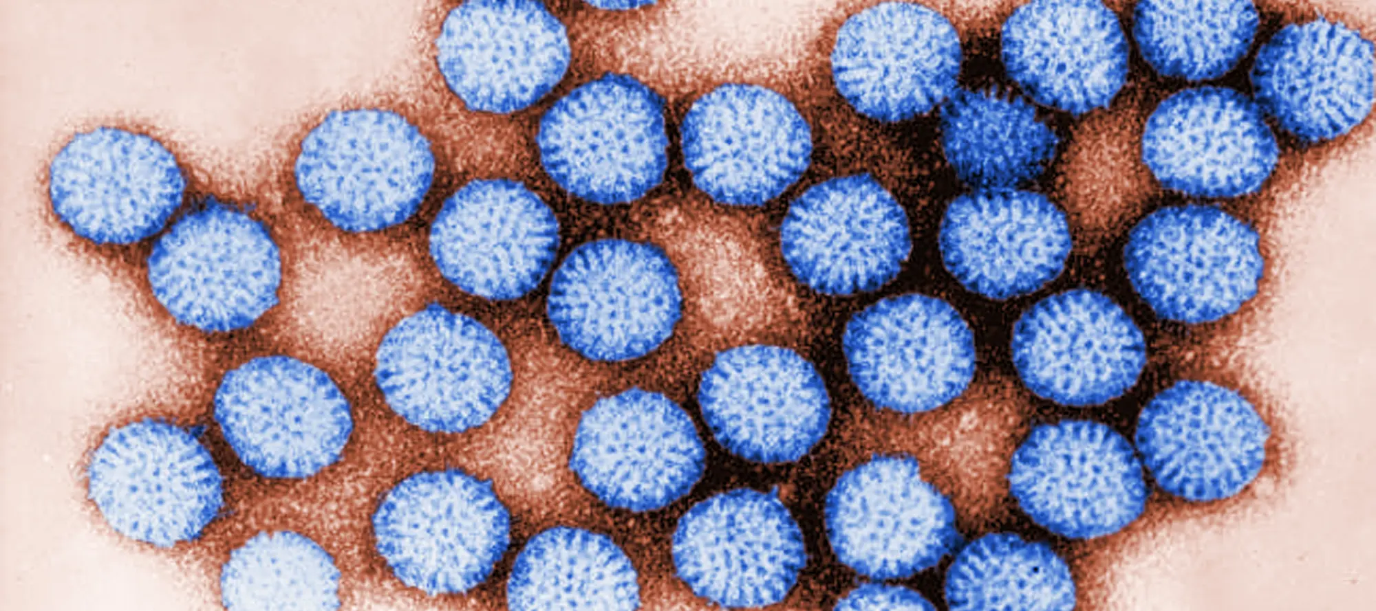
close-up of rotovirus cells

Overview

The viral vector core facility at Sanford Burnham Prebys develops state-of-the-art viral vector-based gene delivery technology. Its portfolio ranges from lentivirus, retrovirus, adenovirus, AAV, VSV, sindbis virus, and Zika virus products as well as customized “The Works” viral vector construction and swapping service package. There are a variety of ready-to- transduce viral vector collections including constitutively expressed or inducible fluorescent proteins for nuclear or membrane localization and bioluminescence reporters for 3D drug screening. The viral core has integrated CRISPRs/Cas technology in the plasmid-free viral vector platform, providing scientists with choices of combined technologies to manipulate genes using CRISPR gene disruption, editing, inhibition, and activation. In addition, we extend technical expertise in nanoparticle research including exosome purification, analysis, and engineering. More importantly, the core actively participates and supports translational research to fight human diseases.


Services

The Viral Vector Core Facility Focuses on the Following Services:

  1. Custom large scale rAAV production and titration. Serotypes include 1-9, 3B, 7M8, shH10, DJ, DJ/8, PHP.B, PHP.eB, PHP.S, B1, rh10, Anc80, and Retrograde. Ready-to-transduce rAAV-CMV-eGFP and CAG-NLS-eGFP/mCherry viruses are available upon request.
  2. Custom large scale lentiviral [HIV-1, integrase defective lenti-virus (IDLV), and FIV] and retroviral (MSCV and MMLV) production and titration is based on vector plasmid DNA provide by the user. Titration is offered for virus ubiquitously expressing fluorescence or puromycin selection marker.
  3. Ready-to-transduce lentiviral preps include: (1) doxy-inducible or constitutively expressed cytoplasmic fluorescent reporters w/out mammalian selection markers (2) nuclear or membrane localized fluorescent reporters (3) secreted or cytoplasmic bioluminescent reporters (4) doxy-inducible or constitutively expressed non-silencing control (scramble) shRNA (5) cell cycle and autophagy reporters.
  4. Custom large scale adenoviral particles (Ad5) production and titration from user-supplied construct or virus include: (1) adenovirus recovery (2) high purity adenoviral preparation using CsCl gradient purification (3) titration via plaque assay. Ready-to-transduce pAd5-eGFP, NLS/CAAX fused eGFP/mCherry, firefly or secreted gaussian/cypridina luciferase, and Cre recombinase viruses are available upon request.
  5. “The Works” custom lentiviral-adenoviral-rAAV vector construction and swapping include:
    1. Doxy-inducible or constitutively expressed cDNA or shRNA
    2. Promoter / enhancer reporter (3) microRNA sponges
  6. siRNA to lenti shRNA vector conversion with choice of doxy-inducible H1 or constitutive hU6 promoter as well as puromycin or neomycin selection marker. Ready-to-transduce non-targeted siRNA converted or universal scramble shRNA lentiviruses are available upon request.
  7. Custom packaged Sindbis virus production and titration. Ready-to-transduce mCherryCAAX-eGFP virus, pSINREP5-based vector construction, packaging, and flow cytometry-based titration are available upon request.
  8. Custom VSV vector construction, production, and titration. Pseudotyped VSV-ΔG-eGFP with Ebola virus glycoprotein (EboV-GP) Zaire strain (Kissidougou-C15, 2014) or wild-type VSV glycoprotein (VSV-G) are available upon request.
  9. Custom Zika virus production and titration are available for African, Asian, and Brazilian (2015-2016 outbreak) strains.

Equipment & Resources

The Core is fully equipped with all the necessary equipment for production of consistent quality viral vectors including state-of-the art cell culture hoods and incubators under appropriate biosafety conditions, as well as an ultracentrifuge, microscopes and a dedicated -80°C freezer for storage of vectors. Producer cell lines 293T and 293A cells are routinely tested for the presence of mycoplasma using Lonza detection kit and discontinued at passage 20.

The Institute is in a partnership program with Sigma-Aldrich and Open Biosystems. Both companies provide affordable shRNA constructs. The Core also currently collaborates with Dr. Terskikh and Dr. Mercola labs to increase ready-to-transduce virus inventory.

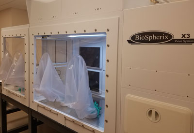
Our lab uses the Cytocentric Xvivo System from BioSpherix Ltd to aseptically control O2, CO2, and temperature levels during the incubation, handling, and analysis of live cells without the disturbances which routinely occur in traditional cell culture incubators. This platform allows our core users to more accurately recapitulate physiologic, in vivo-like conditions during in vitro experiments with living cells. More information can be found at: biospherix.com/xvivo-system-model-x3


Price List

For a Price List, please call (858) 646-3100 ext. 4353 or email us.


Leadership

Chun-Teng Huang
Associate Director

Contact

Chun-Teng Huang
Room 7112
(858) 646-3100 ext. 3803
 

Ching-Lien Fang
Room 7112
(858) 646-3100 ext. 3803

Contact the Facility

Genomics and Spatial Transcriptomics

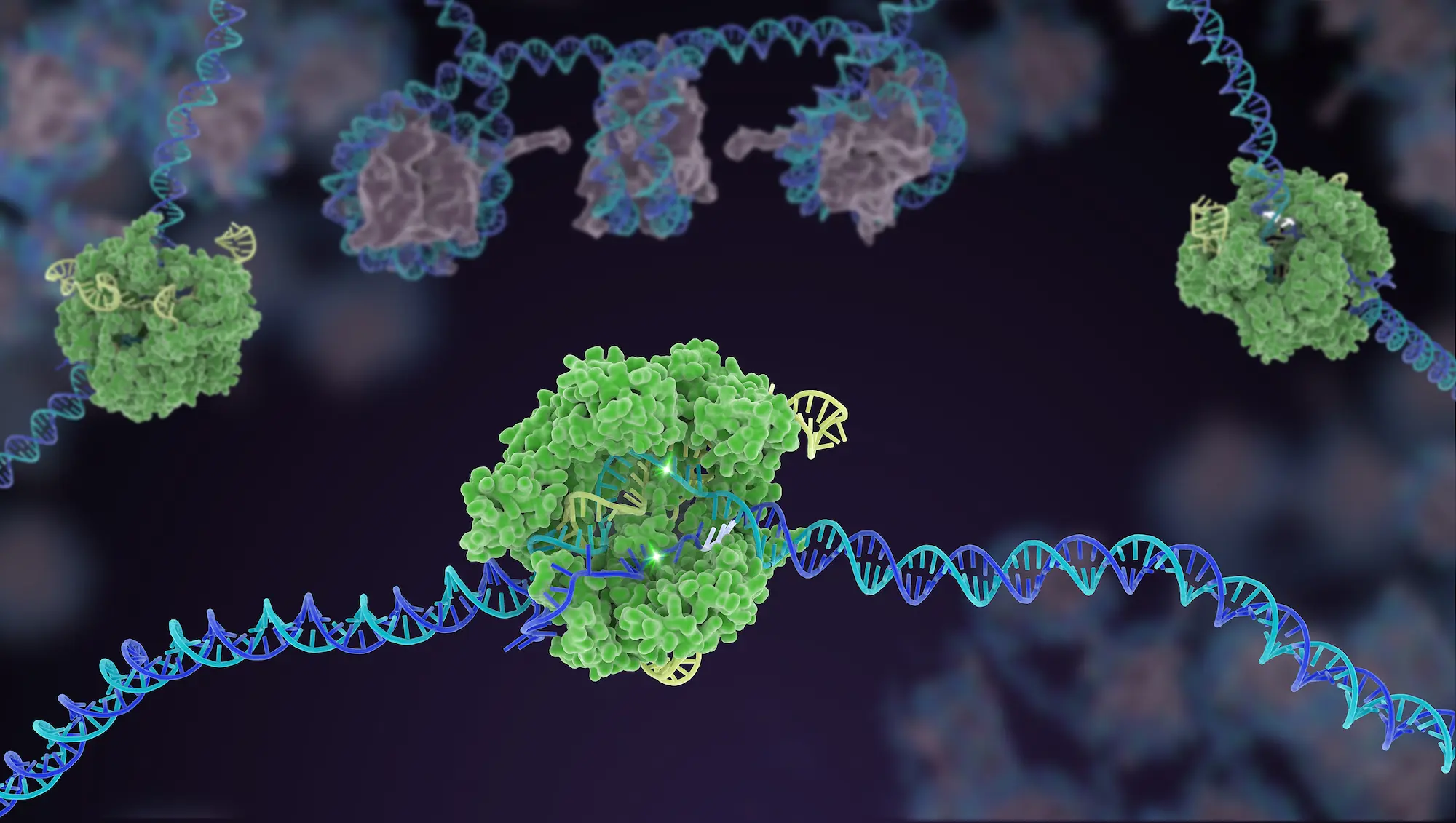
image of cells and dna

Overview

The Sanford Burnham Prebys Genomics Core Facility offers full-service library preparation and Next Generation Sequencing on the Element Biosciences AVITI sequencer, single cell library preparation using the 10X Genomics Chromium X system, multiplex gene expression analysis with the NanoString nCounter System, and assessment of DNA and RNA quality.

We are now also heavily focused on Spatial Transcriptomics, providing analysis on the GeoMx Digital Spatial Profiler and the CosMx Spatial Molecular Imager from NanoString. 

We also provide scientific advising for experimental design and analysis as well as Bioinformatics analysis performed in collaboration with the Bioinformatics Core.

For all inquiries, please contact core director Rebecca Porritt


Services

Next Generation Sequencing

The core provides full-service library preparation and sequencing on the Element Biosciences AVITI sequencer. The AVITI produces exceptionally high-quality sequencing data, with industry leading accuracy, and highly competitive pricing. It is equipped with dual independent flow cells, each with 1B read capacity, enabling high throughput sequencing for RNA and DNA libraries. Bulk RNA and DNA libraries made within the core incorporate Element Biosciences Elevate adaptors with unique dual indexes enabling a streamlined library preparation and sequencing workflow. The AVITI is also compatible with Illumina sequencing libraries through a simple conversion step (included with sequencing services), and thus supports a wide range of applications.

Flow cell options:

  • 2x75bp (500M and 1B reads)
  • 2x150bp (250M, 500M and 1B reads)
  • 2x300bp (100M and 300M)

NGS Library Preparation

We provide QC and library preparation for a range of sequencing applications:

  • Poly-A RNA-seq, Whole transcriptome RNA-seq, Low-input RNA-seq
  • Targeted (hybridization capture and amplicon sequencing)
  • Whole Genome Sequencing, Whole Exome Sequencing
  • ChIP-Seq
  • Indexing, Molecular Barcoding

Single Cell Library Preparation

The genomics core provides single cell library preparation from user provided single-cell or single-nuclei suspensions. We have two systems for single cell library preparation. These include the new 10x Genomics Chromium X system which supports a wide range of applications (see below) and the Fluent Biosciences PIP-seq system for 3’scRNA-seq.

10x Genomics Chromium X supports:

  • Single-cell RNA-seq
  • Single-cell ATAC-seq
  • Single-cell multiomics (RNA-seq and ATAC-seq)
  • Single-cell immune profiling
  • Feature barcoding (such as CITE-seq and Cell Multiplexing)
  • Targeted library preparation

Multiplex Gene Expression Analysis

We provide the NanoString nCounter Analysis System for multiplex profiling of mRNA, microRNA, non-coding RNA or DNA isolated from a range of cells and tissues including fresh, frozen and FFPE samples. The system can count hundreds of sequences in a single reaction, allowing for fast, high-plex analysis of samples. This is ideal for a more targeted approach to gene expression analysis, such as biomarker validation or analysis of specific pathways.

DNA and RNA Quality Assessment

We provide three instruments for assessment of DNA and RNA concentration and quality including the TapeStation (Agilent), BioAnalyzer (Agilent) and Qubit (ThermoFisher).

The TapeStation will be used for QC for all NGS library preparations to measure concentration and fragment size.

Spatial Transcriptomics

We offer two spatial transcriptomic technologies from NanoString; the GeoMx Digital Spatial Profiler and the CosMx Spatial Molecular Imager.

The GeoMx Digital Spatial Profiler is used to spatially resolves RNA (whole transcriptome) or proteins (up to 96) within multiple regions of interest on FFPE or FF tissue sections. A region of interest (ROI) can be defined by first staining the tissue sections with antibodies that identify various tissue compartments or cell types of interest. This allows in-depth NGS-based analysis of transcriptomic profiles in specific cell populations with morphological and spatial context.

The CosMx Molecular Imager allows for true single cell transcriptomics using in-situ high-plex technology. FFPE or FF tissue sections can be analyzed for RNA (up to 1000) or protein (up to 64) expression using unique barcoding and imaging technology. This allows for analysis of all cell types across whole tissue sections.

Cell Line Authentication

We provide STR-based human cell line authentication. The GenePrint 10 System from Promega is used to perform short tandem repeat (STR) analysis on genomic DNA submitted to the core. Samples can be submitted as purified genomic DNA, or cells can be spotted directly on FTA cards in media or trypsin (FTA cards are available in the core).

Training

We offer instrumentation training for personnel who want to reserve, and use select instruments on their own.


Equipment & Resources

NGS Library Preparation and Sequencing

Element Biosciences AVITI sequencer
The AVITI produces exceptionally high-quality sequencing data, with industry leading accuracy. It is equipped with dual independent flow cells, each with 1B read capacity, enabling high throughput sequencing for RNA and DNA libraries. Bulk RNA and DNA libraries made within the core incorporate Element Biosciences Elevate adaptors with unique dual indexes, enabling a streamlined library preparation and sequencing workflow. The AVITI is also compatible with Illumina sequencing libraries through a simple conversion step (included with sequencing services), and thus supports a wide range of applications.

Eppendorf epMotion 7075 Liquid handling system
The epMotion 7075 is used for high throughput production of RNA and DNA libraries for sequencing on the NextSeq 500.

Single Cell Library Preparation

10X Genomics Chromium X
A single cell profiling system that encapsulates single cells or nuclei into GEMs for unique barcoding. The chromium X is compatible with all 10x Genomics single cell assays. It allows for large-scale studies with high-throughput single cell gene expression and immune profiling. The high-throughput chips can process up to 20,000 cells per channel.

Fluent Biosciences PIP-seq equipment
Fluent Biosciences PIP-seq is a highly flexible and cost-effective platform that uses vortexing to partition single cells or nuclei with RNA capture beads for 3’scRNAseq. PIP-seq kits come in a range of sizes from 2,000 – 100,000 cells per sample.

Bio-Rad ddSeq Single-Cell Isolator
The ddSeq is an alternative single library preparation system well suited for pilot studies with less than 1000 cells per sample.

Spatial Transcriptomics

NanoString GeoMx Digital Spatial Profiler 
The GeoMx allows whole tissue sections (fresh frozen or FFPE) to be imaged and stained for RNA or protein. Researchers can then select specific tissue regions or cell types of interest for profiling. The high-plex system can support whole transcriptome analysis for in depth transcriptomic profiling of specific cell populations with morphological and spatial context.

NanoString CosMx Spatial Molecular Imager
The CosMx is a high-plex in situ analysis platform allowing quantification and visualization of up to 1000 RNA or 64 protein analytes than can be resolved down to the subcellular level, allowing for true single cell spatial omics. This system is compatible with fresh frozen and FFPE slide-mounted tissue sections.

Curio Biosciences, Curio seeker
Curio Seeker is a high resolution, whole transcriptome spatial analysis technology. It is compatible with fresh frozen tissues, which are sectioned onto a tile composed of 10uM beads with barcoded mRNA capture probes. Through sequencing of barcodes and mRNA, transcript expression can be spatially resolved. Tile sizes available include 3mm x 3mm and 1cm x 1cm.

DNA and RNA assessment

Agilent TapeStation 4200
The TapeStation system provides sizing, quantitation and quality control of DNA and RNA. It is primarily used for assessing integrity of RNA samples for NGS workflows.

Agilent BioAnalyzer
The BioAnalyzer is a chip-based system that provides sizing, quantitation and quality control of DNA and RNA.

Qubit Fluorometer
The Qubit gives an extremely accurate measurement of small amounts of DNA, RNA or protein.

NanoDrop Spectrophotometer
A microvolume (1µl) spectrophotometer to measure nucleic acid concentration.

Common-use Instruments in the Core

qPCR
  • Roche LC480 real time QPCR instrument (interchangeable 96 or 384 well)
  • Roche LC96 real time QPCR instrument
  • Stratagene Mx3000p real-time QPCR instrument (96 well)
  • ABI 7900HT real time QPCR (interchangeable 96 or 384 wells)

Plate Reader

ThermoFisher Scientific Varioskan™ LUX multimode microplate reader
Supports a range of measurements including Absorbance, Fluorescence Intensity (top or bottom), Luminescence (with 2 dispensers), AlphaScreen, and Time-Resolved Fluorescence.


Price List

For a complete list of services, please call (858) 646-100 ext. 5130 or email us.


Leadership

Rebecca Porritt, PhD
Leader, Spatial Omics

Peter D. Adams, PhD
Scientific Director

Contact

Rebecca Porritt, PhD
(858) 646-3100 ext. 5130
Email Rebecca Porritt

Please call (858) 646-3100 ext. 5130 or use the button below to send us an email.

Contact the Facility

Functional Genomics

A scientist works with lab equipment

Overview

The Functional Genomics Core facility provides infrastructure for unbiased Target Discovery by means of genome-wide genetic screening in cultured cells. The Core’s primary effort is dedicated to loss-of-function screening via RNAi and CRISPR-CAS9 technologies although other services such as micro-RNA (miR) screening and cell population barcoding for clonal analysis are also available. Our range of expertise covers from project feasibility assessment, all the way through verification of identified targets. In addition to screens, the Core produces CRISPR-engineered cell lines to serve as models for screening or to aid the follow-up process, and can provide reagents tailored to the needs of each project such as sh/siRNAs, ORF clones, CRISPR reagent aliquots. Our personnel develop both plate reader and image-based cellular assays, and adapt them to high-throughput screening in arrayed format in conjunction with the High Content Screening Core. CRISPR-CAS9 screens are performed in pooled format using either off-the shelf or custom made lentiviral libraries, and results are de-convoluted in our Genome Analysis shared resource.

The viral vector core facility at Sanford Burnham Prebys develops state-of-the-art viral vector-based gene delivery technology. Its portfolio ranges from lentivirus, retrovirus, adenovirus, AAV, VSV, sindbis virus, and Zika virus products as well as customized “The Works” viral vector construction and swapping service package. There are a variety of ready-to- transduce viral vector collections including constitutively expressed or inducible fluorescent proteins for nuclear or membrane localization and bioluminescence reporters for 3D drug screening. The viral core has integrated CRISPRs/Cas technology in the plasmid-free viral vector platform, providing scientists with choices of combined technologies to manipulate genes using CRISPR gene disruption, editing, inhibition, and activation. In addition, we extend technical expertise in nanoparticle research including exosome purification, analysis, and engineering. More importantly, the core actively participates and supports translational research to fight human diseases.


Services

  1. siRNA library preparation and arrayed screen:

The core has genome-wide human siRNA library ON TARGET-Plus (OTP) library (18,301 genes, Dharmacon (Dh) / Thermo Scientific (TS)), formatted as SMARTpools and 4 single siRNAs per gene. Seventeen collections of pre-spotted pathway-focused libraries include (1) druggable genome (7740 genes, Applied-Biosystems (AB) / Life Technologies (LT)); (2) epigenetics / transcriptional-regulators (1362 genes, AB/LT); (3) epigenetics (882 genes, created by SBP in-house); (4) ubiquitinome (1170 genes, Qiagen and 1432 genes, SBP); (5) kinome (704 genes, AB/LT and 831 genes, SBP); (6) cancer pathway (252 genes, Dh/TS); (7) phosphatases (231 genes, Dh/TS and 314 genes, SBP); (8) G protein coupled-receptor (GPCR) (Dh/TS); (9) metabolome (160 genes, AB/LT);  (10) miniscreen random subset (Dh/TS); (11) autophagy (1601 genes, SBP); (12) innate immunity (96 genes, SBP); (13) B cell activation (127 genes, SBP); (14) T cell activation (287 genes, SBP); (15) ion channel (380 genes, SBP); (16) apoptosis (823 genes, SBP). Customized siRNA cherry-pick service is available upon request. In addition, CRISPR/Cas9 arrayed screening with (17) kinome gRNA library (789 genes, TS) is available.

  1. miRNA mimic and antagonist library arrayed screen:

The core has human miRNA mimic library (875 miRNAs from miRBase sequence database Version 10.1., AB/LT) and miRVana library (2565 miRNAs, TS). Libraries of miRNA antagonist include human miRIDIAN miRNA inhibitor (885 anti-miRNAs from miRBase 13.0 release, Dh/TS), Exiqon miRCURY LNA™ Inhibitor and microRNA Family Inhibitor (Qiagen). Collections of pre-spotted autophagy pathway-focused libraries include miR mimics (259 mimics, SBP) against autophagy-related genes and miR antagonists (255 antagonists, SBP) targeting autophagy-related miRNAs.

  1. Lentiviral shRNA and open reading frame (ORF) overexpression library arrayed screen:

The core has RNAi Consortium (TRC) lentiviral kinome shRNA library (SIGMA) covering 672 kinases and regulators with an average of 11 shRNAs per target. 2440 sequence verified ORFs from the Ultimate ORF Collection (LT) are available as sub-libraries including secreted protein (858 ORFs), apoptosis (476 ORFs), kinase (373 ORFs), and innate immunity (172 ORFs).

  1. CRISPR/Cas genome-wide / sub- library preparation and pooled screen:

The core has CRISPR lentiviral pooled libraries including two human genome-wide (GW) knock-out (KO) (Root and Doench, Zhang labs), one mouse GW KO (Teichmann lab), three human GW activation (Root and Doench, Weissman, Zhang labs), and two human GW inhibition libraries (Root and Doench, Weissman labs). Human pathway-focused pooled libraries contain (1) CRISPR/Cas9 KO sub-libraries of kinases; cell cycle proteins; nuclear proteins; unknown function; control targets (Sabatini and Lander lab) and (2) CRISPR/dCas9 inhibition sub-libraries of kinases, phosphatases, and drug targets; stress and proteostasis; mitochondria, trafficking, and motility; gene expression; membrane proteins (Weissman lab) as well as (3) SBP unpublished CRISPR/Cas9&Cas12 KO sub-libraries of kinome; epigenetics; ubiquitome; autophagy; phosphatase; ion channel; cancer pathway; innate immunity developed in-house. In addition, the core offers custom pooled single, dual (Cas9), triple (Cas12a), and quadruple (3 Cas12 + 1 Cas9 or 4 Cas12) gRNA(s) library construction services.

  1. Custom CRISPR/Cas gene disruption and editing:

The core provides custom creation of isogenic cell lines including gene knock-out, mutation and repair, as well as reporter insertion. Approaches to express CRISPR include lentiviral transduction and transient transfection with in vitro transcribed (IVT) RNA. Moreover, custom genetic modified mouse creation is accomplished via Rosa26 loci knock-in on C57BL/6 C2 and 129 mESCs.

  1. High-activity CRISPR/Cas stable cell line enrichment:

To avoid clonal line screening, the core has developed multiple fluorescent reporters capable of bulk-sorting to enrich lentiviral-transduced cells with high-efficiency SpCas9 and EnAsCas12a for CRISPRko; SpnCas9-ABE, CBE and A&CBE for CRISPR DNA adenine (A) and cytosine (C) base-editing (BE); dCas9-effectors for CRISPRon/off.

  1. Custom gene knock-down and overexpression via CRISPR inhibition, activation, on and off:

Repurposing CRISPR/Cas9 for promoter/enhancer regulation allows any gene and particularly long non-coding RNA (lncRNA) to be (1) inhibited through CRISPRi-dCas9-Krab (Weissman lab) or CRISPRoff-dCas9-Krab-DNMT3 (Gilbert lab) and (2) activated via three available CRISPRa-dCas9-VP64 systems, including SAM (Zhang lab), suntag (Weissman lab), and split fluorescent protein (Huang lab) as well as CRISPRon-TETv4 (Gilbert lab).

  1. NGS-ready amplicon library preparation and quality control:

The core is equipped with Dual Index Primers Set (NEB) which is compatible with Illumina NGS platform and capable of multiplexing up to 96 pooled amplicon libraries. Multiple quality control procedures are integrated to ensure the library complexity and quality during the amplification.

  1. Automated mini-scale production:

The robotic high throughput mini-scale services include clonal cell line creation, mammalian genomic DNA extraction, plasmid DNA isolation, DNA quantification and normalization, as well as lenti-/retro-viral production in 96-well plate format.

  1. Cell population barcoding:

Retrievable barcode technology such as CloneTracer barcoding lentiviral pooled library (Stegmeier lab) is available to create individually barcoded cell populations for linage tracing. Monitoring the unique barcode and number of individual barcode provides a quantitative analysis to study the clonal origin and heterogeneity in cell proliferation, differentiation, as well as resistance to the drug treatment.

  1. Viral Vectors-based gene delivery technology

Equipment & Resources

  • See Services for a list of libraries
  • Benchcell work station with Bravo liquid handling platform (Agilent)
  • STAR liquid handling station (Hamilton)
  • Tissue culture facility
  • WellMate liquid dispenser (Matrix Technologies)
  • Micro Flo Liquid Dispenser (BioTek)
  • ELx 405 Plate washer (BioTek)
  • High-Throughput Microscopes and Plate Readers are utilized via the High Throughput Screening and High Content Screening

Price List

For a Price List, please call (858) 646-3100 ext. 3537 or email us.


Leadership

Ani Deshpande, PhD
Scientific Director

Chun-Teng Huang
Associate Director

Contact

Please call (858) 646-3100 ext. 4353 or use the button below to send us an email.

Contact the Facility