Shared Resource Archives - Page 2 of 3 - Sanford Burnham Prebys

Functional Genomics

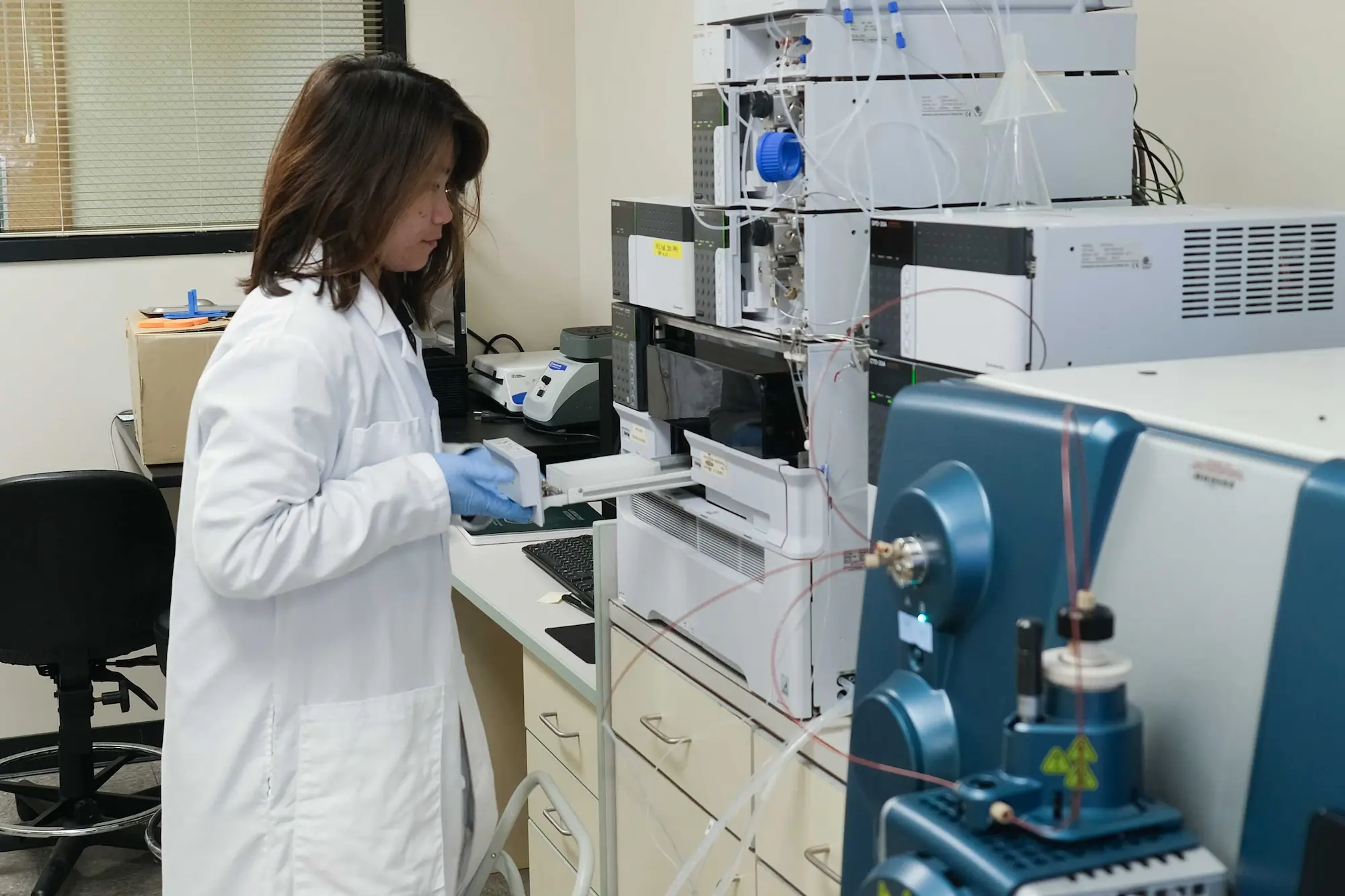
A scientist works with lab equipment

Overview

The Sanford Burnham Prebys Functional Genomics and Viral Vector Shared Resources provide comprehensive infrastructure and expertise for high-throughput genomic screening and gene delivery. The facility supports cell-based gain-of-function (ORF, CRISPRa/CRISPRon) and loss-of-function (siRNA, miRNA, shRNA, CRISPR-KO, CRISPRi/CRISPRoff) studies, encompassing assay development, reporter cell line engineering, large-scale genome-wide or pathway-specific library screening (arrayed or pooled formats), and validation of identified targets. Advanced capabilities include CRISPR gene editing, activation, and repression, as well as nucleotide base editing and tiling approaches to interrogate gene function, protein domains, and drug-target interactions.

High-throughput automation enables DNA purification, plasmid quantification, virus production, and custom assays in 96- and 384-well formats using our NIH S10-funded Biomek i7 Hybrid Automated Workstation (OD036254). Complementing these capabilities, the Viral Vector Facility offers a broad range of gene delivery platforms, including lentivirus, retrovirus, adenovirus, adeno-associated virus, and other viral systems, along with customizable vector design services. The core produces high-titer viral particles for in vitro and in vivo applications and provides over 50 ready-to-use reporter viruses for imaging and 2D/3D assays. Additional expertise includes non-viral nanoparticle technologies, such as extracellular vesicle and exosome purification, analysis, and engineering.

Together, these integrated resources enable scalable functional genomics studies and advanced gene delivery approaches to accelerate discovery of gene function, mechanisms, and therapeutic targets.


Services

  1. siRNA library preparation and arrayed screen
    The core has genome-wide human siRNA library ON TARGET-Plus (OTP) library (18,301 genes, Dharmacon (Dh) / Thermo Scientific (TS)), formatted as SMARTpools and 4 single siRNAs per gene. Seventeen collections of pre-spotted pathway-focused libraries include (1) druggable genome (7740 genes, Applied-Biosystems (AB) / Life Technologies (LT)); (2) epigenetics / transcriptional-regulators (1362 genes, AB/LT); (3) epigenetics (882 genes, created by SBP in-house); (4) ubiquitinome (1170 genes, Qiagen and 1432 genes, SBP); (5) kinome (704 genes, AB/LT and 831 genes, SBP); (6) cancer pathway (252 genes, Dh/TS); (7) phosphatases (231 genes, Dh/TS and 314 genes, SBP); (8) G protein coupled-receptor (GPCR) (Dh/TS); (9) metabolome (160 genes, AB/LT); (10) miniscreen random subset (Dh/TS); (11) autophagy (1601 genes, SBP); (12) innate immunity (96 genes, SBP); (13) B cell activation (127 genes, SBP); (14) T cell activation (287 genes, SBP); (15) ion channel (380 genes, SBP); (16) apoptosis (823 genes, SBP). Customized siRNA cherry-pick service is available upon request. In addition, CRISPR/Cas9 arrayed screening with (17) kinome gRNA library (789 genes, TS) is available.
  2. miRNA mimic and antagonist library arrayed screen
    The core has human miRNA mimic library (875 miRNAs from miRBase sequence database Version 10.1., AB/LT) and miRVana library (2565 miRNAs, TS). Libraries of miRNA antagonist include human miRIDIAN miRNA inhibitor (885 anti-miRNAs from miRBase 13.0 release, Dh/TS), Exiqon miRCURY LNA™ Inhibitor and microRNA Family Inhibitor (Qiagen). Collections of pre-spotted autophagy pathway-focused libraries include miR mimics (259 mimics, SBP) against autophagy-related genes and miR antagonists (255 antagonists, SBP) targeting autophagy-related miRNAs.
  3. Lentiviral shRNA and open reading frame (ORF) overexpression library arrayed screen
    The core has RNAi Consortium (TRC) lentiviral kinome shRNA library (SIGMA) covering 672 kinases and regulators with an average of 11 shRNAs per target. 2440 sequence verified ORFs from the Ultimate ORF Collection (LT) are available as sub-libraries including secreted protein (858 ORFs), apoptosis (476 ORFs), kinase (373 ORFs), and innate immunity (172 ORFs).
  4. CRISPR/Cas genome-wide / sub- library preparation and arrayed/pooled screen
    The Core has acquired a portion of the CRISPRa T. gonfio library, including kinase, phosphatase, and drug target genes (2,012 genes), from Dr. Aguzzi’s group at UZH and has generated arrayed bacterial, plasmid DNA, and lentiviral libraries. Additionally, the core has obtained CRISPR lentiviral pooled libraries from Addgene, including two human genome-wide (GW) knock-out (KO) (Root and Doench, Zhang labs), one mouse GW KO (Teichmann lab), three human GW activation (Root and Doench, Weissman, Zhang labs), and two human GW inhibition libraries (Root and Doench, Weissman labs). Human pathway-focused pooled libraries contain (1) CRISPR/Cas9 KO sub-libraries of kinases; cell cycle proteins; nuclear proteins; unknown function; control targets (Sabatini and Lander lab) and (2) CRISPR/dCas9 inhibition sub-libraries of kinases, phosphatases, and drug targets; stress and proteostasis; mitochondria, trafficking, and motility; gene expression; membrane proteins (Weissman lab) as well as (3) SBP unpublished CRISPR/Cas9&Cas12 KO sub-libraries of kinome; epigenetics; ubiquitome; autophagy; phosphatase; ion channel; cancer pathway; innate immunity developed in-house. Furthermore, the core offers custom pooled single, dual (Cas9), triple (Cas12a), and quadruple (3 Cas12 + 1 Cas9 or 4 Cas12) gRNA(s) library construction services.
  5. Custom CRISPR/Cas gene disruption and editing
    The core provides custom creation of isogenic cell lines including gene knock-out, mutation and repair, as well as reporter insertion. Approaches to express CRISPR include lentiviral transduction and transient transfection with in vitro transcribed (IVT) RNA. Moreover, custom genetic modified mouse creation is accomplished via Rosa26 loci knock-in on C57BL/6 C2 and 129 mESCs.
  6. High-activity CRISPR/Cas stable cell line enrichment
    To avoid clonal line screening, the core has developed multiple fluorescent reporters capable of bulk-sorting to enrich lentiviral-transduced cells with high-efficiency SpCas9 and EnAsCas12a for CRISPRko; SpnCas9-ABE, CBE and A&CBE for CRISPR DNA adenine (A) and cytosine (C) base-editing (BE); dCas9-effectors for CRISPRon/off.
  1. Custom gene knock-down and overexpression via CRISPR inhibition, activation, on and off
    Repurposing CRISPR/Cas9 for promoter/enhancer regulation allows any gene and particularly long non-coding RNA (lncRNA) to be (1) inhibited through CRISPRi-dCas9-Krab (Weissman lab) or CRISPRoff-dCas9-Krab-DNMT3 (Gilbert lab) and (2) activated via three available CRISPRa-dCas9-VP64 systems, including SAM (Zhang lab), suntag (Weissman lab), and split fluorescent protein (Huang lab) as well as CRISPRon-TETv4 (Gilbert lab).
  1. NGS-ready amplicon library preparation and quality control
    The core is equipped with Dual Index Primers Set (NEB) which is compatible with Illumina NGS platform and capable of multiplexing up to 96 pooled amplicon libraries. Multiple quality control procedures are integrated to ensure the library complexity and quality during the amplification.
  1. Automated mini-scale production
    The robotic high throughput mini-scale services include clonal cell line creation, mammalian genomic DNA extraction, plasmid DNA isolation, DNA quantification and normalization, as well as lenti-/retro-viral production in 96-well plate format.
  1. Cell population barcoding
    Retrievable barcode technology such as CloneTracer barcoding lentiviral pooled library (Stegmeier lab) is available to create individually barcoded cell populations for linage tracing. Monitoring the unique barcode and number of individual barcode provides a quantitative analysis to study the clonal origin and heterogeneity in cell proliferation, differentiation, as well as resistance to the drug treatment
  1. Viral Vectors-based gene delivery technology

Equipment & Resources

See Services for a list of libraries

  • Benchcell work station with Bravo liquid handling platform (Agilent)
  • STAR liquid handling station (Hamilton)
  • Tissue culture facility
  • WellMate liquid dispenser (Matrix Technologies)
  • Micro Flo Liquid Dispenser (BioTek)
  • ELx 405 Plate washer (BioTek)
  • High-Throughput Microscopes and Plate Readers are utilized via the High Throughput Screening and High Content Screening
  • Biomek i7 Hybrid Automated Workstation (Beckman Coulter), funded by S10 OD036254
  • CloneSelect Imager (Molecular Devices)

Price List

For a Price List, please call (858) 646-3100 ext. 3537 or email us.


Leadership

Ani Deshpande, PhD
Scientific Director

Chun-Teng Huang
Associate Director
cthuang@sbpdiscovery.org

Contact

Please call (858) 646-3100 ext. 4353 or use the button below to send us an email.
Contact the Facility

Cryo-EM

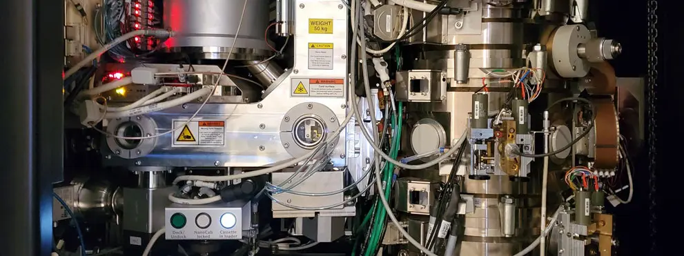
cryo-em microscope

Overview

The newly revamped Cryo-EM core facility located at Sanford Burnham Prebys in La Jolla, California, San Diego area, offers cryo-EM services and instrumentation to both academic and commercial users. State-of-the-art instruments include a ThermoFisher Titan Krios with a Gatan K3 camera, a Tecnai T12 transmission electron microscope, as well as a Vitrobot Mark IV for sample vitrification and preparation. The facility is currently optimized for single particle analysis (SPA) workflows and offers cryo-EM solutions for basic research as well as drug discovery in collaboration with the Conrad Prebys Center at Sanford Burnham Prebys.

Please contact us if you are interested in learning more about our services!

Contact the Facility


Services

  • Full imaging services
    You supply the sample and relevant information. Experienced facility staff will prepare negative stain or vitrified samples, carry out screening, and collect data. Image processing can be done by you or our facility staff.
  • Instrument access only
    You are trained on proper instrument procedures. After that, you can schedule time on the different instruments independently (excluding the Krios).
  • Flexible services
    For example, you can use our sample vitrification system (Vitrobot Mark IV) to prepare cryo-EM sample grids, then facility staff carries out data collection. 


Please contact us to discuss options.

Contact the Facility


Equipment & Resources

Microscopes

  • Titan Krios with Gatan K3 direct electron detector
    This 300kV cryo-electron microscope with a 3-condenser lens system is primarily used to collect high-resolution SPA data.
  • Tecnai T12 with Eagle 4K CCD
    This 120kV electron microscope is primarily used for imaging negative stained samples. This is an important step to get samples ready for Cryo-EM or can sometimes be used as the final imaging result.

Sample Preparation

  • Vitrobot Mark IV
    The Vitrobot is a semi-automated vitrification system for Cryo-EM samples. It’s control of process parameters like humidity and temperature can help with preparing reproducible sample grids.

Price List

Full Imaging Service
(facility staff does the imaging etc. for you)
External
Non-Profit
Commercial
Krios data collection per 24 hours$1,462.05$2,848.29
Krios screening per hour$132.36$257.85
T12 imaging per hour$120.15$234.07
Sample grid preparation using Vitrobot per hour$103.95$202.51
neg. staining grid preparation per hour$85.73$167.01
Usage Rates
(user does the imaging etc. themselves)
External
Non-Profit
Commercial
Krios per 24 hours$1,140.58$2,222.02
T12 per hour$34.43$67.07
Vitrobot per hour$18.23$35.51
Consultation per hour$87.73$167.01
Training per hour$128.59$250.51
Suppliesat costat cost

Contact the Facility


Leadership

Jianhua Zhao, PhD
Scientific Director

Laura Koepping, MS
Facility Manager
lkoepping@sbpdiscovery.org

Contact

Please call Laura Koepping at (858) 646-3100 ext 5097 or use the button below to send an email.

Contact the Facility

Protein Analysis

protein structure

Overview

The Protein Analysis Core provides a variety of analytical services focused on biophysical characterization of structural and functional properties of proteins in solution, under native, non-denaturing conditions. We perform quality control of protein samples (folding, stability, aggregation); measure molecular weight of proteins, protein complexes, oligomers and assemblies; characterize protein conformation and shape in solution; determine oligomeric state of protein (including stoichiometry and Kd for self-association) and measure protein binding to proteins, peptides, small molecules, compounds, metals, nucleotides and other ligands (including determination of equilibrium (Kd) and kinetic rate (kon, koff) constants, stoichiometry, binding enthalpy and entropy).


Services

  • Analytical ultracentrifugation (AUC)
  • Differential scanning calorimetry (DSC)
  • Grating-Coupled Interferometry (GCI)
  • Isothermal titration calorimetry (ITC)
  • Microscale Thermophoresis (MST)

Equipment & Resources

  • Analytical ultracentrifuge, ProteomeLab™ XL-I (BeckmanCoulter)
    In AUC experiment, the effects of G-force on macromolecules (proteins, protein complexes and assemblies, organelles, nanoparticles, etc) are monitored in real time via two optical detection systems. Depending on centrifugation speed and molecular weight, macromolecules either form a concentration gradient in the AUC cell, or slowly sediment towards the bottom of the cell. Applications of AUC include molecular weight measurement for proteins and protein complexes; protein to protein binding including Kd determination for self- and hetero-association, characterization of protein shape, oligomerization, aggregation and heterogeneity of protein samples.
  • Differential Scanning Calorimeter, VP Capillary DSC (Microcal/Malvern)
    In DSC experiments, thermal unfolding of proteins is monitored via changes in the heat capacity. These changes are believed to originate from the disruption of the forces stabilizing native protein structure (such as van der Waals, hydrophobic and electrostatic interactions, hydrogen bonds) and the hydration of the exposed residues. Therefore, protein denaturation profiles and thermodynamic parameters obtained from DSC experiments, such as calorimetric enthalpy (H) and denaturation temperature (Tm) are sensitive to the structural state of proteins. Applications of DSC include evaluation of protein folding and effects of mutations and modifications on protein structural properties, optimization of buffer formulations for protein storage and experiments, quality control of protein samples, detection of protein binding to compounds, other proteins, peptides, ligands, etc.
  • Grating-Coupled Interferometer, WAVEdelta (Creoptix/Malvern)
    Newest microfluidic method, similar to SPR (e.g. Biacore) based on GCI technology. The protein is immobilized on the chip, which may affect protein properties or its interaction with ligands. However, advantages of GCI include high sensitivity, small amount of protein required, broad detection range and ability to measure ON and OFF kinetic rates and Kd for a wide range of ligands.
  • Isothermal titration calorimeter, Affinity ITC (TA Instruments/Waters)
    ITC is used to measure affinity for interactions of proteins with other proteins, peptides, lipids, nucleotides, saccharides, metal ions, inhibitors, compounds and other ligands. In ITC experiment aliquots of a titrant are injected into the cell containing protein solution. Upon each titration the amount of heat released or absorbed is measured. In addition to the equilibrium dissociation constant (Kd), number of binding sites, binding enthalpy and entropy are determined from each titration. ITC is a truly equilibrium solution method where Kd is measured for native proteins. No labeling or immobilization is required.
  • Microscale Thermophoresis Monolith NT.115 (NanoTemprer Technologies)
    The Monolith NT.115 allows measuring every interaction type, ranging from the formation of huge protein complexes to the binding of single metal ions. As there are no prerequisites with respect to the buffer composition, even such complex liquids as serum or lysates can be used for interaction studies. The instrument is equipped with one Nano detector that has two different (blue and red) fluorescence channels.

Price List

For a complete list of services, please call (858) 646-1000 ext. 3515 or email us.

Protein Analysis ServicesInternal
Subsidized
InternalExternal
Non-Profit
External
For-Profit
ITC titrationeach$102.85$116.88$126.23$245.91
MST timeminute$0.37$0.43$0.46$0.89
DSC scaneach$71.50$81.25$87.75$170.95
AUC timeminute$0.11$0.13$0.14$0.26
AUC sample handling, per celleach$60.50$68.75$74.25$144.65

Leadership

Andrey Bobkov, PhD
Facility Manager

Contact

Andrey A. Bobkov

Andrey A. Bobkov, Manager of Protein Analysis Facility, performs all analytical services. He has received a MS in Biochemistry from Moscow State University (Russia) and a PhD in Biochemistry from the Bach Institute of Biochemistry of Russian Academy of Sciences. He did his postdoctoral training at UCLA Chemistry and Biochemistry Dept. Andrey has more than 30 years of experience and over 60 publications in the field of Biophysical Analysis.

Please call (858) 646-3100 ext. 3515 or use the button below to send us an email.

Contact the Facility

Stem Cells

close-up of stem cells

Overview

The Institutional Stem Cell Core has been discontinued, with the functions of the former Core now split into two separate operations. 

  • iPSCs: The generation and characterization of induced Pluripotent Stem Cells (iPSCs) is now being performed on a collaborative basis for both internal and external investigators with the Snyder lab. For information regarding iPSC projects, contact Dr. Evan Snyder.
  • Shared Laboratory: A well-equipped shared laboratory dedicated to the culture and analysis of stem cells is available to SBP investigators. This shared stem cell lab is managed by Chun-Teng Huang, who also manages the adjacent Viral Vectors Core.

Leadership

Evan Snyder, MD, PhD
Professor and Director
Center for Stem Cells and Regenerative Medicine

Chun-Teng Huang
Associate Director
Viral Vectors
cthuang@sbpdiscovery.org

Contact

Please use the button below to send us an email.
Contact the Facility

Histology

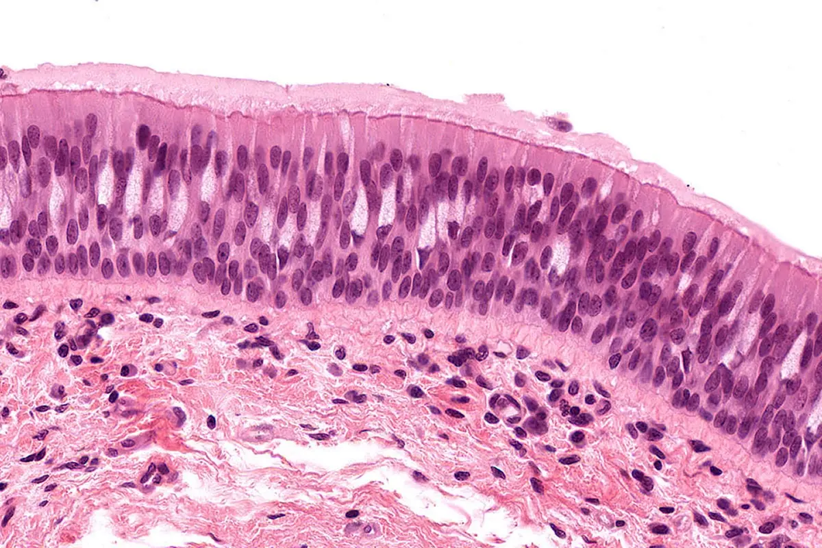
cellular image

Overview

The Histology Core Facility provides standard and customized research-specific services, including tissue processing, fixed sectioning, and frozen cryosectioning. We offer routine and special histological stains, as well as advanced molecular labeling via immunohistochemistry (IHC), immunofluorescence (IF), and RNA-ISH. Finished slides are returned directly to researchers or digitized using high-throughput brightfield and fluorescent Aperio ScanScopes, which offer remote web access and powerful quantitative image analysis tools.

Additionally, we provide expert training and open access to common-use equipment (paraffin microtomes, cryostats, cytospins). Our staff offers specialized hands-on training for basic histological techniques, including user training for IHC assay optimization.


Services

Conventional Histology

  • Aperio high-resolution slide scanning and optional image analysis for bright field and fluorescence samples
  • Routine and custom sectioning
  • Paraffin and frozen samples
  • Standard and special stain (H&E, PAS, Oil Red O , trichome, PSR and more.

Immunohistochemistry

  • Double labeling of antibodies and RNA probes
  • Immunofluorescence
  • Development of custom protocols (overlay assays, competition assays)
  • Titration and staining of commercial and custom antibodies

Equipment & Resources

  • Leica BOND-RX: Fully automated IHC, IF, and ISH staining.
  • Leica Autostainer XL: Automated H&E and special stains.
  • Tissue-Tek VIP 5: Automated paraffin tissue processing.
  • Aperio AT2 ScanScope: High-throughput brightfield slide scanning.
  • Aperio FL ScanScope: Fluorescent slide scanning with remote access.
  • CellCut Laser Capture Microscope: Laser-guided single-cell isolation.
  • Leica CM 3050S Cryostat: High-quality frozen tissue sectioning down to -40°C.
  • Leica Paraffin Microtomes: High-precision sectioning of wax blocks.
  • Tissue-Tek Embedding Station: Paraffin embedding and block preparation.
  • Leica HistoCore PERMA: Automated high-resolution slide labeling.
  • Shandon Cytospin 3: Centrifugal preparation of liquid cytology samples.
  • ImageScope Software: Digital slide viewing and quantitative analysis.

Price List

For a complete list of services, please call (858) 646-3100 ext. 3552 or email us.

Histology ServicesInternal
Subsidized
InternalExternal
Non-Profit
External
For-Profit
Small Combo/Embed/H&E/5unstainedeach$30$37.50$40.50$78.90
Large Combo/Embed/H&E/10unstainedeach$55$68.75$74.25$144.65
Additional Sections/Paraffinper slide$5.50$6.88$7.43$14.47
Embedding Only/Paraffineach$6.50$8.13$8.78$14.47
Immuno Staining (IHC or IF)per slide$35$43.75$47.25$92.05
Aperio 20X scanper slide$12$15$16.20$31.56
Aperio 40X scanper slide$23$28.75$31.05$60.49
Aperio Fluorescence Scanper slide$30$37.50$40.50$78.90
Common Cryostatper hour$22.20$27.60$30$58.20

Contact

Guillermina (Gia) Garcia is an American Society for Clinical Pathology (ASCP) Certified Histotechnician with 20 years of laboratory experience. She manages the day-to-day handling and processing of research samples at the facility. Gia is highly skilled in a wide range of specialized histological staining methods and immunohistochemistry (IHC) protocols. She serves as a primary contact for investigators and is available to answer any questions regarding your active or upcoming research projects.

Please call (858) 646-3100 ext. 3556 or use the button below to send us an email.
Contact the Facility

Cellular Imaging

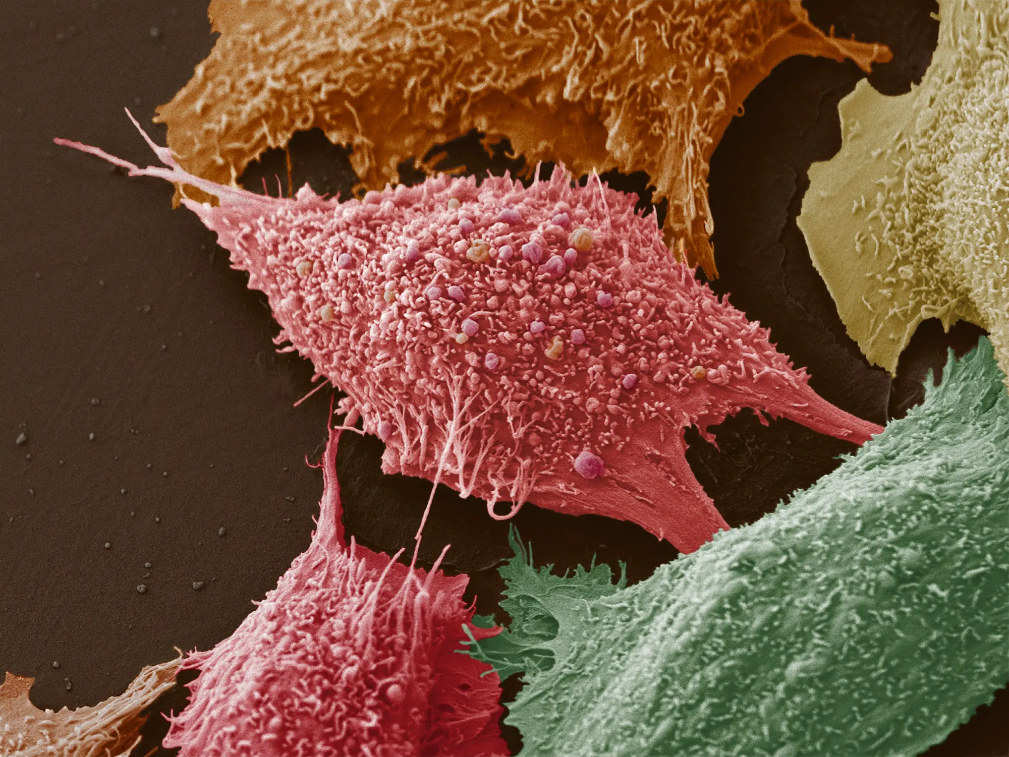
cancer cells

Overview

The Cellular Imaging Facility broadly supports research programs by providing access to sophisticated microscopes for digital imaging, as well as training, assistance and guidance. The core facility offers expertise, training and assistance in advanced biological microscopic imaging techniques and use of complex image processing software, use of well-maintained, aligned, and calibrated microscopic equipment, and troubleshooting of equipment and experimental problems.


Services

  • Light Sheet Microscopy
  • Super Resolution microscopy
  • Single and multiphoton confocal microscopy
  • Live cell imaging
  • Training and consultation
  • Wide field microscopy:
    • Bright field and dark field microscopy
    • Phase and Nomarski differential interference contrast
    • Polarization and epi-fluorescent microscopy
  • Image analysis:
    • Morphometry
    • 3D and 4D rendering
    • Deconvolution

Equipment & Resources

Advanced Microscopy

  • Milteny Blaze Light Sheet Ultra Microscope
    • Objectives: 1.1X, 4X, and 12X with tube lens zoom: 0.66, 1.0, 1.6, and 2.5. This covers the total magnification range of 0.66X up to 30X. 1X is (0.66–2.75x), 4X is (2.4–10x), and 12X is (7.2–30x).
    • 5 Lasers: 405nm, 488nm, 561nm, 639nm, and 785nm
    • Tor imaging multiple or very large samples with subcellular resolution
    • Transparent/Cleared 3D samples up to 4.4 cm × 7.0 cm × 1.7 cm (x,y,z), organoids, tissue sections, biopsies, tumors, whole organs, embryos, larvae
    • 3D-IF antibodies validated for staining large samples, the MACS® Clearing Kit for tissue clearing, easy-to-follow immunostaining and tissue clearing protocols
    • Smart software solutions, from data acquisition to image processing. Data can be opened and analyzed in MACS® iQ View – 3D Large Volume, Fiji/ImageJ, and Imaris software packages

Confocal Microscopy

  • Zeiss LSM 980 AiryScan Microscope System
    The Zeiss LSM 980 Airyscan2 microscope is optimized for spectral multiplexing by simultaneous spectral detection of multiple labels. Adapted illumination and detection schemes allows imaging of most challenging three-dimensional samples with high framerates and beyond the diffraction limit, while still being gentle to sensitive samples
    • 4-line laser launch with 405nm, 488nm, 561nm, and 640nm lasers.
    • light-efficient beam path with up to 36 simultaneous channels and full spectral flexibility from UV into the near infrared (NIR) range
    • 34-channel spectral detector comprised of 1 UV + 32 Visible + 1 Far-red detectors and the Airyscan2 super-resolution detector system
    • AirtScan2 part:
      • 32 detector elements allowing for super resolution quantitative results
      • optimized for spectral multiplexing
      • shorter acquisition times (up to 10x faster)
      • capture of larger view fields or dynamic processes
    • Incubation chamber for live imaging (Co2/Temp/Humidity)
    • Fully automated XY stage with piezo-Z stage for fast scans of slides, dishes, and well plates
  • Nikon N-SIM E Super-Resolution / A1 ER Confocal Microscope System
    • Structured illumination super-resolution microscope combined with enhanced resolution point scanning confocal
    • Nikon’s N-SIM E is a super-resolution system that provides double the resolution of conventional optical microscopes. Combining N-SIM E and the A1 ER confocal microscope allows you the flexibility to select a location in the confocal image, and easily switch to view it in super-resolution, enabling the acquisition of more detail
    • The new system has 10X, 20X, 40X oil, 60X oil, and Super-Res 100X oil objective lenses. N-SIM-E laser lines include 488nm, 561nm, and 640nm. The A1R Hybrid confocal has a 6-line laser launch with 405nm, 445/488/515nm, 561nm, and 640nm lasers
  • LSM 710 NLO Zeiss Multiphoton Laser Point Scanning  Confocal Microscope
    • Four Single Photon Lasers are generating six excitation lines: Ar-ion gas laser produces 457, 488, 514 nm lines and three solid state lasers produce 561, 594 & 633 nm
    • Multi-photon Mai-Tai Laser HB- DeepSee complex system (690-1024nm) dedicated for excitation of multiple fluorophores, including UV excited dyes, such as DAPI
    • CO2 & Thermo Controlled Time lapse system
    • The Microscope operates by ZEN-2011 acquisition/processing package
  • Fluoview-1000 Olympus Laser Point Scanning Confocal Microscope
    • Four Single Photon Lasers provide six excitation lines: diode laser emits 405nm line, Ar-ion laser produces 457, 488, 514 nm, DPSS laser – 561 nm and He-Ne laser – 633 nm
    • Dual SIM Scanner for simultaneous stimulation & registration fluorophores
    • Olympus software provides modules for Spectral Deconvolution; FRAP, FRET and Photo-activation applications
    • CO2 & Thermo Controlled Time lapse system
  • Yokogawa Spinning Disk Laser Confocal Microscope
    • XY and Piezo-Z object stage (ASI 2000-Piezo)
    • Single Photon Kr-Ar laser excites 488, 568 and 647 nm (Melles Griot)
    • Cooled Monochrome CCD Quant EM Camera (Photometrics)
    • MetaMorph (Mol. Devices) software (v.7.7.7)
    • CO2 & Thermo Controlled Time lapse system

Wide Field Microscopy

  • EVOS® FL Auto Imaging System
    • Time-lapse imaging, Image stitching, and Automated cell counting
    • Environmental chamber enabling precise control of temperature and humidity Automated X/Y scanning stage with interchangeable vessel holders for slides, multiwell plates, 35 mm dishes, 60 mm petri dishes, and T-25 flasks
    • Dual cameras: Monochrome: high-sensitivity interline CCD; Color: high-sensitivity CMOS; 3.1 megapixels
    • 4X, 10X, 20X, and 40X air objectives as well as a 60X oil lens
    • Epifluorescence and transmitted light (bright-field and phase-contrast)
    • 7 light cubes (DAPI, GFP, RFP, Texas Red, Cy5, Cy5.5, Cy7) for a wide range of fluorescent antibodies
  • Inverted IX81 Olympus Wide Field and Fluorescence Microscope (FRET)
    • DG4 UV source galvanometer equipped with 300W Xenon lamp, capable for sequential acquisition of 4 excitation channels & 10 position fast (30msec) Emission Filter Wheel (Sutter)
    • FRET & Ca2+ Imaging, DIC
    • XY-linear encoded objective stage ASI 2000(ASI Inc.); Z-IX81 step-motor
    • ZDC ( Zero Drift focus Control, Olympus)) attachment 
    • Cooled Monochrome CCD Cascade 512B FT Camera. (Photometrics Inc.)
    • MetaMorph software, v. 7.7.7 (Mol. Devices)
    • CO2 & Thermo Controlled Time lapse system
  • Inverted IX81 Olympus Wide Field and Fluorescence Microscope (TIRF)
    • Excitation illumination system presented by combination of X-cite 120 Metal halide UV source (EXFO) for the epi-fluorescent microscopy and single photon lasers combiner containing three solid state lasers (488, 561 and 640 nm)
    • XY and Piezo-Z object stage ASI 2000 (ASI Inc.)
    • Excitation & Emission Filter Wheels (Sutter)
    • Cooled, Monochrome CCD QuantEM Camera (Photometrics Inc.)
    • CO2 & Thermo Controlled Time lapse system
  • Inverted IX81 Olympus Wide Field and Fluorescence Microscope
    • XY-Martzhauser Tango objective stage (Martzhauser Inc.); Z-IX81 step-motor
    • Cooled, Color CCD SPOT RT-3 Camera (Diag.Instruments)
    • X-cite 120 Metal Halide UV source (EXFO)
    • Excitation & Emission Filter Wheels (Sutter)
    • CO2 & Thermo Controlled Time lapse system
  • Inverted TE300 Nikon Wide Field, Fluorescence Microscope #1
    • Multiple Fluorescence; Phase and Nomarski DIC contrast.
    • Cooled, Color CCD SPOT RT Camera (Diagnostic Instruments Inc.)
  • Inverted TE300 Nikon Wide Field, Fluorescence Microscope #2
    • Multiple Fluorescence; Nomarski (DIC) contrast
    • XYZ object stage ASI 2000 (ASI Inc.)
    • Cooled Color CCD SPOT RT Camera(Diagnostic Instruments Inc)
    • Thermo Controlled Time lapse system
  • Upright BX50 Olympus Wide Field, Fluorescence Microscope
    • Transmitted light and Triple Fluorescent imaging
    • Cooled Monochrome ORCA ER (Hi Res) camera (Hamamatsu)
  • Software packages
    • Image Pro Plus (Media Cybernetics)
    • Volocity 4D Rendering (Improvision, PerkinElmer)
    • MetaMorph, MetaFluor packages (Mol. Devices)
    • Spot RT Acquisition / Processing Software (Diagnostic Instruments Inc.)
    • ImageJ and IrfanView (free sources imaging packages)

Price List

For a Price List, please call (858) 795-5455 or email us.


Leadership

Maximiliano D’Angelo, PhD
Scientific Director

Leslie Boyd, BS
Facility Manager
lboyd@sbpdiscovery.org

Contact

Please call (858) 795-5455 or use the button below to send us an email.
Contact the Facility

Model Organisms

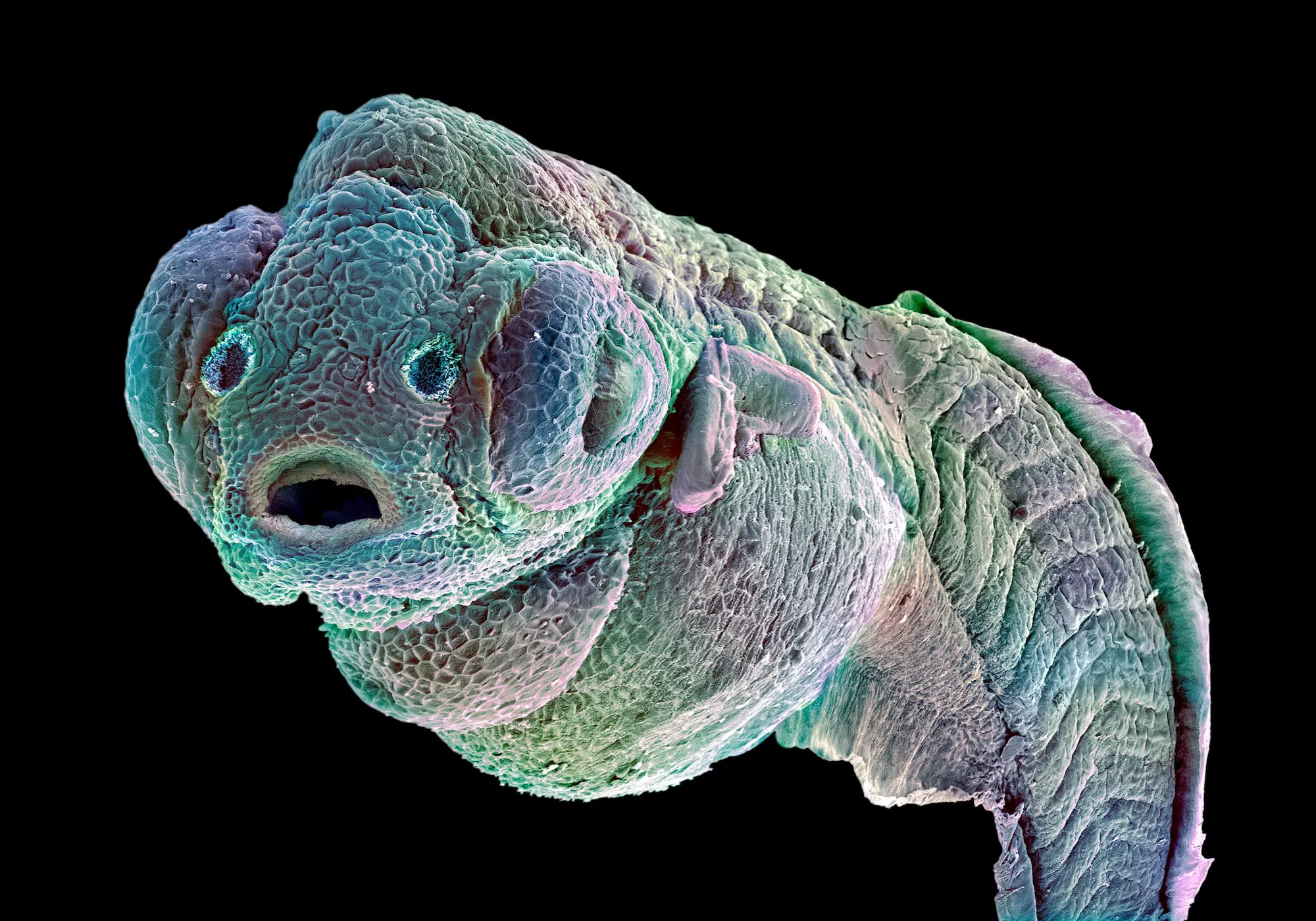
zebrafish larva

Overview

The Facility provides Drosophila melanogaster and Caenorhabditis elegans as model systems. The client interested in finding out if their gene or protein of interest has a phenotype can use the Facility to discuss their ideas and how to design the experiments. The Facility also provides training in how to manipulate the organisms, use equipment and provides technician assistance.


Services

  • Provides D. melanogaster and C. elegans as model systems
  • Consultation and assistance for design of experiments
  • Training to handle the organisms and how to use equipment
  • Technician assistance
  • Ordering of flies and worms (RNAi-lines, mutants etc)
  • Provides fly food
  • Provides long time storage of flies and worms

Equipment & Resources

  • Dissecting Microscope Leica MZ16F
  • Apotome Optical Sectioning
  • Time lapse Zeiss Imager .M1

Price List

For a Price List, please call (858) 646-3100 ext. 4186 or email us.


Leadership

Rolf Bodmer, PhD
Scientific Director
rolf@sbpdiscovery.org

Contact

Please call (858) 646-3100 ext. 4186 or use the button below to send us an email.
Contact the Facility

Animal Facility

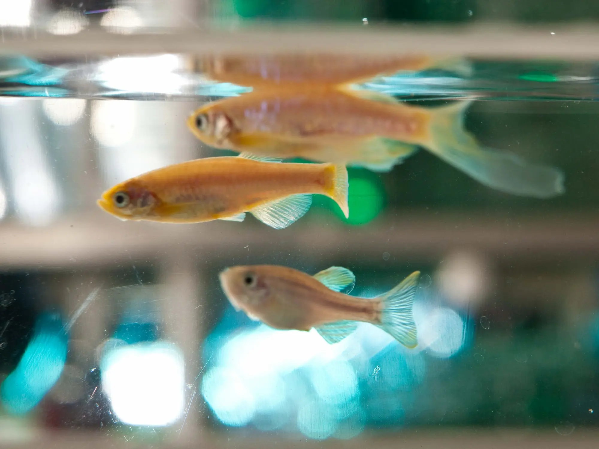
Zebrafish

Overview

The laboratory animal facility encompasses over 25,722 sq. ft. of dedicated animal space in several buildings on our La Jolla campus. The animal facility provides housing for specific pathogen free (SPF) rodents and Zebrafish. Trained animal care technicians provide expertise in animal husbandry, transgenic and knockout mouse breeding colony maintenance and assistance with routine technical procedures. 

Our animal research program is accredited by AAALAC International (the Association for Assessment and Accreditation of Laboratory Animal Care International. Participating in the AAALAC accreditation program and meeting their rigorous standards demonstrates our commitment to humane and responsible animal research and our dedication to good science.


Services

Boarding

Boarding animals for outside users may be approved by Sanford Burnham Prebys IACUC on a case-by-case basis. Animals must be shipped directly from an approved commercial breeder facility. Please call or email the Animal Facility Director at (858) 795-5419 email Kenny Venegas, to discuss your animal housing needs. Access to the animal facility may be restricted to Monday through Friday, 7:30am-3:30pm. Husbandry services include cage changing, cage sanitation, feeding standard diets, water bottle exchange, daily health checks. Breeding colony services are also included in the cage per diem rate, and include breeder set-up, weaning of litters, ear notch identification, tissue collection for genotyping, breeding colony database management and colony maintenance.

Technical Procedures

Trained animal technicians are available to assist researchers with routine technical procedures. Assistance with procedures must be scheduled in advance through the Training Manager, Buddy Charbono. Assistance is available Monday through Friday, 8:00am-3:00pm.

Technical Procedure Services Include:

  • Feeding of special diets and supplemental diets: (research lab must supply all special diets)
  • Drug administration and additives to water bottles: (research lab must supply water additives, including antibiotics, chemicals and drugs)
  • Cell and animal irradiations: schedule procedure using iLab
  • Oral gavage/dosing
  • Injections: SC, IP, IV, IM, ID
  • Weighing animals
  • Surgery pre- and post-op care: (surgery set-up, anesthesia, recovery monitoring, fluid administration)
  • Imaging and Analysis: (see the Animal Imaging & Analysis core for imaging instruments and services)
  • Blood collection: variable routes

Equipment & Resources

General Equipment

  • Oxycycler (BioSpherix, Ltd.)
    Atmosphere control system with full range oxygen control (0.1% to 99.9%) and/or CO2 control (0.1% to 20.0%) as well as other bioactive gas control such as CO, NO, and O3. Hypoxia stress can model components of many severe diseases such as heart attacks, strokes, asthma, epilepsy, sleep apnea, or pulmonary infections.
  • Anesthesia machines
  • Cage changing stations and biosafety hoods
  • Stoelting Stereotaxic instrument: frame and microinjection equipment: for performing injections into precise locations in the CNS.
  • Metabolism cages: Four individual cages for monitoring nutrient input and waste output.

Motor and Sensory Analysis

  • Hotplate Analgesia Meter (San Diego Instruments)
    The SDI Hotplate Analgesia Meter performs rapid, precise screening of the effects of narcotic type analgesic drugs (morphine, codeine, etc.) in test subjects.
  • Animal Treadmill (Columbus Instruments)
    Measure mouse locomotor coordination for fear of falling experiments. 6 lanes with shocker for mice.
  • 5 Station Rota-Rod Treadmill (Med Associates)
    ENV-575M USB-mouse. Measure motor coordination and fatigue.
  • Grip-strength Device

Price List

For a Price List, please call (858) 795-5419 or email us.


Leadership

Alessandra Sacco, PhD
Scientific Director

Kenny Venegas
Interim Director, Animal Resources
kvenegas@sbpdiscovery.org

Buddy Charbono
Compliance and Training Manager
bcharbono@sbpdiscovery.org

Contact

Please call (858) 795-5419 or use the button below to send us an email.
Contact the Facility

Animal Imaging and Analysis

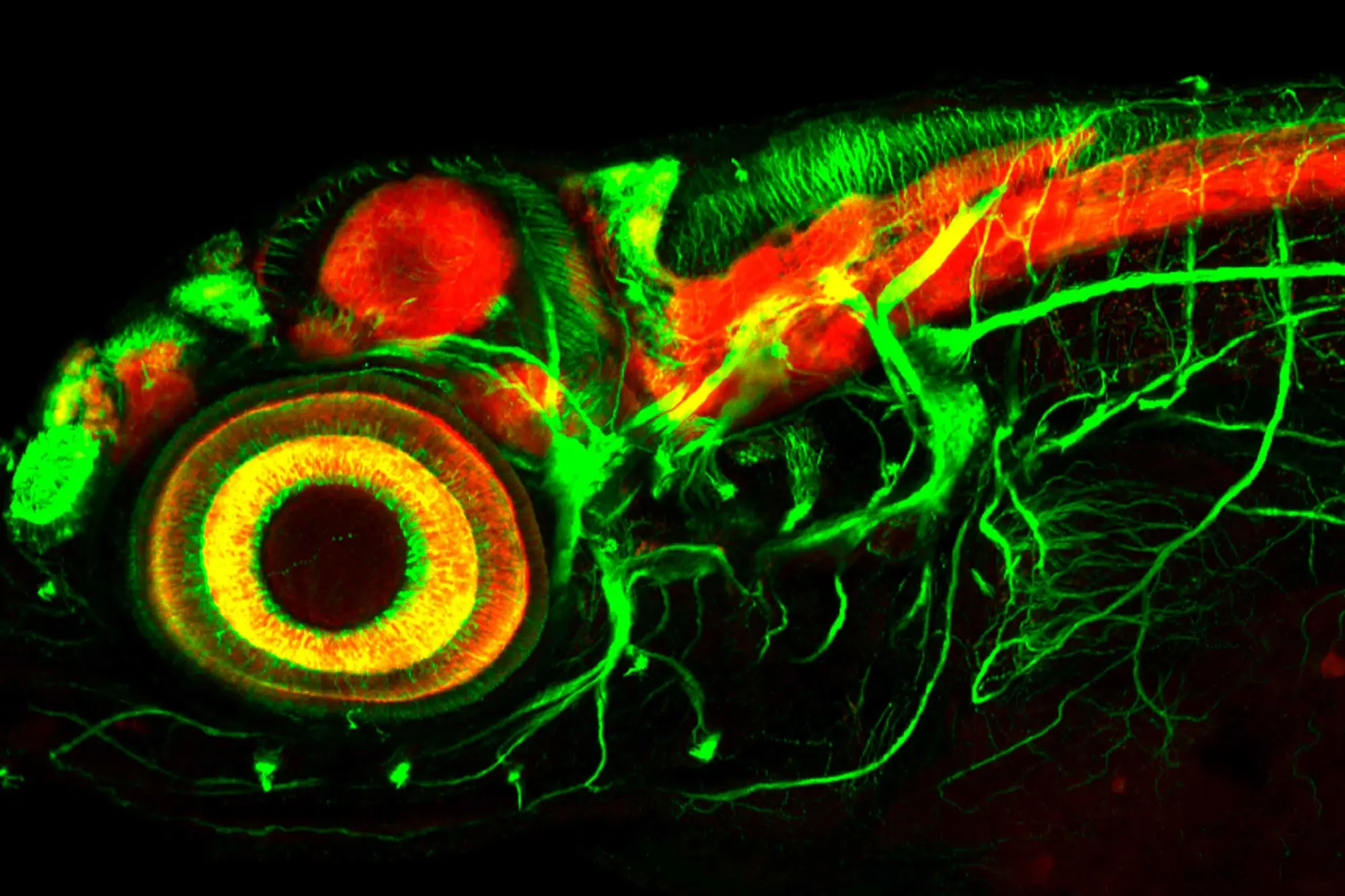
zebrafish larva

Overview

The animal imaging and analysis lab provides state-of-the-art imaging and analytical services for Sanford Burnham Prebys investigators. The facility can perform in vivo non-invasive luminescence and fluorescence imaging for xenograft tumor growth and metastasis studies. Analytical equipment supports complete blood cell counts (CBC) and analysis of serum components revealing metabolic or organ stress from small samples of mouse blood.


Services

Imaging services can be performed by Sanford Burnham Prebys animal facility specialists. Please schedule your procedures through iLab. We can also provide Luciferin for use with the Xenogen IVIS Spectrum Imaging system and Blood Analysis services, CBCs and blood chemistries.

Appropriate animal health information is needed for external investigators wishing to utilize the imaging facility.

Please contact the Facility Manager, Judy Wade, (858) 646-3100 ext. 3285, to inquire about specific tests that can be performed.


Equipment & Resources

Imaging Lab

  • IVIS Spectrum Imager (Xenogen)
    For non-invasive in vivo imaging of luminescence or fluorescence. Anesthesia system allows analysis of live mice. We use luciferase labeled tumor cells or transgenic reporter mice. We have 9 luciferase labeled cell lines, please contact Judy Wadefor their availability.
  • VisualSonics Vevo 770 (VisualSonics)
    High resolution in vivo ultrasound imaging system includes: Pulsed Wave Doppler, M-Mode, Contrast Software Mode, and 3D Acquisition and Visualization Package. Accessories: Ultrasound Guided Injection System, Anesthesia System and Advanced Physiological Monitoring System. The ultrasound is useful for visualization of embryos in early stage pregnancy; visualization of embryos for organ specific injection in utero; volume measurement of mouse mammary tumors for drug studies with fewer animals; volume measurements of mouse prostate tumors to evaluate tumor development without killing of the animals; potential to image pancreatic tumors; and with the potential for measuring tumor blood flow with contrast agents.
  • Pearl Imager (LI-COR)
    The Pearl Impulse is a near-infrared fluorescent imaging system for small animals. With the Pearl Impulse, researchers can visualize imaging agents in mice dynamically with real-time imaging. The rapid one-button acquisition simultaneously detects 700 nm and 800 nm channels. Please click the links below for more information.
  • Faxitron MX-20 (Faxitron X-ray Corporation)
    X-ray instrument for performing bone densitometry and body composition measurements using a dual-function digital and film imaging system.
  • Hologic Trident Specimen Radiography System (Hologic Inc)
    A mobile medical imaging platform designed to provide rapid, high-resolution X-ray verification of tissue samples directly inside the operating room or biopsy suite.

Nearby access to MRI, PET/SPECT and CT Imaging through the Molecular Imaging Center at Sanford Consortium.

Analysis lab

  • VetScan HM5 Hematology System (Zoetis): 22-parameter blood count & clinical chemistry analyzer
  • VetScan VS2 (Zoetis): blood chemistry, electrolytes, blood gas & immunoassay
  • IDEXX Catalyst One Chemistry: blood & urine 
  • CODA-6 (Kent Scientific): tail-cuff blood pressure system
  • Oxycycler (BioSpherix, Ltd.): for oxygen consumption, CO2 production & hypoxia studies
  • Stereotaxic instrument: microinjection equipment

Price List

For a Price List, please call (858) 646-3100 ext. 3262 or email us.


Leadership

Alessandra Sacco, PhD
Scientific Director

Judy Wade
Facility Manager
jwade@sbpdiscovery.org

Contact

Please call (858) 646-3100 ext. 3262 or use the button below to send us an email.
Contact the Facility

Flow Cytometry

Scientist works with lab equipment

Overview

The facility has four analytical flow cytometers (BD LSRFortessa and Fortessa X20, Agilent NovoCyte 3000, and a Cytek Aurora full spectrum analyzer) capable of analyzing over 40 colors, all equipped with plate loaders. A BD FACSAria cell sorters with up to 16-color capability is housed in a biosafety enclosures, as are the X20 and Aurora analyzers. The Core also has an imaging flow cytometer (Amnis ImageStreamX MarkII) with up to 10 colors. Cell sorting is performed by expert Core staff with analytical cytometry available either as a service or for use 24/7 by trained investigators. The Core also offers a validated 27-marker antibody panel for spectral profiling of the murine tumor immune microenvironment. FlowJo analysis software is available on workstations in the core and through an institutional site license.

Scientists planning a flow cytometry experiment are encouraged to consult facility staff for assistance with protocols, fluorochrome selection or other aspects of experiment design. Sorter and analyzer optical configurations can be viewed here.


Services

High Speed Cell Sorting

  • Sorting into tubes, 96 or 384-well plates for culture or single cell sequencing
  • Expertise in calcium flux, cell cycle, side population, and sorting of IPSC and ESC, tumor progenitor and tissue stem cells
  • Performed by expert facility personnel
  • Simultaneous sorting of up to 4 populations
  • BSL2 biosafety enclosure

Analytical Flow Cytometry

  • Consultation
  • Experiment design
  • Hardware and software training
  • Do-it-yourself 24hr/day, or assisted by appointment up to 30 colors with plate loader
  • Data analysis and interpretation
  • Publication quality figures
  • Assistance with methods sections
  • Pre-publication manuscript review

Validated Antibody Panels

  • Murine Tumor Immune Profile (mTIME) Panel
    • Core-developed 27-color spectral antibody panel for profiling the murine tumor immune microenvironment
    • 27 cell surface markers, titrated reagents, validated staining protocol
    • Resolves B cells, T cells (CD4, CD8, activation, Treg, inhibitory receptors), NK, NKT, pDC, cDC1 and cDC2, monocytes, transitional monocytes, macrophages (alternative activation)
    • Six spectral drop-in zones for project-specific customization
    • Compatible with cells expressing GFP, YFP, TdTomato, mCherry, or BFP
    • Available in 10-test increments complete with all necessary buffers.

Equipment & Resources

Cell Sorters

  • BD FACSAriaIIu 16-color high-speed cell sorter
    • Four solid state lasers (405 nm, 488 nm, 561 nm, 640 nm)
    • BSL2 containment
    • Sort into tubes, 96 or 384-well plates

Flow Cytometry Analyzers

  • Cytek Aurora full spectrum cytometer
    • Collects cellular fluorescence across the full visible spectrum
    • Allows for highly multiplexed panels with far more colors than a traditional cytometer
    • Autofluorescence can be unmixed to improve sensitivity for highly autofluorescent cells
  • Acea NovoCyte 3000 13-channel analytical flow cytometer
    • 488nm, 405 nm and 640 nm lasers
    • Multi format autosampler
  • BD LSR Fortessa X20 18-color analytical flow cytometer
    • Five solid state lasers (488nm, 637nm, 405nm, 561nm and 355nm)
    • BSL2 containment
    • HTS plate loader for 96 or 384-well plates
  • BD LSRFortessa 14-color analytical flow cytometer
    • HTS plate loader for 96 or 384-well plates
    • Four solid state lasers (405 nm, 488 nm, 561 nm, 640 nm)

Imaging Flow Cytometers

  • Amnis ImageStreamX MarkII imaging flow cytometer
    • 12 imaging channels (10 colors and 2 brightfield channels)
    • 405 nm, 488 nm and 642 nm laser lines
    • 20x, 40x and 60x objectives
    • 96-well plate loader
    • Extended Depth of Field (EDF) option for spot counting
    • Inspire acquisition software
    • Ideas analysis software

Computing

  • FlowJo site license
  • Computer workstations with large monitors and data analysis software
    • FlowJo
    • ModFit LT
    • SpectroFlo
    • IDEAS

Price List

For a complete list of services, please call (858) 646-3106 or email us.

Flow Cytometry ServicesInternal
Subsidized
InternalExternal
Non-Profit
External
For-Profit
FACSAria Sorting, 1st houreach$164$205$221$431
FACSAria Sorting, additional hourshour$99$124$134$260
ImageStreamX II + Staff Operatorhour$85$107$115$224
LSRFortessa Independent Usehour$35$44$47$92
LSRFortessa + Staff Operatorhour$96$120$130$252
Aurora + Staff Operatorhour$108$135$146$284
mTIME Panel10 tests$650$813$878$1710
Data Analysishour$84$105$113$221

Leadership

Maximiliano D’Angelo, PhD
Scientific Director

Yoav Altman
Facility Director
yoav@sbpdiscovery.org

Contact

The flow cytometry core staff has decades of experience in NCI-designated Cancer Center flow cytometry core labs.

Yoav Altman received his BA degree in Integrative Biology from the University of California at Berkeley. He started working with flow cytometry in 2001 at La Jolla Institute for Allergy and Immunology and joined Sanford Burnham Prebys Medical Discovery Institute in 2002 as Manager of the Flow Cytometry Shared Resource. He was promoted to Director in 2009, and oversees lab operation. Yoav has over 20 years of experience sorting a variety of cell types including IPSCs, hESCs, immune cells, disaggregated tumors and solid tissues such as cardiomyocytes, hepatocytes, intestinal epithelium, brain tissue, and skeletal muscle. His experience includes a variety of single cell assays including side population, cell cycle, apoptosis determination, high parameter immunophenotyping, calcium flux, mitochondrial membrane potential, ROS, fluorescent proteins, and FRET. He is also well versed in optimizing sorting protocols for single cell NGS sequencing using 10x or similar platforms. His current interests include imaging flow cytometry for detection and evaluation of extracellular vesicles and cell-cell interactions. Yoav is available to assist with reagent selection, experiment design, data analysis, protocol development, grant preparation, and writing methods sections for publications

Please call Yoav at (858) 646-3106 or use the button below to send us an email.
Contact the Facility