- Google Scholar Kevin Yip Google Scholar Profile
- H1 Connect F1000 Kevin Yip Faculty Profile
- Bioinformatics micro-modules Watch | Kevin Yip’s Channel for Learning Bioinformatics Concepts, Algorithms and Data Structures
Related Disease: Cancer
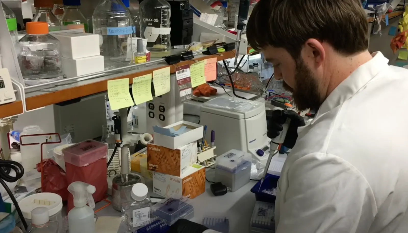
A career history of fundamental discovery and translational research in immunology has guided Dr. Ware to identify new drug targets and develop novel therapeutics. Dr. Ware’s career in immunology and virology began in 1982 when he became a Professor at the University of California, Riverside’s Division of Biomedical Sciences. In 1996, he joined the La Jolla Institute for Immunology in San Diego as Head of the Division of Molecular Immunology. Professor Ware joined Sanford Burnham Prebys Medical Discovery Institute in 2010, serving as the Director of the Infectious and Inflammatory Disease Center and Adjunct Professor of Biology at the University of California at San Diego. He is currently the Director of the Laboratory of Molecular Immunology, which focuses on discovering and designing immunotherapeutics.
As an educator, he taught medical students immunology and virology. He trained over 60 postdoctoral fellows and graduate students who chose careers in research in academic and pharmaceutical science, patent law, or teaching.
Dr. Ware advises scientific panels and review boards for the National Institutes of Health and serves on the scientific advisor boards for the Allen Institute for Immunology and the Arthritis National Research Foundation. Scientific advisor with several biotechnology and pharmaceutical companies on immunotherapy for cancer and autoimmune diseases using innovative approaches to target discovery and drug development.
Dr. Ware’s research program is dedicated to unraveling the intricate intercellular communication pathways that govern immune responses. His work, which centers on cytokines in the Tumor Necrosis Factor (TNF) Superfamily, particularly those that regulate cell survival and death in response to viral pathogens, spans the domains of cancer,autoimmune and infectious diseases.
At Sanford Burnham Prebys, Dr. Ware is pivotal in promoting the translation of the faculty’s scientific discoveries. His efforts have led to the Institute’s reputation as a productive and preferred partner in collaborations with Pharma, including multi-year research and drug development projects with Eli Lilly and Avalo Therapeutics. His success translating fundamental knowledge into rational drug design has led to three novel therapeutics targeting inflammatory pathways, currently in clinical trials.
Education
1981-1982: T cell Immunology, Dana-Farber Cancer Institute of Harvard Medical School in Boston, MA. Tim Springer and Jack Strominger, advisors.
1979-1981: Biochemistry of Complement, University of Texas Health Science Center, San Antonio, TX. W Kolb, advisor
1974-1979: PhD in Molecular Biology and Biochemistry from the University of California, Irvine; Gale Granger, PhD mentor.
Honors and Recognition
Distinguished Fellow, American Association of Immunologists
Honorary Lifetime Membership Award International Cytokine and Interferon Society
Hans J. Muller-Eberhardt Memorial Lecture
Biotech All Star, San Diego Padres Awar
“Pillars of Immunology” discovery of the Lymphotoxin-b Receptor, published in Science
Outstanding Alumnus, Ayala School of Biological Sciences, University of California, Irvine
National MERIT Award R37 (10 years), National Institute of Allergy and Infectious Disease, NIH
National Research Service Award, NIH Postdoctoral Fellowship
Related Disease
Arthritis, Breast Cancer, Cancer, Crohn’s Disease (Colitis), Infectious Diseases, Inflammatory Bowel Disease, Inflammatory/Autoimmune Disease, Inherited Disorders, Leukemia/Lymphoma, Myeloma, Pathogen Invasion, Psoriasis, Systemic Lupus Erythematosus, Type 1 Diabetes
Research in the Laboratory of Molecular Immunology is directed at defining the intercellular communication pathways controlling immune responses. Our work is focused on the Tumor Necrosis Factor (TNF)-related cytokines in regulating decisions of cell survival and death, especially in responses to viral pathogens. Translational research is redirecting the communication networks of TNF superfamily to alter the course of autoimmune and infectious disease and cancer.
Carl Ware’s Research Report
Discovery of the TNF-LIGHT-LTαβ Network
The molecular elements of this cellular communication network were revealed with our discovery of Lymphotoxin-β and its formation of the trimeric heterocomplex with LTαβ1 and its signaling receptor, Lymphotoxin-β Receptor2. The LTβR revealed a new inter-cellular communication pathway that provides a key mechanism underlying the development and homeostasis of lymphoid organs. A second ligand we discovered, LIGHT (TNFSF14), is a novel ligand for the herpesvirus entry mediator (HVEM; TNFRSF14), and surprisingly, the LTβR3 heralding the concept that TNF, LTαβ, and LIGHT form an integrated signaling network thru distinct receptors controlling inflammation and host defense.4
LTβR Signaling in Host Defense and Immune Evasion
Our investigations into the mechanisms of virus evasion of the immune system revealed an essential role of the LTβR pathway in regulating the type 1 interferon response to cytomegalovirus5 and now recognized as a major defense against other pathogens. LTβR signals differentiate macrophages and stromal cells into IFN-producing cells. LTβR transcriptomics and proteomics datasets we generated revealed a novel constellation of anti-viral host defense mechanisms6. Importantly the role of the LTβR pathway to alter tissue microenvironments by differentiation of specialized stromal cells has implications for promoting effective immune responses to cancer.
Discovery of the HVEM-BTLA Immune Checkpoint

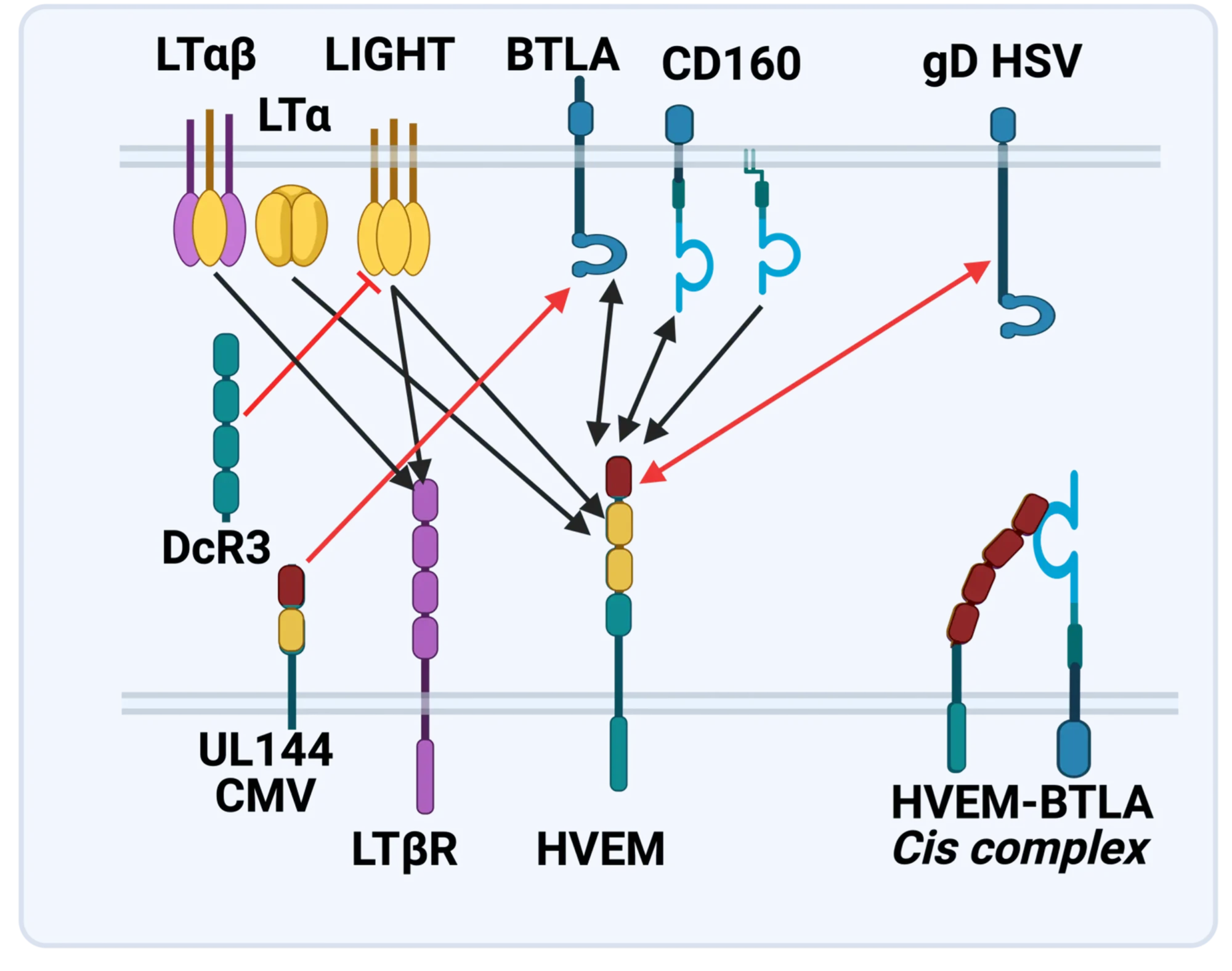
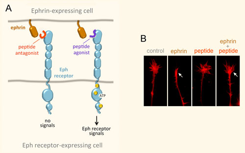
The LTBR-HVEM-BTLA Network in the TNF Superfamilies. Arrows indicate ligand-receptor binding (black), inhibitors (red arrow) and bidirectional signaling (dual arrowheads); HSV, herpes simplex virus; CMV, human cytomegalovirus. BTLA and CD160 are Ig Superfamily proteins. BTLA is an inhibitory checkpoint receptor; DcR3, decoy receptor 3 inhibits LIGHT binding to HVEM and LTBR.
The discovery that HVEM is a coreceptor for the immune checkpoint, B and T lymphocyte attenuator (BTLA), an Ig superfamily member, established a new paradigm in TNF Receptor signaling pathways 7. Additional investigations revealed the importance of the HVEM-BTLA system in limiting immune responses, including T cell help for B cell clonal expansion, antibody maturation, and secretion8. HVEM-BTLA also regulates control of the intestinal microbiome, limiting invasion of pathogenic bacteria and enhancing Treg cell homeostasis 9. The diverse roles of this pathway are seen in the loss of BTLA signaling from mutations in HVEM frequently present in B cell lymphomas10. Additional layers of immune regulators, CD160 and DcR3, control the LIGHT-HVEM-BTLA pathways, revealing this network as a key mechanism controlling immune homeostasis.
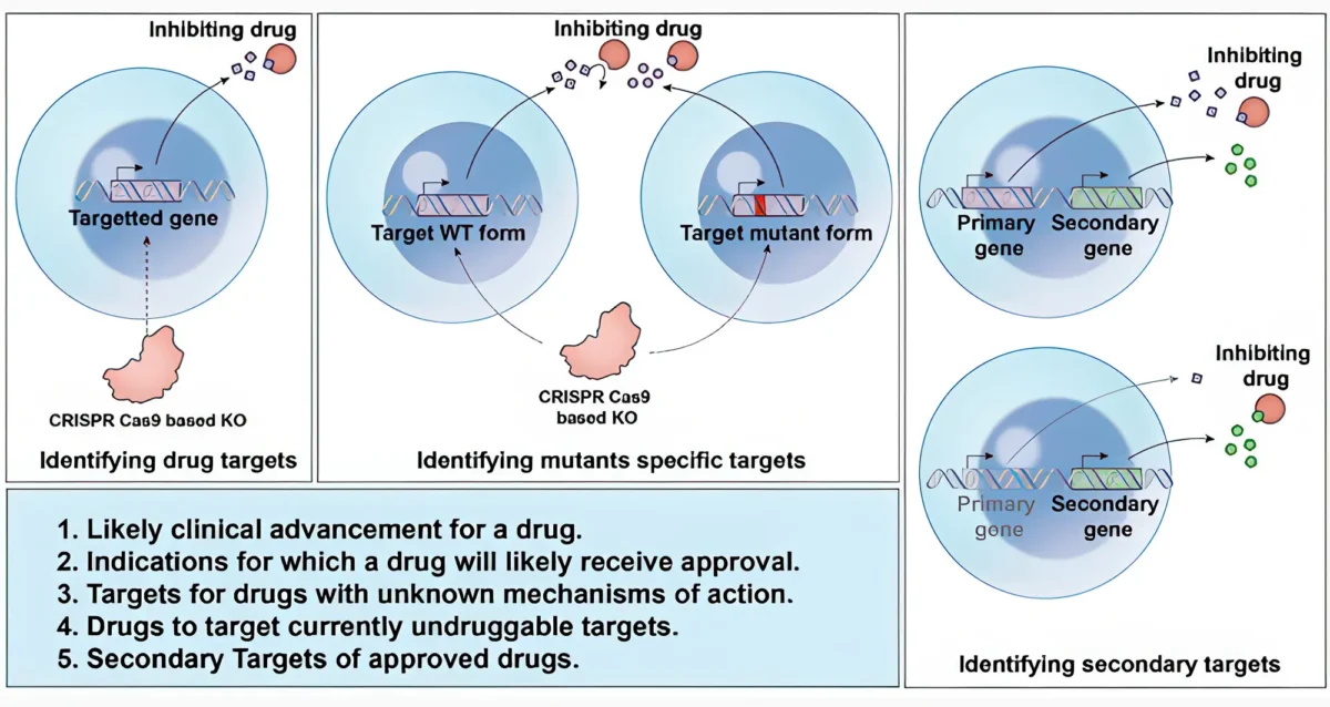
Appreciating the fundamental features of the TNF-LIGHT-LTab Network in effector and homeostasis mechanisms presents a target-rich resource for therapeutic intervention in autoimmunity, infection, and cancer11, 12.
Translational research and Immunotherapy
- 2021-current: Lead Scientific Advisor, Avalo Therapeutics
A neutralizing, fully human mAb (quisovalimab) to the proinflammatory cytokine LIGHT (TNFSF14) completed Phase I with an excellent safety profile and a Phase II trial establishing efficacy in COVID-19 pneumonia (NCT0441205)13. We identified elevated serum levels of LIGHT in hospitalized patients with COVID1914 spurring a randomized, double-blind, multicenter, proof-of-concept trial with adults hospitalized with COVID-19-associated pneumonia and mild to moderate ARDS15. The results established efficacy with a significant proportion of patients remaining alive and free of respiratory failure through day 28 after receiving quisovalimab, most pronounced in patients >60 years of age (76.5% vs. 47.1%, respectively; P = 0.042). These results and animal models validated LIGHT as a target for non-COVID inflammatory conditions, clinical trials ongoing in asthma (NCT05288504)12. - 2021-current: Principal Investigator, Avalo Therapeutics – Sanford Burnham Prebys collaboration
Bioengineered a first-in-class checkpoint agonist targeting BTLA immune checkpoint16 in preclinical development - 2019-current: Director and Principal Investigator, Fair Journey Biologics – Sanford Burnham Prebys collaboration
Immunotherapy for TNBC and PANC, in preclinical development - 2015-2022: Director and Lead Principal Investigator, LILLY-Sanford Burnham Prebys Collaboration in Autoimmunity
Collaborative research partnership with Eli Lilly involved target discovery and therapeutic development directed at immune regulators for autoimmune diseases. The collaboration produced three novel biologics currently in Phase I/2 trials (NCT03933943). The collaboration included a target discovery platform for T cell effector memory and NK cell immunomodulators. - 2015-2020: Lead Principal Investigator, Sanford Burnham Prebys – Capella Biosciences collaboration
Created a fully human mAb specific for membrane LIGHT (CBS001); phase I initiated (NCT05323110). - 2016-2020: Lead Scientific Investigator, Boehringer Ingelheim – Sanford Burnham Prebys Collaboration
Target discovery collaboration in inflammatory and fibrotic diseases6 - 2012-2014: Pfizer Innovation Center, Principal Investigator Bioengineering TNFR Superfamily in Autoimmune disease
- 1. Browning, J.L. et al. Lymphotoxin beta, a novel member of the TNF family that forms a heteromeric complex with lymphotoxin on the cell surface. Cell 72,847-856 (1993).
- 2. Crowe, P.D. et al. A lymphotoxin-beta-specific receptor. Science 264,707-710 (1994).
- 3. Mauri, D.N. et al. LIGHT, a new member of the TNF superfamily, and lymphotoxin alpha are ligands for herpesvirus entry mediator. Immunity 8,21-30 (1998).
- 4. Ward-Kavanagh, L.K., Lin, W.W., Sedy, J.R. & Ware, C.F. The TNF Receptor Superfamily in Co-stimulating and Co-inhibitory Responses. Immunity 44,1005-1019 (2016).
- 5. Schneider, K. et al. Lymphotoxin-mediated crosstalk between B cells and splenic stroma promotes the initial type I interferon response to cytomegalovirus. Cell Host Microbe 3,67-76 (2008).
- 6. Virgen-Slane, R. et al. Cutting Edge: The RNA-Binding Protein Ewing Sarcoma Is a Novel Modulator of Lymphotoxin beta Receptor Signaling. J Immunol 204,1085-1090 (2020).
- 7. Sedy, J.R. et al. B and T lymphocyte attenuator regulates T cell activation through interaction with herpesvirus entry mediator. Nat Immunol 6,90-98 (2005).
- 8. Mintz, M.A. et al. The HVEM-BTLA Axis Restrains T Cell Help to Germinal Center B Cells and Functions as a Cell-Extrinsic Suppressor in Lymphomagenesis. Immunity 51,310-323 e317 (2019).
- 9. Stienne, C. et al. Btla signaling in conventional and regulatory lymphocytes coordinately tempers humoral immunity in the intestinal mucosa. Cell reports 38,110553 (2022).
- 10. Sedy, J.R. & Ramezani-Rad, P. HVEM network signaling in cancer. Adv Cancer Res 142,145-186 (2019).
- 11. Croft, M., Benedict, C.A. & Ware, C.F. Clinical targeting of the TNF and TNFR superfamilies. Nat Rev Drug Discov 12,147-168 (2013).
- 12. Ware, C.F., Croft, M. & Neil, G.A. Realigning the LIGHT signaling network to control dysregulated inflammation. J Exp Med 219 (2022).
- 13. Zhang, M., Perrin, L. & Pardo, P. A Randomized Phase 1 Study to Assess the Safety and Pharmacokinetics of the Subcutaneously Injected Anti-LIGHT Antibody, SAR252067. Clin Pharmacol Drug Dev 6,292-301 (2017).
- 14. Perlin, D.S. et al. Levels of the TNF-Related Cytokine LIGHT Increase in Hospitalized COVID-19 Patients with Cytokine Release Syndrome and ARDS. mSphere 5 (2020).
- 15. Perlin, D.S. et al. Randomized, double-blind, controlled trial of human anti-LIGHT monoclonal antibody in COVID-19 acute respiratory distress syndrome. J Clin Invest 132 (2022).
- 16. Sedy, J.R. et al. A herpesvirus entry mediator mutein with selective agonist action for the inhibitory receptor B and T lymphocyte attenuator. J Biol Chem 292,21060-21070 (2017).
- Mar 25, 2025
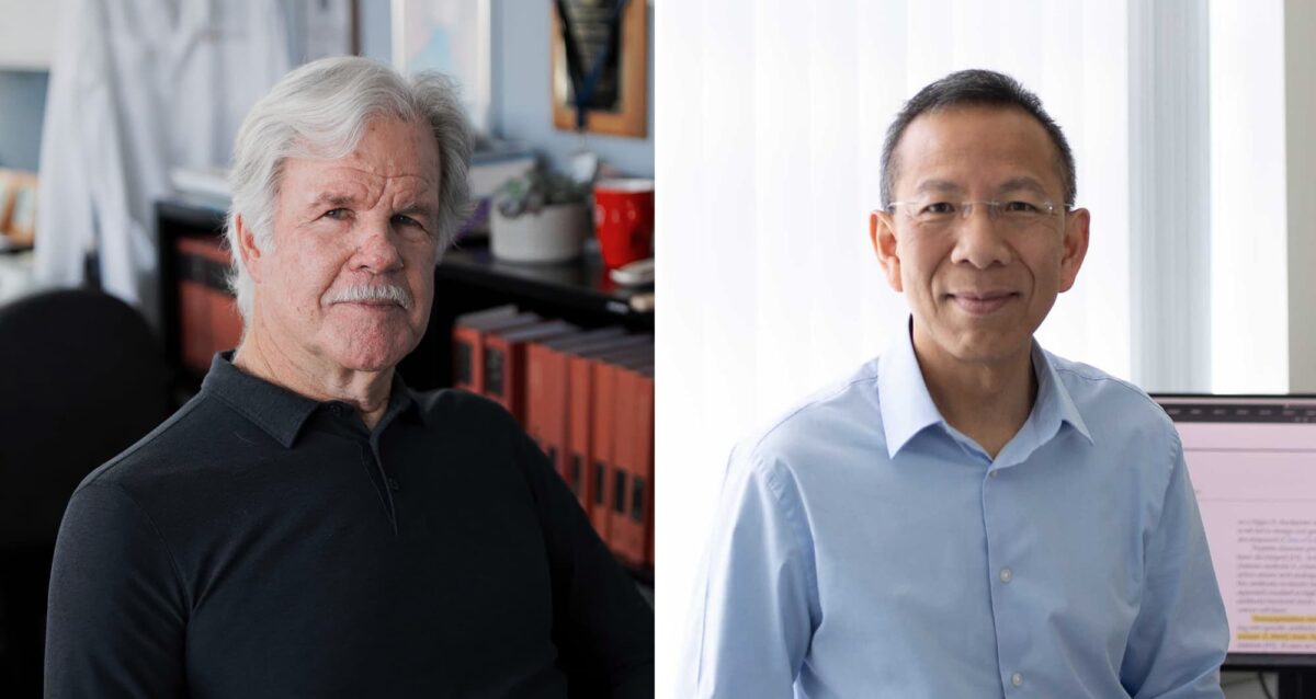
Engineering antibodies with a novel fusion protein
Mar 25, 2025Fusing two immune system proteins leads to a new method of generating antibodies and may advance drug discovery.
- Nov 19, 2024
Protein superfamily crucial to the immune system experiences Broadway-style revival
Nov 19, 2024San Diego scientists note a resurgence of interest in research on protein family to find new drug candidates.
- Jun 1, 2023
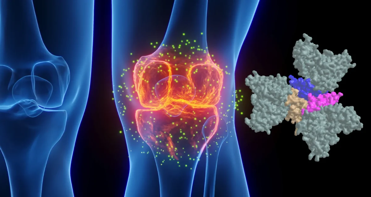
Pumping the brakes on autoimmune disease
Jun 1, 2023New study describes the science behind an autoimmune disease treatment in a Phase 2 clinical trial Researchers at Sanford Burnham…
- Nov 2, 2022
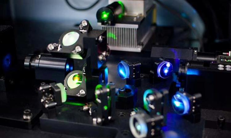
Seeing the immune system in full color
Nov 2, 2022A new flow cytometer at the Institute will help researchers study the immune system with unprecedented resolution and speed. The…
- Apr 6, 2022
How our immune system controls gut microbes
Apr 6, 2022And how this relationship could help fight autoimmune diseases
- Jan 20, 2022
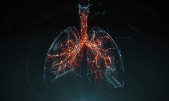
New COVID-19 drug passes phase 2 clinical trial
Jan 20, 2022The new treatment, developed by Avalo Therapeutics with Sanford Burnham Prebys researchers, can mitigate lung damage and improve survival in…
Select Publications
Showing 2 of 2Realigning the LIGHT signaling network to control dysregulated inflammation.
Ware CF, Croft M, Neil GA
J Exp Med 2022 Jul 4 ;219(7):Randomized, double-blind, controlled trial of human anti-LIGHT monoclonal antibody in COVID-19 acute respiratory distress syndrome.
Perlin DS, Neil GA, Anderson C, Zafir-Lavie I, Raines S, Ware CF, Wilkins HJ
J Clin Invest 2022 Feb 1 ;132(3):Eric has a broad background in chemical biology, with specific training and expertise in kinase inhibitors and targeted protein degradation, an emerging modality in which small molecules recruit E3 ligase complexes to target proteins to induce their ubiquitination and subsequent proteasomal degradation. He also has experience in pharmacological modulation of immune cells to improve anti-tumor immunity.
He received his PhD from the University of California San Francisco and postdoctoral training at the Dana-Farber Cancer Institute.
Education and Training
2021: Postdoctoral Fellow, Dana-Farber Cancer Institute / Harvard Medical School
2015: PhD, University of California San Francisco
2009: BS, Duke University
Fellowship
Damon Runyon Cancer Research Foundation Fellowship
Related Disease
Cancer, Immune Disorders, Molecular Biology
Phenomena or Processes
Adaptive Immunity, Cancer Biology, Cell Biology, Disease Therapies, Tumor Microenvironment
Techniques and Technologies
Chemical Biology, Drug Discovery
Dysregulation of transcriptional circuits is a common hallmark of disease, and in particular is found in both tumor and host immune cells in cancer. Eric’s lab is focusing on developing and using chemical tools to modulate the activity of key transcriptional regulators of both tumor cells and host immune cells, with a long-term goal of identifying new therapeutic approaches.
Select Publications
Showing 1 of 1CDK4/6 Inhibition Augments Antitumor Immunity by Enhancing T-cell Activation.
Deng J, Wang ES, Jenkins RW, Li S, Dries R, Yates K, Chhabra S, Huang W, Liu H, Aref AR, Ivanova E, Paweletz CP, Bowden M, Zhou CW, Herter-Sprie GS, Sorrentino JA, Bisi JE, Lizotte PH, Merlino AA, Quinn MM, Bufe LE, Yang A, Zhang Y, Zhang H, Gao P, Chen T, Cavanaugh ME, Rode AJ, Haines E, Roberts PJ, Strum JC, Richards WG, Lorch JH, Parangi S, Gunda V, Boland GM, Bueno R, Palakurthi S, Freeman GJ, Ritz J, Haining WN, Sharpless NE, Arthanari H, Shapiro GI, Barbie DA, Gray NS, Wong KK
Cancer Discov 2018 Feb ;8(2):216-233Kristiina Vuori earned her MD and PhD at University of Oulu, Finland. After completion of internship and residency, she received postdoctoral training at the Institute and was appointed to faculty in 1996. Dr. Vuori was selected as a PEW Scholar in the Biomedical Sciences in 1997. She has been co-Director of the Conrad Prebys Center for Chemical Genomics, housed at Sanford Burnham Prebys, since its inception in 2005. She was appointed Deputy Director of the Institute’s NCI-Designated Cancer Center in 2003, and Director of the Cancer Center in 2006. In 2008, she was appointed Executive Vice President for Scientific Affairs at Sanford Burnham Prebys. She was President of the Institute from 2010 to 2022.
Related Disease
Brain Cancer, Breast Cancer, Cancer, Leukemia/Lymphoma, Lung Cancer, Ovarian Cancer, Prostate Cancer
Dr. Vuori’s research is aimed at unraveling the cell mechanisms of the most life-threatening aspect of cancer, which is cancer metastasis. Metastasis is responsible for nearly all deaths in cancer patients, and understanding of the mechanisms that turn a cancer from a locally growing tumor into highly metastatic cancer cells will provide clues how to prevent this step in cancer progression. All cells in our body stick to one another and to the packaging material, or extracellular matrix, around them. This adhesion is essential for cell survival; if cells become detached from their microenvironment, they will die through a process known as apoptosis. This phenomenon, which is called adhesion dependency of survival, is one of the safeguards that maintain the integrity and normal function of tissues, and prevent cells from becoming cancerous. Normal cells cannot detach from their tissue and establish themselves somewhere else, because they will die on the way. Yet cancer cells somehow get around this requirement; they trespass aggressively into other tissues and metastasize to distant sites in the body without dying. Dr. Vuori’s work is aimed at identifying the molecular mechanisms that in normal cells makes them adhesion-dependent; false action of the very same mechanisms is likely to be the key step in allowing cancer cells to metastasize.
- Feb 1, 2024
New genome mapping tool may uncover secrets for treating blood cancers
Feb 1, 2024The outlook for patients with acute myeloid leukemia (AML), a deadly set of blood cancers that is difficult to treat,…
- Nov 14, 2017
Cancer Center Open House Showcases SBP Scientists
Nov 14, 2017SBP’s Cancer Center Open House on November 9, 2017 enlightened visitors from the community on the topic of “The Science…
- Oct 18, 2017
Spectacular 2017 SBP annual Gala celebrates “Sights Set on Discovery”
Oct 18, 2017Friends and supporters of Sanford Burnham Prebys Medical Discovery Institute (SBP) gathered under the stars on Harbor Island in downtown…
- Dec 13, 2016
Reena Horowitz honored at Sanford Burnham Prebys Medical Discovery Institute
Dec 13, 2016During a special end-of-the-year gathering, Reena Horowitz was honored for her hard work and dedication to Sanford Burnham Prebys Medical…
- Sep 1, 2015
The Mayor of San Diego visits SBP in La Jolla
Sep 1, 2015On Friday, August 28, the Mayor of San Diego, Kevin Faulconer, visited SBP to learn more about how our Institute is…
- Jun 24, 2015
A $100 million gift and a new name
Jun 24, 2015We are beyond excited to announce that Sanford-Burnham has received a gift of $100 million from prominent San Diego developer, philanthropist, and
Select Publications
Showing 3 of 3Sceptrin, a marine natural compound, inhibits cell motility in a variety of cancer cell lines.
Cipres A, O’Malley DP, Li K, Finlay D, Baran PS, Vuori K
ACS Chem Biol 2010 Feb 19 ;5(2):195-202Critical role for caspase-8 in epidermal growth factor signaling.
Finlay D, Howes A, Vuori K
Cancer Res 2009 Jun 15 ;69(12):5023-9Cell biology. Two lipids that give direction.
Côté JF, Vuori K
Science 2009 Apr 17 ;324(5925):346-7Dr. Xiao Tian participates in the Degenerative Diseases Program and the Cancer Genome and Epigenetics Program at Sanford Burnham Prebys. He started his lab in 2024 to understand the fundamental biology of aging and its contribution to age-related diseases. He joined the Institute after his postdoctoral research in Dr. David Sinclair’s lab at Harvard Medical School where he co-wrote the Information Theory of Aging. He obtained his BS from Shandong University and his PhD from the University of Rochester where he worked with Dr. Vera Gorbunova.
Education
2018-2023: Postdoc, Harvard Medical School
2016-2018: Postdoc, University of Rochester
2010-2016: PhD, Biology of Aging, University of Rochester
2005-2009: BS, Microbial Technology, Shandong University
Honors and Awards
2020-2026: K99/R00 Pathway to Independence Awards, NIH/NIA
2019-2020: NASA Postdoctoral Fellowship, NASA Ames Research Center
2017: Outstanding Dissertation Award for the Natural Sciences, University of Rochester
2015: Messersmith Dissertation Fellowship, University of Rochester
2014: Award for Outstanding Self-Financed Students Abroad, China Scholarship Council
2010-2014: Holtfreter Fellowship, University of Rochester
2007: Weichai Power Scholarship, Shandong University
2006-2008: Excellent Student Scholarship, Shandong University
Related Disease
Aging-Related Diseases, Alzheimer’s Disease, Cancer
Phenomena or Processes
Aging, Epigenetics, Genomic Instability, Neurodegeneration
Research Models
Computational Modeling, Mouse, Naked Mole Rat, Primary Human Cells
Techniques and Technologies
Bioinformatics, Epigenomics, Gene Expression, Gene Knockout (Complete and Conditional), High-Throughput/Robotic Screening, Mouse Behavioral Analysis
The Tian lab studies the fundamental mechanisms of aging and their roles in the onset of age-related diseases. Our recent research in epigenetic reprogramming and aging clocks indicates that the progressive loss of epigenetic information over time is a key driver of aging. The current research of the lab focuses on understanding how the epigenetic landscape is set up and maintained and investigating why the maintenance system fails which leads to aging and related diseases including cancer and neurodegeneration. Building on this, our ultimate goal is to develop safe and effective rejuvenation strategies to counteract aging.
Xiao Tian’s Research Report
Below are my major contributions to the field of aging research during my PhD and postdoc work:
Uncovering the first anti-cancer and longevity mechanism of the naked mole rat
We identified high-molecular-mass hyaluronan (HMM HA) as a critical mechanism for the cancer resistance of the naked mole-rat cells. Furthermore, we found that this anti-cancer and longevity mechanism from naked mole rats can be integrated into other species, highlighting the significant implications of identifying new mechanisms from long-lived species. Our goal is to decipher how long-lived organisms control the pace of aging and deter age-related diseases.
Discovering enhanced DNA double-strand break (DSB) repair in long-lived species
Using a group of 18 species, we revealed that long-lived species possess efficient DNA double-strand break repair, mediated by potent SIRT6 activities. Future directions include deciphering how long-lived species address different types of cellular stress to maintain genomic and epigenomic stability.
Epigenetic reprogramming as a strategy to counteract aging and age-related diseases
We illustrated that epigenetic information loss as a result of DNA damage repair is a key driver of aging. One interventional strategy is to recover the lost epigenetic information, such as through reprogramming. We revealed that epigenetic reprogramming is an effective strategy to counteract aging and even potentially modify the trajectory of age-related diseases including glaucoma. We are currently testing other disease settings.
- Oct 24, 2024
A Conversation About Aging and Cancer at Sanford Burnham Prebys
Oct 24, 2024The Institute recently welcomed members of the community for an engaging afternoon focused on the intersection of aging and cancer….
- Feb 29, 2024
Time to talk about aging research
Feb 29, 2024Hundreds of scientists gather in San Diego and virtually to share knowledge on the science of aging
- Feb 13, 2024
With the help of naked mole rats, Xiao Tian seeks to expose and explain the epigenetic drivers of aging
Feb 13, 2024Getting older is inevitable, but aging may be optional, or at least a treatable condition. Increasingly, researchers are parsing the…
- Nov 2, 2023
La Jolla’s Sanford Burnham Prebys adding six scientists in hopes of launching new era of collaboration
Nov 2, 2023The newcomers, brought in with help from a $70 million donation, are versed in different technologies that can be applied…
- Aug 2, 2023
Big Data. Bigger Opportunities.
Aug 2, 2023New and rapidly developing technologies, such as cryo-electron (cryo-EM) and artificial intelligence, are providing the tools to revolutionize biomedical research,…
Select Publications
Showing 1 of 1High-molecular-mass hyaluronan mediates the cancer resistance of the naked mole rat.
Tian X, Azpurua J, Hine C, Vaidya A, Myakishev-Rempel M, Ablaeva J, Mao Z, Nevo E, Gorbunova V, Seluanov A
Nature 2013 Jul 18 ;499(7458):346-9Xueqin (Sherine) Sun seeks to better understand the genetic and epigenetic underpinnings of cancers, using genome editing technologies, animal and patient-derived models, and other tools to develop more effective cancer therapies.
“My lab is interested in studying how DNA or the machinery that interprets it leads to the transformation of normal cells into cancerous cells and concurrently, their specific vulnerabilities. Identifying these intrinsic vulnerabilities and targeting them properly is profoundly important to developing effective cancer therapies.”
Another aspect of Sun’s work is understanding how cancer cells and tumors change their circumstances and environment to improve survival, including hiding from or repressing the immune system.
“Changes to DNA itself and the way how DNA is interpreted by cells can transform normal cells into cancer cells. And transformed cells propagate by enhancing the misinterpreted DNA information, which in turn becomes the Achilles’ heel of cancer cells. Our goal is to find out how DNA information is misinterpreted in different ways and how to correct it to halt cancer.”
At Sanford Burnham Prebys, Sun and colleagues will employ a host of leading-edge tools and approaches, including functional genomics, artificial intelligence, structural biology, large-scale drug screening, and advanced imaging/spatial technologies.
Sun conducted her postdoctoral fellowship at Cold Spring Harbor Laboratory under the guidance of Alea Mills, PhD, a professor at the National Cancer Institute-designated cancer center at Cold Spring Harbor.
She received her PhD from Wuhan University in China.
Related Disease
Aging-Related Diseases, Brain Cancer, Cancer, Childhood Diseases, Immune Disorders, Inflammatory/Autoimmune Disease, Leukemia/Lymphoma
Phenomena or Processes
Adapter Proteins, Adult/Multipotent Stem Cells, Aging, Angiogenesis, Apoptosis and Cell Death, Bcl-2 Family, Cancer Biology, Cancer Epigenetics, Cell Adhesion and Migration, Cell Biology, Cell Cycle Progression, Cell Differentiation, Cell Motility, Cell Proliferation, Cell Signaling, Cell Surface Receptors, Cellular Senescence, Chromosome Dynamics, Combinatorial Therapies, Cytokines, Development and Differentiation, Disease Therapies, DNA Damage Checkpoint Function, Embryonic/Pluripotent Stem Cells, Epigenetics, Gene Regulation, Genomic Instability, Growth Factors, Hematopoiesis, Host Defense, Host-Pathogen Interactions, Inflammation, Innate Immunity, Kinase Inhibitors, Metastasis, Neurogenesis, Oncogenes, Phosphorylation, Posttranslational Modification, Receptor Tyrosine Kinases, Serine/Threonine Kinases, Signal Transduction, TNF-Family, Transcription Factors, Transcriptional Regulation, Tumor Microenvironment, Tumorigenesis, Tyrosine Kinases, Ubiquitin, Ubiquitin Protease System and Ubiquitin-like Proteins
Anatomical Systems and Sites
Brain, General Cell Biology, Hematopoietic System, Immune System and Inflammation, Nervous System
Research Models
Bacteria, Cultured Cell Lines, Human Adult/Somatic Stem Cells, Human Cell Lines, Mouse, Mouse Cell Lines, Mouse Embryonic Stem Cells, Mouse Somatic Stem Cells, Primary Cells, Primary Human Cells
Techniques and Technologies
3D Image Analysis, 3D Reconstructions, Biochemistry, Bioinformatics, Cell Biology, Cellular and Molecular Imaging, Chemical Biology, Computational Biology, Confocal Microscopy, Correlative Light and Electron Microscopy, Drug Delivery, Drug Discovery, Drug Efficacy, Electron Microscopy, Fluorescence Microscopy, Fragment-Based Drug Design, Gene Expression, Gene Knockout (Complete and Conditional), Gene Silencing, Genetics, Genomics, High Content Imaging, High-Throughput/Robotic Screening, In vivo Modeling, Live Cell Imaging, Live Imaging, Mass Spectrometry, Microscopy and Imaging, Molecular Biology, Molecular Genetics, Nucleic Acid Synthesis, Protein-Protein Interactions, Protein-Small Molecule Interactions, Proteomics, Rational Drug Design, RNA Interference (RNAi), Scanning Cytometry, Small Molecule Compounds, Transgenic Organisms, Transplantation
We seek to understand why cancer occurs and what is the Achille’s heel of cancer, and to develop effective therapeutic interventions.
The successful treatment of any disease requires a good understanding of the mechanisms at work. Cancer is fundamentally caused by aberrant gene expression, which reflects the misinterpretation of DNA information at both genetic and epigenetic levels. We are interested in uncovering DNA-related alterations that drive cancer-favored transcriptional programs, identifying cancer-specific vulnerabilities, and developing effective therapeutic interventions for cancer treatment.
Xueqin Sun’s Research Report
Precise gene expression (the interpretation of DNA) is essential for almost all biological processes, and understanding gene regulation is one of the most pivotal frontiers in biological research under both health and disease circumstances. Gene expression is mainly regulated at genetic (with changes of DNA sequence) and epigenetic (without changing DNA sequence) levels. And gene dysregulation can lead to various health conditions and diseases, including developmental disorders, aging, and cancer. The overarching goal of Sun Lab is to uncover driving genetic and epigenetic alterations involved in cancer, to understand how developmental pathways and aging process impact cancer progression, and to identify mechanisms of action for developing more effective therapeutic strategies.
We are an interdisciplinary lab particularly focused on the following research directions:
- The EP400 chromatin remodeling complex
The EP400 complex is an evolutionarily conserved SWR1-class ATP-dependent chromatin remodeling complex encompassing ~17 components, with a total molecular mass of ~1.5 mega-dalton. The EP400 complex plays critical roles in diverse cellular processes, including chromosome stability, transcription, DNA recombination, DNA damage repair, embryonic stem cell renewal/development, and oncogenesis. The EP400 complex can incorporate histone variants, such as H2AZ and H3.3, into the genome to regulate gene expression. Our recent work discovers BRD8—one of the core subunits of the EP400 complex—as a unique vulnerability of p53 wildtype glioblastoma (GBM), the most prevalent and devastating type of brain cancer. BRD8-driven EP400 complex highjacks H2AZ at p53 target loci to block p53-mediated transactivation and tumor suppression (Nature, 2023). The bromodomain of BRD8 plays the key role in this process. Bromodomain is a druggable domain as evidenced by a number of successful small molecules targeting diverse bromodomains encoded by the human genome across multiple cancer types. Furthermore, findings from others and us suggest that the EP400 complex is involved in different cancers. Thus, we seek to unravel the roles of the EP400 complex in health and disease, and to better understand how to target the EP400 complex for developing effective therapeutic interventions. - The NuRD chromatin remodeling complex
The NuRD complex is also a highly conserved class of ~ 1 MDa multi-subunit chromatin remodeling complexes that consume energy derived from ATP hydrolysis to remodel the configuration of chromatin to control gene transcription programs, with a primary role in gene silencing. Chromatin remodeling is vital for efficiently framing the cellular response to both intrinsic and extrinsic signals and has enormous implications for determining cellular states. NuRD complex is unique in combining ATP-dependent chromatin remodeling, protein deacetylase activity, and recognition of methylated DNA and histone modifications, and has multifarious roles in chromatin organization, transcription regulation, and genome maintenance; thereby, largely impacts health and disease. The NuRD complex has been in the central stage of brain development studies, and is significantly related to brain disorders/diseases. Interestingly, NuRD complex re-assembles by exchanging the chromatin remodeling subunits CHD3/4/5 to achieve specific regulation of an array of genes required for generating distinct cell types in a highly organized manner, especially over brain development. Amongst the genes encoding NuRD complex components, CHD5 is located in human chromosome 1 short arm (1p36), a region that is frequently hemizygously deleted in diverse cancers. Besides genetic deletion, CHD5 is also often silenced in cancer cells due to epigenetic mechanisms, such as promoter hypermethylation, aberrant expression of other chromatin regulators, and microRNAs-mediated translational repression and/or mRNA instability. Our current work seeks to determine whether and how CHD5-driven NuRD complex is involved in tumorigenesis (In preparation, 2024). We will further understand how NuRD complex is involved in both development and tumorigenesis, and identify mechanism of action to develop rational therapeutic strategies. - Novel genetic and epigenetic underpinnings in GBM
GBM is notorious for being a highly complex and plastic cancer type. However, at the genetic level, GBM harbors a relatively low genetic alteration burden compared to the majority of other cancers from pan-cancer profiling studies. This indicates the largely undocumented epigenetic mechanisms that interplay with genetic alterations and co-reprogram transcriptional networks essential for GBM development. Epigenetic changes are usually reversible by nature, as evidenced by numerous successes in targeting epigenetic regulators using small chemical compounds. As actionable therapeutic targets for GBM have been scarce, we are keen to uncover novel epigenetic pathways underlying gliomagenesis under different genetic backgrounds, which will potentially provide promising therapeutic opportunities for GBM treatment. - Novel GBM mouse models
Despite decades of effort, our knowledge about GBM biology is still very limited. GBM harbors a number of genetic alterations. However, among these recurrent genetic lesions, only several have been implicated in gliomagenesis, with most being undocumented. Moreover, the mechanisms by which these genetic alterations are involved in establishing GBM-favored epigenetic landscapes and transcription programs during GBM progression are still largely elusive. The lack of efficient approach to establish mouse models for investigating gene function in gliomagenesis and the limit of current mouse models to recapitulate clinical GBM features in brain is the prime reason that hinders GBM biological research. To this end, we have developed an engineered neural stem cells (NSCs)-based strategy to rapidly generate highly aggressive GBM with desired genetic lesions (genotypes) in mouse brain. Therefore, we will further optimize this strategy to establish a series of novel mouse models possessing recurrent combinations of genetic alterations (genotypes) in GBM, in order to systematically study whether and how these genetic lesions are involved in gliomagenesis and identify genotype-specific dependencies. - Crosstalk between GBM cells and tumor microenvironment
GBM exhibits highly diffuse and infiltrative nature, which contributes to therapeutic resistance and tumor relapse after surgical removal, resulting in dismal prognosis. A better understanding of gliomagenesis involving not only malignant cells themselves, but also the holistic bidirectional interactions of malignant cells with a variety of proximal and distal cells within the organism, is profound for developing novel effective therapies to improve GBM prognosis. Individual invasive GBM cells intermingle with normal brain cells and often cause relapse in brain areas essential for patient survival. Emerging evidence indicates that glioma cells highjack normal brain cells to thrive, and even transform them. However, how gliomagenesis reshapes ecological composition/landscape in host brain and how brain microenvironment affects gliomagenesis are still largely unclear. By using our novel highly invasive mouse models that recapitulate the multiforme diffuse topographies of GBM in brain, we seek to understand the interactions between GBM cells and brain microenvironment, and identify extrinsic pathways that are essential for GBM progression and migration.
Our lab is focused on both fundamental questions in cancer biology and translation of promising therapeutic strategies.
To achieve these, we work together with many fantastic collaborators to develop and leverage cutting-edge technologies, including but not limited to, high-throughput functional genomics (CRISPR/Cas9 screens, exon tiling scan, targeted mutagenesis, etc.), cell and molecular biology, genomics, epigenomics, proteomics, biochemistry, microscopy (2D/3D, time-lapse, two-photon, light sheet, etc.), automated large-scale drug synthesis/screening, structural biology, single cell and spatial multi-omics, artificial intelligence, and bioinformatics. We also establish novel patient-derived models and novel mouse models to facilitate our research programs. Our ultimate goals are to better understand fundamental genetic and epigenetic apparatuses involved in cancer-specific transcriptional networks, provide more effective therapeutic opportunities, and contribute to shifting the paradigms in cancer treatment and precision medicine.
- Aug 19, 2024
Women in Science event at Sanford Burnham Prebys examines how female faculty members navigate research careers
Aug 19, 2024Topics at the event included work/life balance, caregiving and family obligations, and gender disparities in academic rank at research and…
- Mar 13, 2024
Xueqin Sun seeks to illuminate the underlying causes of cancer
Mar 13, 2024New Sanford Burnham Prebys scientist investigates the mutational powers of cancer cells — and their vulnerabilities
- Dec 19, 2023
Sanford Burnham Prebys continues unprecedented recruitment of early-career scientists
Dec 19, 2023Continuing its rapid and dramatic recruitment of emerging, top-tier researchers, Sanford Burnham Prebys has hired two more highly regarded early-career…
Select Publications
Showing 2 of 2BRD8 maintains glioblastoma by epigenetic reprogramming of the p53 network.
Sun X, Klingbeil O, Lu B, Wu C, Ballon C, Ouyang M, Wu XS, Jin Y, Hwangbo Y, Huang YH, Somerville TDD, Chang K, Park J, Chung T, Lyons SK, Shi J, Vogel H, Schulder M, Vakoc CR, Mills AA
Nature 2023 Jan ;613(7942):195-202ANP32A regulates histone H3 acetylation and promotes leukemogenesis.
Yang X, Lu B, Sun X, Han C, Fu C, Xu K, Wang M, Li D, Chen Z, Opal P, Wen Q, Crispino JD, Wang QF, Huang Z
Leukemia 2018 Jul ;32(7):1587-1597Charles Spruck earned his BS in Biology at UCLA and PhD in Molecular Biology at the University of Southern California. He worked as a postdoctoral fellow at The Scripps Research Institute in La Jolla and was recruited to the Sidney Kimmel Cancer Center in San Diego as an Assistant Professor in 2003. He joined Sanford Burnham Prebys in 2010.
Education and Training
2003: Post-doc, The Scripps Research Institute
1986: PhD, University of Southern California
1995; BS, University of California at Los Angeles
Prestigious Funding Awards / Major Collaborative Grants
NIH/NCI DoD BCRP CBCRP TRDRP
Honors and Recognition
ACS Scholar
Related Disease
Breast Cancer, Cancer, Lung Cancer, Molecular Biology
Phenomena or Processes
Cancer Biology, Cancer Epigenetics, Cell Biology, Cell Cycle Progression, Cell Signaling, Genomic Instability, Innate Immunity, Metastasis, Posttranslational Modification, Proteolytic Pathways
Research Models
Cultured Cell Lines, Human Cell Lines, Mouse, Mouse Cell Lines
Techniques and Technologies
Cell Biology, Drug Discovery, Gene Knockout (Complete and Conditional), In vivo Modeling
“Despite recent advances in treatment, patients with advanced metastatic cancers have few treatment options. Our lab is focused on developing new effective and non-toxic treatments for these patients.”
Dr. Spruck’s laboratory is focused on developing new, effective, and non-toxic treatments for patients with advanced cancers. The lab focuses on defining the molecular networks that regulate cancer cell division and drive metastasis progression. Recent studies have focused on viral mimicry as a therapeutic approach in cancer, which involves the activation of dormant endogenous retroviruses and retrotransposons in cancer cells to enhance immunogenicity and the effectiveness of immune checkpoint blockade immunotherapy and DNA damaging therapies. The laboratory utilizes various biochemical and molecular approaches, CRISPR gene editing, and animal models of cancer. An emphasis is on studies of breast, lung, prostate, and brain tumors.
Charles Spruck’s Research Report
Developing viral mimicry therapeutic approaches for cancer: Approximately 45% of the human genome is composed of repetitive elements (REs), including endogenous retroviruses and retrotransposons, that are normally transcriptionally silenced in somatic cells. Recent studies suggest that the transcriptional awakening of ERVs/retrotransposons beyond a threshold level of tolerance in cancer cells induces antiviral responses that can enhance the efficacy of certain therapies, including immunotherapy. We recently discovered a novel epigenetic regulatory pathway, FBXO44/SUV39H1, that is essential for ERV/retrotransposon silencing in cancer cells. Preclinical studies showed that FBXO44/SUV39H1 inactivation induces viral mimicry in cancer cells, leading to increased immunogenicity, decreased tumorigenicity, and enhanced the efficacy of immune checkpoint blockade therapy. We are currently exploring therapeutic approaches to target this pathway, and others like it, to prevent tumor growth and enhance immunotherapy response. We are also exploring the role of reactivated REs in human diseases.
Targeting metastatic tumors: Metastasis is a major cause of mortality in cancer. Through genomic screens and biochemical studies, we are identifying novel molecular pathways that drive cancer cell motility, invasion, and metastasis. Recently, we identified a novel molecular axis, FBXO7/EYA2-SCF(FBXW7), that promotes cancer cell motility and cancer stem cell self-renewal and suppresses cancer cell immunogenicity. Targeting this axis prevented metastasis progression, reduced the cancer stem cell population, and stimulated anti-tumor immune responses in preclinical mouse breast cancer models.
- Apr 24, 2023
Charles Spruck awarded $1.7M to advance “ancient virus” treatment for prostate cancer
Apr 24, 2023The approach uses ancient viruses, embedded in our genomes, to trick the body into thinking it has an infection. With…
- Jun 30, 2022
Three Sanford Burnham Prebys faculty receive promotions
Jun 30, 2022The promoted faculty, all from the Institute’s NCI-designated Cancer Center, include Ani Deshpande, PhD, Brooke Emerling, PhD and Charles Spruck,…
- Mar 15, 2022
Tricking the body to treat breast cancer
Mar 15, 2022With the help of two new grants from the National Institutes of Health totaling more than $4.4 million, Charles Spruck,…
- Nov 3, 2021
Boosting immunotherapy in aggressive brain cancer
Nov 3, 2021Institute have collaborated the University of Pittsburgh Cancer Institute to reveal a new approach to enhance the effects of immunotherapy in…
- Feb 11, 2021
Mining “junk DNA” reveals a new way to kill cancer cells
Feb 11, 2021Scientists unearth a previously unknown vulnerability for cancer and a promising drug candidate that leverages the approach Scientists at Sanford…
- Oct 20, 2016
Hearst Foundation’s new fellowship funds innovative research to fight breast cancer
Oct 20, 2016Mark Goldberg, PhD, is working on a potential way to turn cancer stem cells into harmless cells. He and his…
Select Publications
Showing 3 of 3FBXO44 promotes DNA replication-coupled repetitive element silencing in cancer cells.
Shen JZ, Qiu Z, Wu Q, Finlay D, Garcia G, Sun D, Rantala J, Barshop W, Hope JL, Gimple RC, Sangfelt O, Bradley LM, Wohlschlegel J, Rich JN, Spruck C
Cell 2021 Jan 21 ;184(2):352-369.e23A FBXO7/EYA2-SCF(FBXW7) axis promotes AXL-mediated maintenance of mesenchymal and immune evasion phenotypes of cancer cells.
Shen JZ, Qiu Z, Wu Q, Zhang G, Harris R, Sun D, Rantala J, Barshop WD, Zhao L, Lv D, Won KA, Wohlschlegel J, Sangfelt O, Laman H, Rich JN, Spruck C
Mol Cell 2022 Mar 17 ;82(6):1123-1139.e8Transcription Elongation Machinery Is a Druggable Dependency and Potentiates Immunotherapy in Glioblastoma Stem Cells.
Qiu Z, Zhao L, Shen JZ, Liang Z, Wu Q, Yang K, Min L, Gimple RC, Yang Q, Bhargava S, Jin C, Kim C, Hinz D, Dixit D, Bernatchez JA, Prager BC, Zhang G, Dong Z, Lv D, Wang X, Kim LJY, Zhu Z, Jones KA, Zheng Y, Wang X, Siqueira-Neto JL, Chavez L, Fu XD, Spruck C, Rich JN
Cancer Discov 2022 Feb ;12(2):502-521Evan Y. Snyder earned his MD and PhD (in neuroscience) from the University of Pennsylvania in 1980 as a member of NIH’s Medical Scientist Training Program (MSTP). He had also studied psychology and linguistics at the University of Oxford. After moving to Boston in 1980, he completed residencies in pediatrics and neurology as well as a clinical fellowship in Neonatal-Perinatal Medicine at Children’s Hospital-Boston, Harvard Medical School. He also served as Chief Resident in Medicine (1984-1985) and Chief Resident in Neurology (1987) at Children’s Hospital-Boston. In 1989, he became an attending physician in the Department of Pediatrics (Division of Newborn Medicine) and Department of Neurology at Children’s Hospital-Boston, Harvard Medical School. From 1985-1991, concurrent with his clinical activities, he conducted postdoctoral research as a fellow in the Department of Genetics, Harvard Medical School. In 1992, Dr. Snyder was appointed an instructor in neurology (neonatology) at Harvard Medical School and was promoted to assistant professor in 1996. He maintained lab spaces in both Children’s Hospital-Boston and at Harvard Institutes of Medicine/Beth-Israel Deaconess Medical Center. In 2003, Dr. Snyder was recruited to Sanford Burnham Prebys as Professor and Director of the Program in Stem Cell and Regenerative Biology. He then inaugurated the Stem Cell Research Center (serving as its founding director) and initiated the Southern California Stem Cell Consortium. Dr. Snyder is a Fellow of the American Academy of Pediatrics (FAAP). He also received training in Philosophy and Linguistics at Oxford University.
Related Disease
Alzheimer’s Disease, Amyotrophic Lateral Sclerosis (Lou Gehrig’s Disease), Arthritis, Brain Cancer, Brain Injury, Breast Cancer, Cancer, Childhood Diseases, Congenital Disorders of Glycosylation, HIV-Associated Dementia, Huntington’s Disease, Multiple Sclerosis, Muscular Dystrophy, Neurodegenerative and Neuromuscular Diseases, Neurological and Psychiatric Disorders, Parkinson’s Disease, Peripheral Vascular Disease, Skin Cancer and Melanoma, Spinal Cord Injury, Stroke, Traumatic Injury
We believe the study of stem cell biology will provide insights into many areas: developmental biology, homeostasis in the normal adult, and recovery from injury. Indeed, past and current research has already produced data in these areas that would have been difficult or impossible via any other vehicle. We have engaged in a multidisciplinary approach, simultaneously exploring the basic biology of stem cells, their role throughout the lifetime of an individual, as well as their therapeutic potential. Taken together, these bodies of knowledge will glean the greatest benefit for scientists and, most importantly, for patients. All of our research to date has been preformed in animal models with the ultimate goal of bringing them to clinical trials as soon as possible. Stem cells offer an intriguing mix of controversy, discovery, and hope. Politicians are charged with dealing with the controversial facets of stem cells, as we prefer to focus our energy on their potential for discovery and hope.
The Snyder Lab studies stem cell biology, with the goal of understanding normal development, tissue homeostasis, and recovery from injury and disease. A major focus is neural stem cells (NSCs), which can self-renew and differentiate into neurons, astrocytes, and oligodendrocytes. These properties make NSCs ideal for repair of damage due to injury or disease, but they also make them susceptible to transformation into malignant cancers.
- May 28, 2025
Evan Snyder named Fellow of the Child Neurology Society
May 28, 2025Evan Snyder, MD, PhD, has been named a Fellow of the Child Neurology Society, honoring a distinguished career and…
- Nov 11, 2024
Seven questions for FDA advisor Evan Snyder
Nov 11, 2024Sanford Burnham Prebys physician-scientist advises the FDA on cell-based therapeutics, tissue engineering and gene therapies.
- Jul 23, 2024
Mini lungs make major COVID-19 discoveries possible
Jul 23, 2024Scientists infect miniature lung organoids with the virus responsible for COVID-19, revealing new ways in which the infection spreads and…
- Jan 25, 2022
New CIRM grant to fund research internships for underrepresented high school students
Jan 25, 2022Thanks to a new grant awarded to Sanford Burnham Prebys by the California Institute for Regenerative Medicine (CIRM), 57 California…
- Nov 18, 2021
From child neurology to stem cells: An interview with Evan Snyder
Nov 18, 2021What do Evan Snyder and Sigmund Freud have in common? Both radically changed how we see the human brain. In…
- Jul 26, 2021
Biomarker could help diagnosis schizophrenia at an early age
Jul 26, 2021Research could lead to blood-based diagnostic test Scientists at Sanford Burnham Prebys have discovered how levels of a protein could…
Select Publications
Showing 1 of 1Neural stem cells implanted into MPTP-treated monkeys increase the size of endogenous tyrosine hydroxylase-positive cells found in the striatum: a return to control measures.
Bjugstad KB, Redmond DE Jr, Teng YD, Elsworth JD, Roth RH, Blanchard BC, Snyder EY, Sladek JR Jr
Cell Transplant 2005 ;14(4):183-92Dr. Sanju Sinha earned his Bachelor of Technology in Bioengineering at the Indian Institute of Technology in Guwahati, India. He recently completed his postdoctoral research and PhD in computational biology at the National Cancer Institute (NCI) with Dr. Eytan Ruppin with a co-mentorship of Dr. Brid Ryan during his PhD His PhD was earned in a joint University of Maryland and NCI program.
“At the core of my work is the desire to make a lasting impact on patient’s lives, offering patients not just better treatment, but an opportunity to avoid the disease altogether. Sanford Burnham Prebys is renowned for its work in understanding aging and developing new drugs—two areas that are key to my research. This makes it the perfect place for what I’m hoping to achieve.”
Education
2021: PhD, Computational Biology, University of Maryland and National Cancer Institute
2016: B.Tech., Bioengineering, Indian Institute of Technology, Guwahat
Honors and Recognition
2023: Top Five Outstanding NCI Postdoctoral Fellow
2023: Transition to Industry Fellowship
2021: Emerging Leaders of Computational Oncology by MSKCC.
2020: NCI Outstanding PhD award
2020: NCI CCR milestone
2019: NCI Fellows Award for Research Excellence
Related Disease
Cancer
Phenomena or Processes
Aging, Cancer Biology
Techniques and Technologies
Bioinformatics, Computational Biology, Drug Discovery, Machine Learning
Developing cancer preventive therapies using the power of AI.
At the heart of our research is the mission to develop cancer-preventive therapies using the power of artificial intelligence. Our current focus lies in dissecting the role of aging in cancer susceptibility, a crucial factor often overlooked in conventional research. We employ computational tools to analyze multi-omics data to understand the aging-induced alterations in the tissue microenvironment that increase cancer risk. But we don’t stop at understanding these changes. We apply this knowledge in designing preventive therapy candidates that specifically target these alterations. Drawing on my experience in machine learning, drug discovery, and precision oncology, our lab is on a quest to reimagine the drug discovery pipeline.
“As we aim to push the boundaries of cancer prevention research, we are hiring individuals eager to contribute to this mission at multiple levels including postdoctoral researchers, experienced computational biologists, and, PhD students.”
Sanju Sinha’s Research Report
My key contributions to the field of computational oncology during my PhD and postdoctoral tenure are outlined below:
DeepTarget: A Computational Tool for Decoding the Mechanism of Action for Cancer Drugs
DeepTarget, our innovative computational tool, integrates data from genetic and drug screens to intricately understand the mechanism of action of cancer drugs. It identifies both primary and secondary drug targets, paving the way for a comprehensive understanding of drug function, its optimal indications, and clinical potential.
Revealing the Potential Cancer Risks associated with Genetic Editing
Through computational analyses of vast genetic screens, we shed light on the inherent selection potential of specific cancer gene mutations associated with CRISPR-Knockout editing. This work has significantly enhanced our understanding of the risks associated with gene editing.
First Single-cell based Precision Oncology Framework: A Proof-of-concept
Our innovative approach to precision oncology utilizes single-cell transcriptomics to predict patient treatment responses and detect resistance. We demonstrated its efficacy using patient-derived primary cells and three recent single-cell clinical cohorts, providing a powerful tool for the next generation of precision oncology approaches.
Why High Tumor Mutation Burden Biomarker of Immunotherapy is only effective for Certain Cancer Types?
We unveiled the microenvironment context that can determine if the high-tumor mutation burden (TMB) biomarker will be effective in a certain cancer type – High M1 Macrophage levels and Low Resting Dendritic Cells. Based on this, we also predicted the rare tumor types with High-TMB where immunotherapy is most likely to be successful.
Uncovering Therapeutic Prospects for African Americans: A Step Forward in Inclusive Cancer Research
Our work revealed distinct biological differences in tumors across African American and European American patients. Most notably, we identified a higher prevalence of homologous recombination deficiency in African American patients, pointing towards promising, personalized treatment options.
- May 9, 2025
PERCEPTION proves a predictable NCI milestone
May 9, 2025PERCEPTION, an artificial intelligence-based tool, was able to predict tumor response to targeted therapy using single-cell datasets.
- Jan 3, 2025
AI-driven cancer prediction tool makes NIH director’s highlights for 2024
Jan 3, 2025Sanju Sinha, PhD, and colleagues published details about a new artificial intelligence-powered tool called PERCEPTION.
- Dec 9, 2024
How cancer cells change as they metastasize
Dec 9, 2024Most cancer deaths are caused by metastasis, but how cancer cells and tumors modify themselves and spread from their origins…
- Aug 6, 2024
Coding clinic
Aug 6, 2024Rapidly evolving computational tools may unlock vast archives of untapped clinical information—and help solve complex challenges confronting healthcare providers
- Aug 1, 2024
Objective omics
Aug 1, 2024Although the hypothesis is a core concept in science, unbiased omics methods may reduce attachments to incorrect hypotheses that can…
- Jul 30, 2024
Using machines to personalize patient care
Jul 30, 2024Artificial intelligence (AI) and other computational techniques are aiding scientists and physicians in their quest to create treatments for individuals…
- Our Machine Learning Resources A Complete Guide to Studying Machine Learning from YouTube | X
- Almost All The Big Data Resources Available In Cancer Research 10 Resources to Learn Almost All the Big Data Resources Available in Cancer Research | X
- Computational Resources To Study Immune System 11 Resources (Videos And Papers) Covering the Fundamentals and Computational Tools Available to Study the Immune System | X
Select Publications
Showing 2 of 2Higher prevalence of homologous recombination deficiency in tumors from African Americans versus European Americans.
Sinha S, Mitchell KA, Zingone A, Bowman E, Sinha N, Schäffer AA, Lee JS, Ruppin E, Ryan BM
Nat Cancer 2020 Jan ;1(1):112-121A systematic genome-wide mapping of oncogenic mutation selection during CRISPR-Cas9 genome editing.
Sinha S, Barbosa K, Cheng K, Leiserson MDM, Jain P, Deshpande A, Wilson DM 3rd, Ryan BM, Luo J, Ronai ZA, Lee JS, Deshpande AJ, Ruppin E
Nat Commun 2021 Nov 11 ;12(1):6512Elena Pasquale earned her PhD in biology from the University of Parma, Italy. She did postdoctoral work at Cornell University, after which she was appointed Research Assistant Professor at University of Parma. Following a second postdoctoral training period at the University of California in San Diego, Dr. Pasquale was appointed Assistant Research Biologist at that institution. Dr. Pasquale was recruited to Sanford Burnham Prebys in 1990.
Related Disease
Cancer, Neurodegenerative and Neuromuscular Diseases, Skin Cancer and Melanoma
Cancer, Neurodegenerative and Neuromuscular Diseases
Receptor tyrosine kinases of the Eph family and their ligands, the ephrins, represent an important cell communication system that controls a vast array physiological and disease processes. For example, Eph receptors and ephrins take part in the formation of blood vessels, including the blood vessels that feed tumors, and regulate the malignant properties of cancer cells and their interplay with the tumor microenvironment. They also regulate the formation, plasticity and regeneration of neuronal circuits as well as neurodegenerative processes such as those occurring in amyotrophic lateral sclerosis (ALS) and Alzheimer’s disease. The signal transduction mechanisms of Eph receptors are intriguing, and complex, because these receptors engage in multiple modes of signaling. Binding to ephrin ligands on the surface of neighboring cells induces canonical signaling involving receptor clustering, autophosphorylation on tyrosine residues, and kinase activity-dependent downstream signaling. Binding to the Eph receptors can also cause the ephrins, which have a cytoplasmic domain or a GPI-anchor, to transmit signals. This leads to bidirectional signals emanating from Eph receptor-ephrin complexes positioned at sites of cell-cell contact. In addition, at least some Eph receptors can also signal through non-canonical mechanisms that are independent of ligand binding and kinase activity, for example through interplay with other receptor tyrosine kinase families and with serine/threonine kinases.
Our research investigates Eph receptor signaling activities in order to understand their role in normal physiology and in pathological conditions such as cancer and neurodegenerative disorders. This knowledge is useful for the development of disease treatments based on modulating Eph receptor/ephrin activities. Ongoing efforts in our laboratory also focus on the development of agents targeting Eph receptors for research and translational applications.
Elena Pasquale’s Research Report
We discovered several Eph receptors and ephrins, and research in our laboratory is dedicated to the characterization of Eph receptor signal transduction mechanisms and biological functions using biochemical, mass spectrometry, molecular biology and cell biology approaches in conjunction with animal models. We have identified tyrosine and serine/threonine phosphorylation sites of Eph receptors and ephrins using mass spectrometry and investigated the signaling role of these phosphorylation sites. For example, our past work showed that two conserved tyrosine phosphorylation sites in the juxtamembrane segment of the Eph receptors not only mediate association with binding partners but also regulate receptor kinase activity. We also found that the SRC and ABL non-receptor tyrosine kinases and the SHEP1 scaffolding protein are binding partners of the Eph receptors, and we identified signaling connections between Eph receptors and integrins. We also found that EphA4 is highly expressed in the adult brain, where it regulates synaptic connections. More recent work in our laboratory focuses on elucidating signaling pathways that mediate the activities of Eph receptors in cancer cells.
Tumor Suppression and Tumor Promotion by Eph Receptors
Many Eph receptors are highly expressed in tumors, but their role in cancer is incompletely understood and likely depends on the cellular context. Certain Eph receptors and ephrins promote tumor angiogenesis. We showed that the EphA2 receptor is upregulated in the tumor vasculature together with the ephrin-A1 ligand, which suggested a role in tumor angiogenesis that is now well established. We also found that the EphB4 receptor expressed on the surface of breast cancer cells can promote tumor xenograft growth by enhancing blood vessel formation through interactions with its preferred ligand, ephrin-B2, present in tumor endothelial cells. Additional intriguing roles for the Eph receptors in cancer progression have also emerged. We found that canonical signaling by the EphB4 receptor is low in breast cancer cells and that ephrin-induced stimulation of EphB4 kinase activity inhibits breast cancer cell malignancy in culture and tumor growth in vivo (Figure 1A) through inhibition of the CRK proto-oncogene. More recently, we elucidated an additional mechanism of tumor suppression mediated by canonical ephrin-induced EphA2 signaling (Figure 1A), which leads to inhibition of the AKT-mTORC1 oncogenic pathway through interplay of EphA2 with a phosphatase that dephosphorylates the AKT serine/threonine kinase.

Figure 1. Dual activities of Eph Receptors in Cancer Cells. (A) Eph receptor-ephrin binding at cell-cell contact sites results in the dimerization/clustering of Eph receptor-ephrin complexes, and initiation of canonical signals through the receptor cytoplasmic domain. Signals through the ephrins can also be generated. Tyrosine phosphorylation sites (yellow circles) promote Eph kinase activity and also provide binding sites for signaling proteins containing SH2 domains. Other effectors also mediate Eph signals, including PDZ domain-containing proteins. The Eph receptor domains are indicated on the left; LBD, ligand-binding domain. (B) Eph receptors can potentiate the oncogenic effects of other receptors. These activities are independent of ephrin binding and/or kinase activity and their mechanism is not well understood but in some cases depends on Eph receptor phosphorylation on serine/threonine residues (red circle).
There is also evidence that some Eph receptors can increase cancer cell malignancy through non-canonical ephrin-independent and/or kinase-independent signaling activities (Figure 1B), which is the subject of ongoing work. These tumor promoting activities include inducing invasiveness and metastasis, epithelial-to-mesenchymal transition, stem cell-like features and drug resistance.
Eph Receptor Mutations in Cancer
The Eph receptors are frequently mutated in many types of cancer. In particular, genome-wide sequencing studies have detected somatic mutations in one or more Eph receptors in 25%-45% of melanomas, 15%-45% of lung cancers, 25-40% of colorectal cancers and 12%-25% of head and neck and uterine cancers (Figure 2), but very limited information is available on the effects of the mutations. Studies by ours and other groups have shown that a number of EphA2 and EphA3 mutations inactivate Eph receptor canonical signaling by disrupting ephrin binding or kinase activity, consistent with a role of canonical signaling in tumor suppression. Ongoing work in our laboratory focuses on characterizing the functional effects of Eph receptor mutations in cancers such as melanoma, and investigating whether the mutations shift the balance of the Eph receptor signaling activities from tumor suppression to tumor promotion. We are also interested in the interplay of Eph receptor mutations with mutations affecting well-established oncogenes and tumor suppressor genes. Understanding the effects of Eph receptor mutations in cancer cells will help shed light on the role of the Eph receptor/ephrin system in cancer cell transformation, malignant progression and drug resistance.

Figure 2. A large percentage of tumor specimens and cell lines harbor one or more Eph receptor mutations. Groups of bars of the same color represent studies of the same cancer type. The cancers with most Eph receptor mutations are shown; other tumor types have fewer or no Eph receptor mutations. The graph is based on data from cBioPortal for Cancer Genomics (www.cbioportal.org).
Peptides Targeting Eph Receptors
We have identified a number of peptides that bind to Eph receptors and inhibit ephrin binding by using phage display approaches. Collaborating groups have elucidated the structural features of several of these peptides in complex with the ligand-binding domain of Eph receptors, demonstrating that the peptides bind to the ephrin-binding pocket in the ligand-binding domain (Figure 3A). Most of the peptides are antagonists, but the peptides targeting EphA2 are agonists that activate receptor signaling and endocytosis similarly to the natural ephrin ligands. Interestingly, some of the identified peptides are highly specific and bind to only one Eph receptor family member. This is unlike the natural ephrin ligands, each of which promiscuously binds to multiple Eph receptors. Thus, Eph receptor-targeting peptides represent valuable pharmacological tools to study the functional importance of specific Eph receptors in tumors and the nervous system. Furthermore, they could be used as leads to develop therapies against cancer and neurological disorders, and to promote neural repair after nervous system injury (Figure 3B). Finally, our peptides have been used by other groups to deliver conjugated imaging agents, drugs and nanoparticles to Eph receptor-positive tumors. Current work focuses on identifying novel Eph receptor-targeting agents (such as peptides and small molecules) as well as improving the existing ones in collaboration with medicinal chemists and structural biologists, and evaluating them in cell culture and in vivo animal models.

Figure 3. Peptides can target the ephrin-binding pocket of Eph receptors with high affinity and specificity, affecting receptor function. (A) Peptides targeting the Eph receptors can function as antagonists that inhibit ephrin binding and receptor signaling, or in some cases as agonists that mimic the ephrins by activating Eph receptor signaling. Yellow circles indicate tyrosine phosphorylation sites in the activated Eph receptor. (B) An EphA4 peptide antagonist blocks ephrin-induced growth cone collapse in EphA4-expressing axons, suggesting its usefulness for promoting neural repair. The arrow in the second panel marks a growth cone collapsed due to ephrin treatment; the arrow in the fourth panel marks a growth cone that did not collapse following ephrin treatment in the presence of a peptide antagonist.
- Jan 7, 2025
Mutations in protein receptor gene linked to Alzheimer’s disease
Jan 7, 2025New research on four variants in the EPHA1 gene reveals how its genetic typos may contribute to risk of dementia.
- Nov 27, 2023
The “Eph” system may pave the way for novel cancer therapies
Nov 27, 2023Over the past three decades, researchers have been investigating an important cell communication system called the “Eph system,” and the…
- May 13, 2019
Targeting long-sought EphA2 receptor becomes crystal clear
May 13, 2019Scientists have long sought to target a cellular receptor called EphA2 because of its known role in many disorders, including…
- Jul 31, 2017
SBP researchers awarded Padres Pedal the Cause collaborative grants
Jul 31, 2017Sanford Burnham Prebys Medical Research (SBP) is pleased to announce that it has been awarded five collaborative grants with the…
- May 16, 2017
What SBP Scientists are Researching to Battle Skin Cancer
May 16, 2017Skin cancer is one of the most common of all cancers, and melanoma accounts for about 1 percent of skin…
- Oct 29, 2015
The Collaboration 4 Cure Alzheimer’s research awards announced at SBP
Oct 29, 2015On October 28, Mayor Kevin Faulconer, County Supervisor Diane Jacob, San Diego philanthropist Darlene Shiley, and Mary Ball, president and…
Select Publications
Showing 2 of 2A cancer mutation promotes EphA4 oligomerization and signaling by altering the conformation of the SAM domain.
Light TP, Gomez-Soler M, Wang Z, Karl K, Zapata-Mercado E, Gehring MP, Lechtenberg BC, Pogorelov TV, Hristova K, Pasquale EB
J Biol Chem 2021 Jul ;297(1):100876Matrix Rigidity Controls Epithelial-Mesenchymal Plasticity and Tumor Metastasis via a Mechanoresponsive EPHA2/LYN Complex.
Fattet L, Jung HY, Matsumoto MW, Aubol BE, Kumar A, Adams JA, Chen AC, Sah RL, Engler AJ, Pasquale EB, Yang J
Dev Cell 2020 Aug 10 ;54(3):302-316.e7