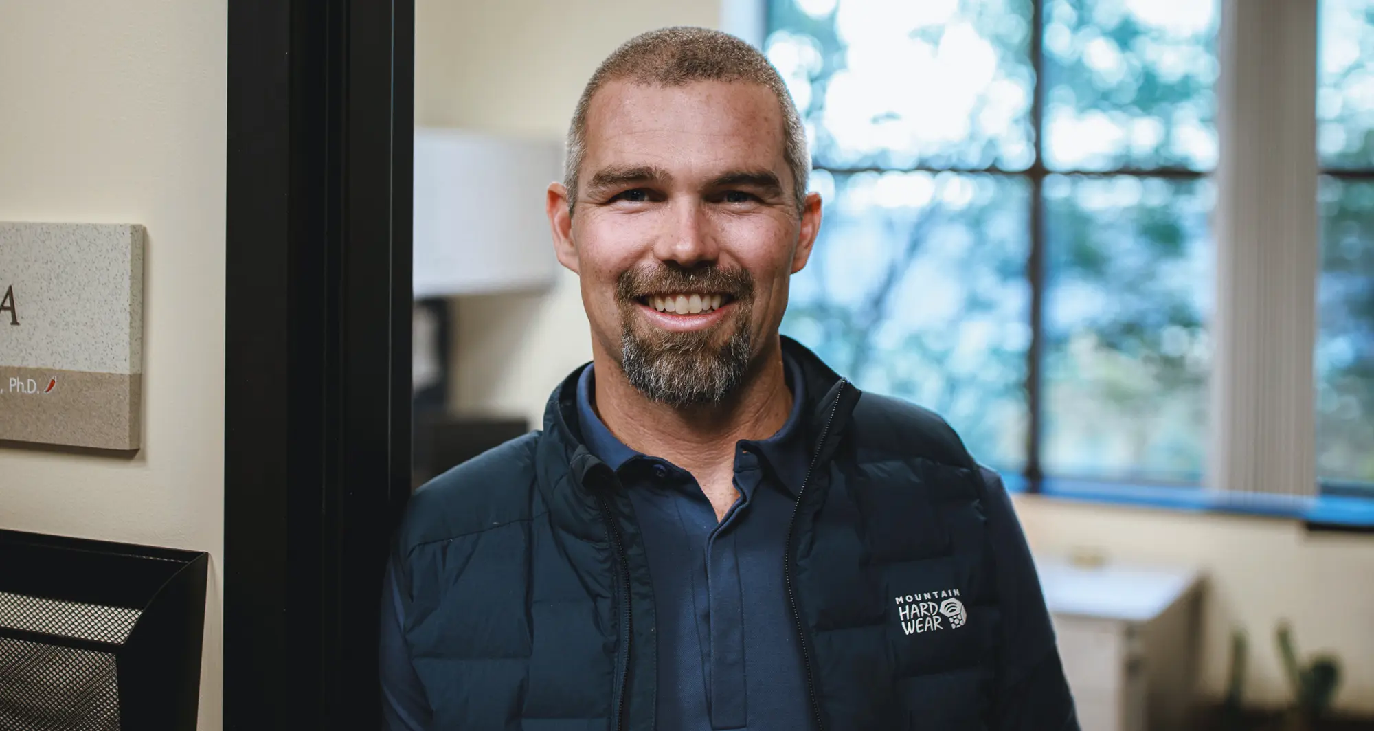
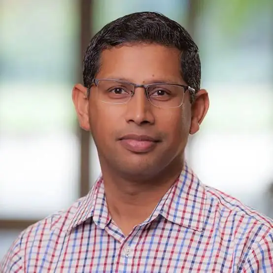
Recipient of the Fishman Awards: Cynthia Schwartz Shenkman Research Excellence Fishman Award Theo Tzaridis discusses his work on pediatric brain tumors, why rigorous preclinical science matters, and how donor support accelerates discoveries.
Established in 2024, the Cynthia Schwartz Shenkman Research Excellence Fishman Award is unique in nature because it recognizes a Sanford Burnham Prebys postdoc for their outstanding biomedical research contributions and demonstrated track record of research excellence.
What’s your current role and focus at Sanford Burnham Prebys?
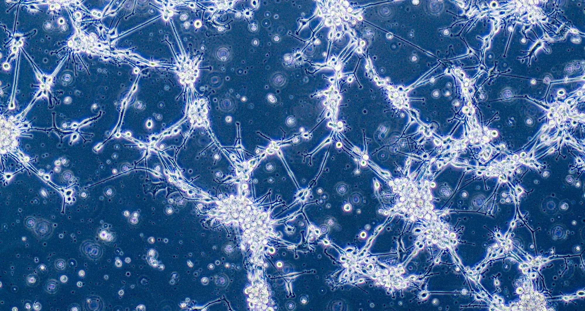
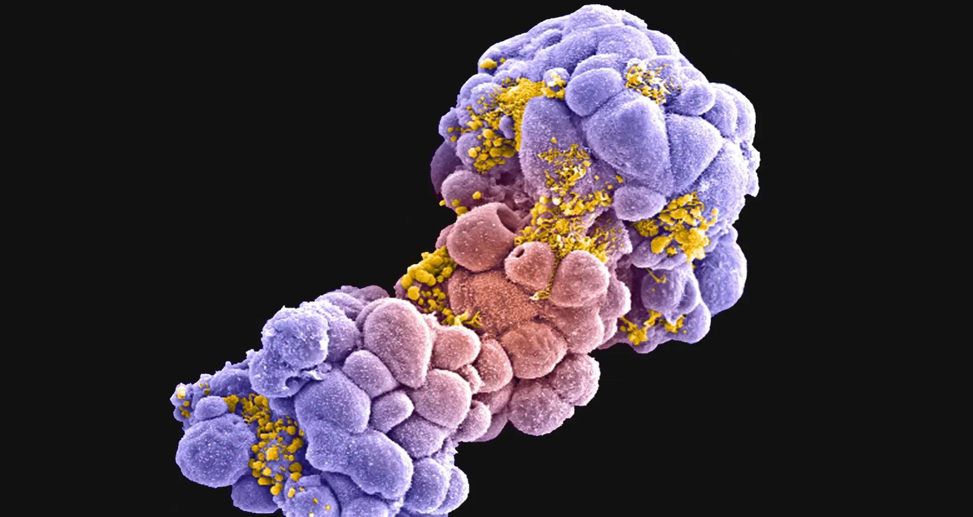
I’m a physician–scientist studying pediatric brain tumors. I focus on diffuse midline glioma (DMG). I joined Rob Wechsler-Reya’s group at the institute and benefited from him as an amazing mentor and his expertise in mouse modeling of brain tumors tremendously. After Rob moved institutions, I joined Peter Adams’s lab. Peter’s aging and cancer perspective gives my immunotherapy work a fresh lens and he is a truly spectacular mentor. We’ve built a DMG “niche” in the lab and I’ve deepened my in vivo skills, which are essential for translating ideas toward the clinic.
What drew you into oncology and neurology?
Even in high school I was fascinated by how a cell can go “crazy”, grow uncontrollably and form a tumor. Medicine let me pair that curiosity with real patient impact. My MD thesis work in Heidelberg, Germany, suggested an old chemotherapy could reactivate a tumor suppressor which paved the way for a clinical trial. During my neurology residency in Bonn, Germany, I helped plan, analyze, and published results from a clinical trial that became the first positive glioblastoma study in 14 years. Those experiences were very rewarding and cemented my focus on translational research.
You mentioned that your approach to immunotherapy starts with “back to basics.” What does that mean?
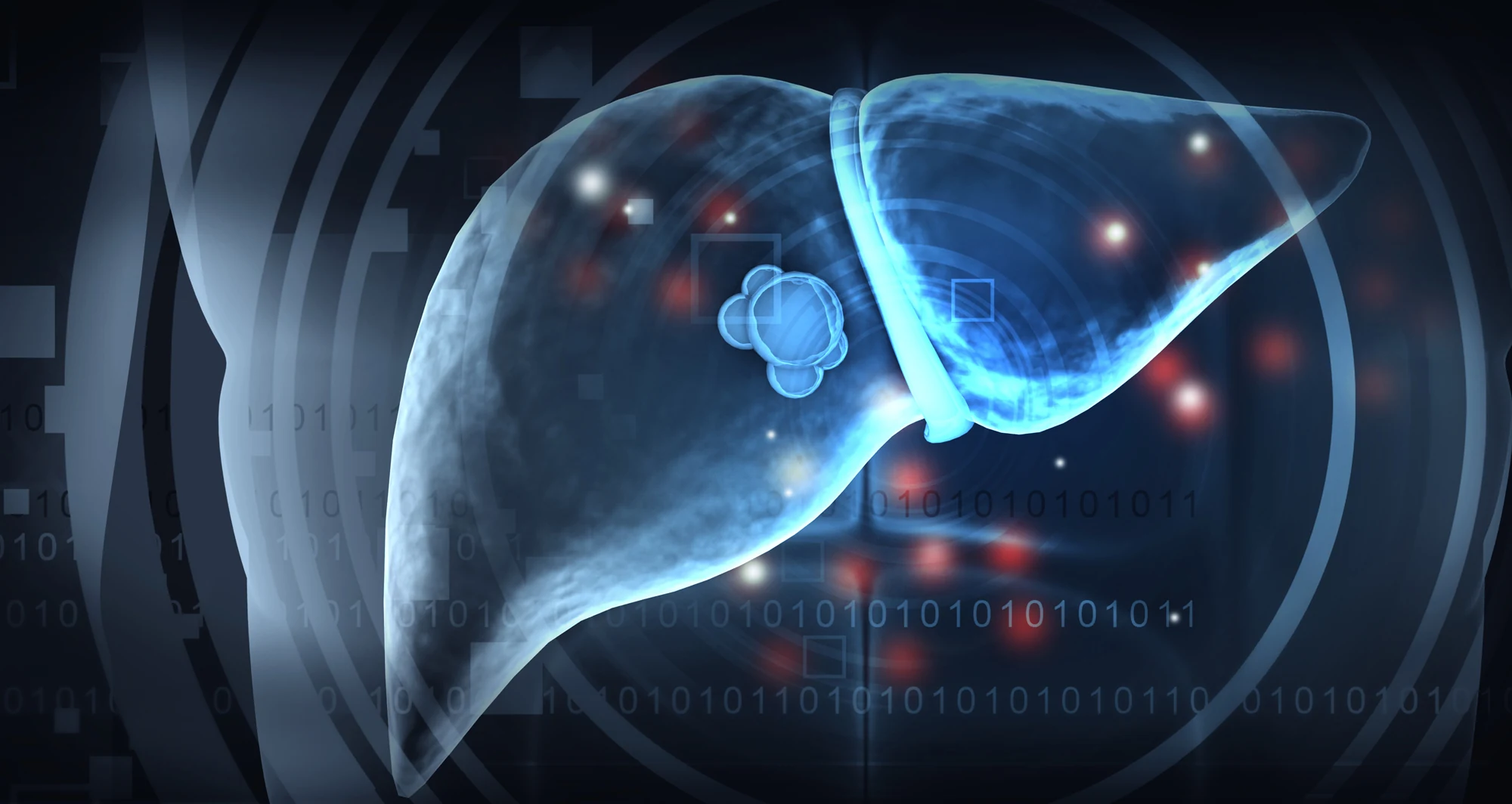
Many brain tumor trials borrowed targets from other cancers without confirming those targets exist in the brain tumor microenvironment. We went back to basics, systematically profiled immune checkpoint molecules present in DMG and found CD155 (also called the poliovirus receptor) consistently expressed across models and patient samples. That points to smarter targeting rather than one-size-fits-all strategies.
How has the Institute’s environment shaped your work?
The culture at Sanford Burnham Prebys is genuinely team oriented. Core facilities (flow cytometry, mouse) are exceptional partners in experimental design. We also engage in a cross-institution “Brain Tumor Club” on the Mesa and contribute data to a molecular tumor board that informs real treatment decisions. In one case, marker data I generated supported a physician’s plan to pursue a personalized immune therapy known as CAR T-cells for a child which was an incredibly meaningful moment.
Any notable collaborations beyond campus?
Yes. Our in vivo expertise enabled joint studies with Emory University, including work on small molecules for pediatric brain tumors. We have also collaborated with Columbia University and the Dana Farber Institute. These multi-site projects help validate findings independently which is critical in pediatrics where patient numbers are limited.
How did the Fishman Awards affect your trajectory?
The Fishman Career Development Award I received in 2023, and the Cynthia Schwartz Shenkman Research Excellence Fishman Award I recently received provided fuel at key moments. The Fishman Career Development Award sent me to the American Association for Cancer Research (AACR) conference in 2024, where I met a company carrying the only clinical-grade antibody to my target; after an MTA, we’re now testing it here. I also attended the La Jolla Immunology Conference and received a best oral presentation award which is validation that stretching into complex immunology is worth it. Importantly, the Fishman Award application process itself which includes writing, presenting, getting feedback, built resilience and sharpened my vision.
Where do you want to take this next?
I aim to lead an independent lab tightly linked to a clinical trials unit. Success requires basic scientists and clinicians at the same table from day one, plus rigorous preclinical “homework” to identify the subgroups most likely to benefit before launching trials. It’s harder, but in the long run it saves precious time and resources and gives patients better odds.
What is life like outside the lab?
I’m a dad of two, so there is hardly time for anything, but we try to do hikes and some beach time. San Diego’s landscapes are a gift. Before kids I did theater; these days, I read when I can, and we take short family adventures (Anza-Borrego is a favorite).
Is there anything you’d like supporters to know?
Your support is more than funding, it’s belief. At a time when the value of science may be questioned, you’re helping researchers communicate clearly, collaborate widely, and move ideas toward children who can’t wait. The Fishman Awards exemplify that: they strengthen science and the storytelling that brings people along. Thank you.