grants Archives - Page 2 of 5 - Sanford Burnham Prebys
Institute News

San Diego Nathan Shock Center announces pilot grant awardees

AuthorMiles Martin
Date

December 21, 2021

The San Diego Nathan Shock Center (SD-NSC) of Excellence in the Basic Biology of Aging, a consortium between Sanford Burnham Prebys, the Salk Institute for Biological Studies and the University of California San Diego, has announced its second-year class of pilot grant awardees. Recipients from six different institutions will receive up to $15,000 to pursue research that advances our understanding of how humans age, with the ultimate goal of extending health span, the number of years of healthy, disease-free life. 

Aging is the biggest risk factor for most human diseases. Individuals age at different rates, and even specific cells and tissues within a person age differently. This depends on intrinsic properties, including genetics and where cells are in the body, and extrinsic factors, like exposure to environmental toxins and pathogens. Understanding this “heterogeneity” and how it contributes to overall human aging, risk for disease or therapeutic responses is the theme of the SD-NSC and the focus of pilot grant awards

“We are excited to support researchers who are working on these innovative, basic biology of aging research projects,” says Salk Institute Professor Gerald Shadel, who directs the SD-NSC. “The findings from this collective group of projects will deepen our understanding of the heterogeneity of aging, which is key to finding interventions to improve human health span.”

The six pilot grant awardees are: 

  • Leena Bharath, assistant professor at Merrimack College, “Human T cell inflammation in aging”
  • Shefali Krishna, staff scientist at the Salk Institute for Biological Studies, “Characterization and function of mitochondrial age mosaicism and heterogeneity” 
  • Gargi Mahapatra, postdoctoral fellow at Wake Forest School of Medicine, “Identifying mediators of bioenergetic decline in peripheral cells of older adults across a spectrum of cognitive abilities”
  • Chiara Nicoletti, postdoctoral fellow at Sanford Burnham Prebys, “Extracellular vesicles as soluble mediators of accelerated aging within the heterogeneous population of muscle-resident cells in Duchenne muscular dystrophy”
  • Anastasia Shindyapina, instructor in medicine at Brigham and Women’s Hospital and Harvard Medical School, “Unraveling heterogeneous biological aging of mouse immune cells at single-cell resolution”
  • Xu Zhang, research associate at the Mayo Clinic, “The dynamics and heterogeneity of cell fates during cellular senescence.” 

Grant recipients will receive subsidized access to the SD-NSC Research Resource Cores (shared research facilities), necessary reagents/supplies, and access to training workshops offered by the center and its core research facilities. They will also be paired with an established aging-research investigator, who will provide career mentoring and guidance to ensure project success.

Research reported in this announcement was supported by the National Institute On Aging of the National Institutes of Health under award number P30AG068635. The content is solely the responsibility of the authors and does not necessarily represent the official views of the National Institutes of Health.

This piece was originally published by the Salk Institute for Biological Studies.

Institute News

This enzyme is one of the hardest working proteins in the body

AuthorMiles Martin
Date

October 21, 2021

Researchers from Sanford Burnham Prebys have shown that a protein they identified plays a major role in the breakdown of hyaluronic acid, a compound found in the scaffolding between our cells. The findings, published recently in the Journal of Biological Chemistry, could have implications for epilepsy, cancer and other human diseases associated with hyaluronic acid and similar compounds.

They also shed light on one of the most active biochemical processes in the body. 

“Our body turns over hyaluronic acid at an extremely rapid rate, far faster than the other compounds surrounding our cells,” says senior author Yu Yamaguchi, MD, PhD, a professor in the Human Genetics Program at Sanford Burnham Prebys.

Hyaluronic acid, a common ingredient in cosmetic anti-aging products, is a one of several large sugar molecules known as glycosaminoglycans (GAGs). These are found naturally in the extracellular matrix, the complex network of organic compounds surrounding our cells that gives structure to our tissues. In addition to its structural role, the extracellular matrix is involved in regulating the immune system and is critical in the early development of connective tissues like cartilage, bone and skin.

“The extracellular matrix is found in every organ and tissue of the body, and malfunctions in its biochemistry can trigger or contribute to a variety of diseases, some of which we don’t even know about yet,” says Yamaguchi. His team studies how GAGs affect childhood diseases including congenital deafness, epilepsy and multiple hereditary exostoses, a rare genetic disorder that causes debilitating cartilage growths on the skeleton.

Hyaluronic acid is also known to be correlated with several health conditions, depending on its concentration in certain tissues. Reduced levels of hyaluronic acid in the skin caused by aging contribute to loss of skin elasticity and reduced capacity to heal without scarring. Levels of hyaluronic acid in the blood dramatically increase in alcoholic liver disease, fatty liver and liver fibrosis. In addition, hyaluronic acid levels have been correlated with increased tumor growth in certain cancers.

“These compounds are literally everywhere in the body, and we continue to learn about how GAG’s influence disease, but there’s also a lot we still don’t know about how these molecules are processed,” says Yamaguchi, “Research like this is about understanding what’s happening at the molecular level so we can later translate that into treatments for disease.” 

For this study, the team focused on a protein called TMEM2, which they had previously found to break down hyaluronic acid by cutting the longer molecule into manageable pieces for other enzymes to process further. Using mice as a research model, they selectively shut off the gene that codes for TMEM2 and were able to successfully measure precisely how much the absence of TMEM2 affects the overall levels of hyaluronic acid.

The answer: a lot.

“We saw up to a 40-fold increase in the amount of hyaluronic acid in the study mice compared to our controls,” says Yamaguchi. “This tells us that TMEM2 is one of the key players in the process of degrading this compound, and its dysfunction may be a key player in driving human diseases.” 

The team further confirmed this role of the TMEM2 protein by using fluorescent compounds that detect hyaluronic acid to determine where the TMEM2 protein is most active. They found the most activity on the surface of cells lining blood vessels in the liver and lymph nodes, which are known to be the main sites of hyaluronic acid degradation. 

“These findings refine our understanding of this critical biochemical process and set us up to explore it further in the interest of developing treatments for human diseases,” says Yamaguchi. “Hyaluronic acid is so much a part of our tissues that there could be any number of diseases out there waiting to benefit from discoveries like these.”

Institute News

Conrad Prebys Foundation provides $3 million for pediatric brain cancer research

AuthorSusan Gammon
Date

April 7, 2021

Conrad Prebys was an extraordinary man and a passionate philanthropist. Today, his generosity extends beyond his life through the Conrad Prebys Foundation.

This year, the Foundation provided $3 million to Robert Wechsler-Reya, PhD, and his team of researchers to advance a potential drug to treat medulloblastoma—the most common malignant brain tumor in children.

Children with medulloblastoma often receive aggressive treatment (surgery, radiation and chemotherapy), but many still die of their disease, and survivors suffer long-term effects from therapy. Safer and more effective therapies are desperately needed.

Wechsler-Reya recently combined forces with Michael Jackson, PhD, senior vice president of Drug Discovery and Development, to find a drug(s) that would inhibit the growth of Group 3 medulloblastoma, the most aggressive form of the disease. Using high-throughput screening technology, they identified a compound that reduces levels of a protein called MYC, which is found at exceptionally high levels in Group 3 medulloblastoma, as well as in cancers of the blood, breast, lung and prostate.

“An effective MYC inhibitor could have a major impact on the survival and quality of life of patients with medulloblastoma,” says Wechsler-Reya. “We identified a compound that reduces levels of MYC in medulloblastoma cells, but now we need to learn how it works to optimize it as an anti-cancer drug and advance studies toward the clinic.

“Historically, pharmaceutical companies and funding agencies have under-invested in childhood cancers, and the majority of drugs currently used to treat these cancers were originally developed for adult cancer,” adds Wechsler-Reya. “We believe that effective drugs for pediatric brain tumors must be developed—and this award from the Foundation will help us achieve this goal.”

“We are profoundly grateful to Conrad for his generosity over the years,” says President Kristiina Vuori, MD, PhD “He has a special legacy at our Institute, which was renamed Sanford Burnham Prebys in 2015 to honor him. We are now thankful to his Foundation for including us in their inaugural grant cycle, and for supporting the critical work we do to benefit children and others suffering from cancer.”

The Conrad Prebys Foundation allocated $78 million in its inaugural grant cycle to fund 121 projects. The awards reflect areas of personal interest to Conrad Prebys—including visual and performing arts, higher education, health care, youth development and animal conservation.

Sanford Burnham Prebys joins a long list of recipients, which included other prominent San Diego institutions such as Rady Children’s Hospital, KPBS, San Diego State University, Scripps Research, Museum of Contemporary Art San Diego and the La Jolla Music Society.

Institute News

Scientists design potential drug for triple-negative breast cancer

AuthorMonica May
Date

February 16, 2021

Drug candidate blocks autophagy, a cellular recycling process that cancer cells hijack as a way to resist treatment

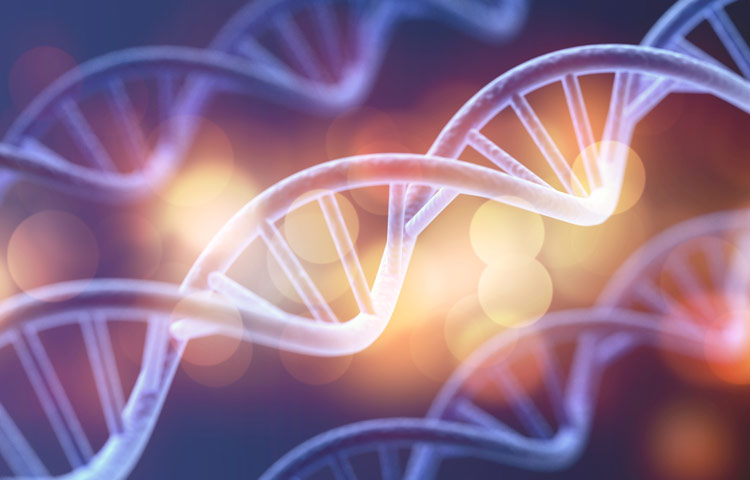
Scientists at Sanford Burnham Prebys Medical Discovery Institute have designed a next-generation drug, called SBP-7455, which holds promise as a treatment for triple-negative breast cancer—an aggressive cancer with limited treatment options. The drug blocks a cellular recycling process called autophagy, which cancer cells hijack as a way to resist treatment. The proof-of-concept study was published in the Journal of Medicinal Chemistry.

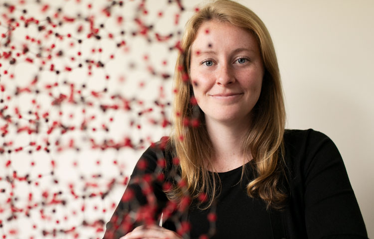
“Scientists are now learning that autophagy is one of the main ways that cancer cells are able to survive, even in the presence of growth-blocking treatments,” says Huiyu Ren, a graduate student in the laboratory of Nicholas Cosford, PhD, at Sanford Burnham Prebys, and first author of the study. “If all goes well, we hope this compound will stop cancer cells from turning on autophagy and allow people with triple-negative breast cancer to benefit from their treatment for as long as possible.”

Cells normally use autophagy as a way to recycle waste products. However, when cancer cells’ survival is threatened by a growth-blocking treatment, this process is often “revved up” so the cancer cell can continue to receive nutrients and keep growing. Certain cancers are more likely to rely on the autophagy process for survival, including breast, pancreatic, prostate and lung cancers.

“While this study focused on triple-negative breast cancer, an area of great unmet need, we are actively testing this drug’s potential against more cancer types,” says Cosford, professor and deputy director in the National Cancer Institute (NCI)-designated Cancer Center at Sanford Burnham Prebys and senior author of the study. “An autophagy-inhibiting drug that stops treatment resistance from taking hold would be a great addition to an oncologist’s toolbox.”

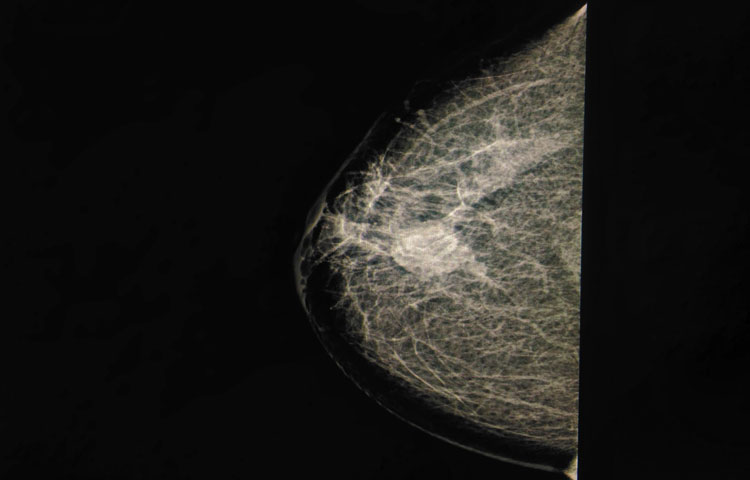
About 15% to 20% of all breast cancers are triple negative, which means they do not respond to hormonal therapy or targeted treatments. The cancer is currently treated with surgery, chemotherapy and radiation, and is deadlier than other breast cancer types. If the tumor returns, other treatments such as PARP inhibitors or immunotherapy are considered. People under the age of 50 are more likely to have triple-negative breast cancer, as well as women who are Black, Hispanic, and/or have an inherited BRCA mutation.

An optimized drug

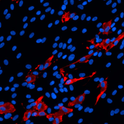
In this study, the scientists optimized a first-generation drug they created in 2015. The result is a compound called SBP-7455 that blocks two autophagy proteins, ULK1 and ULK2. SBP-7455 exhibits promising bioavailability in mice and reduces autophagy levels in triple-negative breast cancer cells, resulting in cell death. Importantly, combining the drug with PARP inhibitors, which are currently used to treat people with recurrent triple-negative breast cancer, makes the drug even more effective.

“We are hopeful that we have found a new potential therapy for people living with triple-negative breast cancer,” says Reuben Shaw, PhD, a study author and professor in the Molecular and Cell Biology Laboratory and director of the NCI-designated Cancer Center at the Salk Institute. “We envision this drug being used in combination with targeted therapies, such as PARP inhibitors, to prevent cancer cells from becoming treatment resistant.”

Next, the scientists plan to test the drug in mouse models of triple-negative breast cancer to confirm that the compound can stop tumor growth in an animal model. In parallel, they will continue optimization efforts to ensure the drug has the greatest chance of clinical success.

“Triple-negative breast cancer is one of the hardest cancers to treat today,” says Ren. “I hope that our research marks the start of a path to successful treatment that helps more people survive this aggressive cancer.”


Additional study authors include Nicole A. Bakas, Mitchell Vamos, Allison S. Limpert, Carina D. Wimer, Lester J. Lambert, Lutz Tautz, Maria Celeridad and Douglas J. Sheffler of Sanford Burnham Prebys; Apirat Chaikuad and Stefan Knapp of the Buchmann Institute for Molecular Life Sciences and Goethe-University Frankfurt; and Sonja N. Brun of the Salk Institute.

This work was supported by the National Institutes of Health (P30CA030199, T32CA211036), Epstein Family Foundation, Larry L. Hillblom Foundation (2019-A-005-NET), Pancreatic Cancer Action Network (19-65-COSF), SGC—a registered charity that receives funds from AbbVie, Bayer Pharma AG, Boehringer Ingelheim, Canada Foundation for Innovation, Eshelman Institute for Innovation, Genome Canada through Ontario Genomics Institute [OGI-196], EU/EFPIA/OICR/McGill/KTH/Diamond, Innovative Medicines Initiative 2 Joint Undertaking (875510), Janssen, Merck KGaA, Merck & Co, Pfizer, São Paulo Research Foundation-FAPESP, Takeda, and Wellcome.

The study’s DOI is 0.1021/acs.jmedchem.0c00873.

Institute News

Mining “junk DNA” reveals a new way to kill cancer cells

AuthorMonica May
Date

February 11, 2021

Scientists unearth a previously unknown vulnerability for cancer and a promising drug candidate that leverages the approach

Scientists at Sanford Burnham Prebys have uncovered a drug candidate, called F5446, that exposes ancient viruses buried in “junk DNA” to selectively kill cancer cells. Published in the journal Cell, the proof-of-concept study reveals a previously unknown Achilles’ heel for cancer that could lead to treatments for deadly breast, brain, colon and lung cancers.

“We found within ‘junk DNA’ a mechanism to stimulate an immune response to cancer cells, while also causing tumor-specific DNA damage and cell death,” says Charles Spruck, PhD, assistant professor in the National Cancer Institute (NCI)-designated Cancer Center and senior author of the study. “This is a very new field of research, with only a handful of papers published, but this has the potential to be a game-changer in terms of how we treat cancer.”

Since the human genome was fully sequenced in 2003, scientists have learned that our DNA is filled with some very strange stuff—including mysterious, noncoding regions dubbed “junk DNA.” These regions are silenced for a reason—they contain the genomes of ancient viruses and other destabilizing elements. An emerging area of cancer research called “viral mimicry” aims to activate these noncoding regions and expose the ancient viruses to make it appear that a cancer cell is infected. The hypothesis is that the immune system will then be triggered to destroy the tumor.

A one-two punch to cancer

In the study, Spruck and his team set out to find the molecular machinery that silences “junk DNA” in cancer cells. Using sophisticated molecular biology techniques, they found that a protein called FBXO44 is key to this process. Blocking this protein caused the noncoding sections of DNA to unwind—but not for long.

“When we revealed noncoding regions, which aren’t meant to be expressed, this caused DNA breakage. This told the cell that something is deeply wrong, and it committed suicide,” explains Spruck. “At the same time, the DNA of the ancient virus was exposed, so the immune system was recruited to the area and caused more cell death. So, we really delivered a one-two punch to cancer.”

The scientists then showed that a drug that targets the FBXO44 pathway, called F5446, shrank tumors in mice with breast cancer. The drug also improved the survival of mice with breast cancer that were resistant to anti-PD-1 treatment, an immunotherapy that is highly effective but often stops working over time. Additional studies in cells grown in a lab dish showed that the drug stops the growth of other tumors, including brain, colon and lung cancers.

The scientists also conducted many experiments to show that this silencing mechanism only occurs in cancer cells, not regular cells. Analysis of patient tumor databases confirmed that FBXO44 is overproduced in many cancers and correlated with worse outcomes—further indicating that a drug that inhibits this protein would be beneficial.

Moving the research toward people

As a next step, the scientists are working with the Conrad Prebys Center for Chemical Genomics to design an FBXO44 pathway-inhibiting drug that is more potent and selective than F5446. This state-of-the-art drug discovery facility is located at Sanford Burnham Prebys.

“Now that we have a compound that works, medicinal chemists can make modifications to the drug so we have a greater chance of success when we test it in people,” says Jia Zack Shen, PhD, staff scientist at Sanford Burnham Prebys and co-first author of the study. “Our greatest hope is that this approach will be a safe and effective pan-cancer drug, which maybe one day could even replace toxic chemotherapy.”


Institute News

What scientists are learning about COVID-19 and the brain

AuthorMonica May
Date

December 8, 2020

We caught up with cell biologist Anne Bang, who recently teamed up with her husband to study how SARS-CoV-2 affects the brain

Brain fog. Memory loss. Dizziness and confusion. Although COVID-19 is primarily thought of as a lung disease, survivors continue to report lingering and highly concerning neurological effects—severe enough to impact their ability to work and live normal lives. Doctors are also seeing a worrisome increase in strokes in younger patients, among other observations.

To learn what scientists know so far about COVID-19 and its effect on the brain, we caught up with Anne Bang, PhD, director of Cell Biology at Sanford Burnham Prebys’ Conrad Prebys Center for Chemical Genomics. Bang recently teamed up with scientists at Penn Medicine and a virologist at Scripps Research—who also happens to be her husband—to investigate whether SARS-CoV-2 infects brain cells. Their findings were published in Cell Stem Cell.

What do scientists know about the brain and COVID-19 so far?

Unfortunately, information is still very limited. There are reports of viral replication in the brain and spinal cord fluid of people with COVID-19 who have neurological symptoms. But as you can imagine, taking brain biopsies from someone who has COVID-19 is not realistic. So we really don’t know a lot yet. For this reason, scientists are turning to systems that can model the human brain, such as brain cells created from induced pluripotent stem cells (iPSCs) and brain organoids, to study SARS-CoV-2’s impact on the brain.

What did you find in your study?

We created several types of brain cells using iPSCs and brain organoids, which we then infected with SARS-CoV-2. We found that SARS-CoV-2 primarily infects a brain cell type called choroid plexus cells—largely bypassing neurons and astrocytes. The choroid plexus is a specialized part of the blood-brain barrier, which controls what can enter your brain and produces cerebral spinal fluid. More research emerges every day, but so far, the consensus in the field seems to align with our findings.

SARS_CoV2_ Infected human choroid plexus cells a type of brain cell

The scientists found that SARS-CoV-2 (red) primarily infects brain cells called choroid plexus cells (blue), which are part of the brain’s protective blood-brain barrier.

How might this finding translate to what we’re seeing in patients?

We know that choroid plexus cells produce high levels of ACE2, which is the receptor that SARS-CoV-2 uses to enter and infect cells. Because the choroid plexus is the “gatekeeper” to the brain, it’s possible that the virus enters the brain by infecting these cells. However, much more research is needed before we can give a definitive answer to this question.

We have more questions than answers right now about COVID-19. What is one question you wish we had the answer to?

How does the virus get from the nose and mouth and spread to other parts of the body? This is a big question for me and the scientific field. Once we know how the virus travels throughout the body, we can potentially stop its spread and control the dangerous symptoms.

What was it like working with your husband? Was this your first time working together?

It was really fun. I found out that he is great to work with. We’ve been together for 30 years, and incredibly, this was the first time we worked together.

Institute News

Top San Diego researchers receive $5 million to study cellular aging

Authorsgammon
Date

September 22, 2020

Professors Peter Adams, PhD, and Malene Hansen, PhD, of Sanford Burnham Prebys will lead key research and development cores

Sanford Burnham Prebys researchers are joining forces with University of California San Diego (UC San Diego) and the Salk Institute to form a world-class San Diego Nathan Shock Center (SD-NSC), a consortium established to study cellular and tissue aging in humans. The center will be funded by the National Institute on Aging (NIA), part of the National Institutes of Health, and is expected to receive $5 million over the next two years.

Professors Peter Adams, Ph.D., and Malene Hansen, PhD, of Sanford Burnham Prebys will lead key research and development cores, along with Professors Rusty Gage, PhD, Martin Hetzer, PhD, and Tatyana Sharpee, PhD, of Salk; and Anthony Molina, PhD, of UC San Diego. Salk Professor Gerald Shadel, PhD, will be the director of the SD-NSC.

“This is a special opportunity for San Diego’s aging research community to share our ideas, skills and technologies to drive innovative research in the basic biology of aging,” says Adams. “We are grateful for this support and will work to create the strongest environment possible to achieve meaningful breakthroughs that will benefit human health.”

Aging is the most significant risk factor for human disease. Human cells and tissues age at different rates depending on their intrinsic properties, where they are in the body and environment exposures. Yet, scientists do not fully understand this variability (“heterogeneity”) and how it contributes to overall human aging, risk for disease or therapeutic responses.

To explore the complex heterogeneity of human aging, the SD-NSC will deploy three cutting-edge Research Resource Cores, including the Human Cell Models of Aging Core, to be led by Gage and Molina; the Heterogeneity of Aging Core, to be led by Hetzer and Adams; and the Integrative Models of Aging Core, to be led by Sharpee.

The cores will allow detailed analysis of human cells and organoids (artificially grown clusters of cells that model tissues), derived from a unique aging cohort at UC San Diego that is annotated for multiple measures of the actual biological age of individuals. In addition, the cores will provide scientific services to the Nathan Shock Centers network and the aging research community at large, including the dissemination of samples, protocols and computational tools to facilitate the study of heterogeneity in aging.

A Research Development Core headed by Hansen will also be established to encourage and support new investigators to enter the field of aging research. Through this core, the SD-NSC will provide pilot research grants, workshops and customized mentoring programs to promote the research and development of young investigators, as well as in-person and virtual trainings to spur collaboration and the sharing of knowledge related to the basic biology of aging.

“For years, my colleagues and I have been organizing successful symposia such as the annual La Jolla Aging Meeting (LJAM), where we share new aging research and discuss opportunities for collaboration,” says Hansen, who also hold the positions of associate dean for Student Affairs and faculty adviser for Postdoctoral Training at Sanford Burnham Prebys. “The San Diego Nathan Shock Center will enable us to broaden the reach and impact of LJAM, as well as take training and mentoring of the next generation of researchers to a new level.”

The SD-NSC builds on Sanford Burnham Prebys’ strengths in fundamental aging research, renowned for its use of model organisms to unravel cell changes associated with normal development and aging. By building, analyzing and probing models of disease, scientists in the Institute’s Aging, Development and Regeneration Program are providing new tools to uncover novel therapeutic targets for heart disease, neurodegeneration, muscle disorders, diabetes, cancer and other debilitating diseases.

The SD-NSC will be one of a network of eight Nathan Shock Centers nationwide, and is funded by NIA grant number P30AG068635.

Institute News

Sanford Burnham Prebys researchers awarded 2020 Padres Pedal the Cause grants

AuthorSusan Gammon
Date

July 9, 2020

We are pleased to announce that Padres Pedal the Cause (PPTC) has awarded three collaborative research grants to Sanford Burnham Prebys, Moores Cancer Center at UC San Diego Health and the Salk Institute. Funding for the research comes from the record setting $3.1 million raised in the 2019 event and brings the lifetime raise for PPTC to over $13 million.

PPTC’s goal is to leverage the strengths of San Diego—home to three nationally recognized NIH cancer institutions and a renowned pediatric hospital. Each grant unites scientists at beneficiary institutions and aims to advance research toward developing therapies to attack and cure cancer.

Congratulations to the recipients!

  • Robert Wechsler-Reya, PhD, (SBP) and John Crawford, MD, (Moores Cancer Center/Rady Children’s) will work on a new approach to treat medulloblastoma—the most common malignant brain tumor in children.
  • Garth Powis, D. Phil., (SBP) Pradipta Ghosh, MD, (Moores Cancer Center) and Michael Bouvet, MD, (Moores Cancer Center) are joining forces to find medical treatments for gastric cancer—a disease for which no therapy exists. 
  • Nicholas Cosford, PhD, (SBP) Hatim Husain, MD, (Moores Cancer Center) and Reuben Shaw, PhD, (Salk Institute) will perform a first-of-its-kind study for lung cancer—the number one cause of cancer-related deaths per year.

The PPTC event featured multiple cycling courses, a 5K run or walk, spin classes and kid-friendly activities. The number of participants reached an all-time high of nearly 3,000 in 2019.

Congratulations to everyone who worked, played and cycled their way to success!

Read the full list of 2020 grants funded by Padres Pedal the Cause.

Institute News

Rett Syndrome Foundation funds a potential cure

AuthorSusan Gammon
Date

April 23, 2020

The research may lead to a major step toward a cure.

Rett syndrome is a neurodevelopmental disorder that affects almost all aspects of a child’s life, from walking to eating to intellectual capability. There is no cure for the disease, which occurs mostly in girls, and treatments are aimed at slowing the loss of abilities and alleviating the debilitating symptoms. 

Jing Crystal Zhao, PhD, associate professor at Sanford Burnham Prebys, has received new funding from the Rett Syndrome Foundation to find ways to reverse the changes in a gene that causes Rett syndrome. The research may lead to a major step toward a cure.

“I’m very grateful to the Rett Syndrome Foundation, and excited to begin this project,” says Zhao. “While Rett syndrome may not be well known among the general public, our research may lead to treatments to improve the lives of patients around the world.”

More than 90% of Rett Syndrome cases are caused by genetic changes in a gene called MECP2. Every female carries two copies of the MECP2 gene. Rett syndrome patients carry both a normal and a mutant copy of MECP2. Unfortunately, in some cells, the normal copy of MEPC2 becomes inactive due to a biological process called X-chromosome inactivation—a process that occurs in females—and this leads to Rett syndrome. 

“Recent studies suggest that reversing X-chromosome inactivation could reactivate the normal copy of the MECP2 gene,” says Zhao. “We have identified an DNA element that plays a key role in X-chromosome inactivation. We are now going to test if we can block this element and restore the silent MECP2 gene, which could be life changing. 

“Our aim is to help individuals regain the skills and abilities stolen by Rett syndrome,” adds Zhao. “This award takes us closer to that goal.” 
 

Institute News

Scientists discover an early sign of type 2 diabetes: Misfolded proinsulin

AuthorMonica May
Date

March 19, 2020

The findings could lead to tests or treatments that help prevent type 2 diabetes.

Misfolded proinsulin—a protein the body normally processes into insulin—is an early sign of type 2 diabetes, according to a study by scientists at Sanford Burnham Prebys and the University of Michigan Medical School. The discovery, published in eLife, could lead to tests or treatments that help prevent people from developing type 2 diabetes.

“Understanding the molecular events that occur as prediabetes progresses to diabetes opens new avenues for us to detect or interrupt these processes,” says Randal Kaufman, PhD, director and professor in the Degenerative Diseases Program at Sanford Burnham Prebys and co-corresponding author of the study. “With this information, we can start to find interventions that might spare millions of people from a serious, lifelong condition.”

More than one in three Americans, or approximately 88 million people, have prediabetes—which is characterized by elevated blood sugar. If left untreated, within four years nearly 40% of people with prediabetes develop type 2 diabetes, which occurs when the body doesn’t use insulin properly. In 2017, the cost of treating diabetes exceeded $327 billion, according to the American Diabetes Association. Due to increasing obesity rates, the number of people with the condition—particularly children—is on the rise.

Identifying the molecular events that occur during progression from prediabetes to full-blown diabetes remains one of the most perplexing problems in diabetes research. In the study, the scientists set out to answer this question by tracking proinsulin folding in the beta cells of humans and mice that are healthy, prediabetic and diabetic.

These studies revealed that instead of undergoing its normal folding process, proinsulin proteins were abnormally linked to each other. Levels of the abnormal proinsulin accumulated as prediabetes progressed to type 2 diabetes. Obese mice in the earliest stages of diabetes had the highest levels of abnormal proinsulin in their beta cells.

“Proinsulin misfolding is the earliest known event that may contribute to the progression from prediabetes to diabetes,” says Kaufman. “Together, these studies show that abnormally linked proinsulin holds promise as a potential measure of how close someone may be to developing type 2 diabetes.”

Now, the researchers are set to uncover more details about this process, such as the proteins that interact with the misfolded proinsulin.

“Understanding the fundamental molecular events that lead to type 2 diabetes is critical as the number of people with prediabetes continues to rise,” says Kaufman. “If we don’t find preventive measures, we will soon have a diabetes epidemic.”


The study’s first author is Anoop Arunagiri, PhD; and the study’s senior author is Peter Arvan, both of the University of Michigan Medical School.

Additional authors include Leena Haataja and Fawnnie Pamenan of the University of Michigan Medical School; Ming Liu of the University of Michigan Medical School and Tianjin Medical University in China; Anita Pottekat and Pamela Itkin-Ansari of Sanford Burnham Prebys; Soohyun Kim of Konkuk University in South Korea; Lori M. Zeltser of Columbia University; Adrienne W. Paton and James C. Paton of the University of Adelaide in Australia; and Billy Tsai of the University of Michigan.

The study’s DOI is 10.7554/eLife.44532.

This work was supported by the National Institutes of Health (R01DK111174, R24DK110973 and R01DK48280) and the Juvenile Diabetes Research Foundation International (2-SRA-2018-539-A-B).