grants Archives - Page 3 of 5 - Sanford Burnham Prebys
Institute News

Padres Pedal the Cause donates $3.1 million to cancer research

AuthorMonica May
Date

February 18, 2020

Garth Powis, who heads our NCI-designated Cancer Center, joined representatives from the beneficiary institutes onstage for the check presentation ceremony.

Padres Pedal the Cause (PPTC) hit it out of the ballpark, revealing that its 2019 event at Petco Park raised a record $3.1 million to accelerate local cancer research. 

The amount, revealed in an evening ceremony held on Thursday, February 6, brings the total raised to $13.2 million. Garth Powis, D. Phil., director of Sanford Burnham Prebys’ National Cancer Institute (NCI)-designated Cancer Center—one of only seven in the nation—joined representatives from the beneficiary institutes onstage for the check presentation ceremony. 

The audience in the packed auditorium gave the news a standing ovation. Most guests, who included hundreds of event participants, San Diego business leaders, top donors and fundraisers, as well as Padres Pedal founders Bill and Amy Koman, have firsthand experience with cancer—the number-one cause of death in San Diego. 

Typically held in November, PPTC features multiple cycling courses, a 5K run or walk, spin classes and kid-friendly activities. The number of participants has steadily increased since the event’s launch in 2013—reaching an all-time high of nearly 3,000 individuals last year. 

The nonprofit’s goal is to leverage the incredible strengths of San Diego—home to three nationally recognized NIH cancer institutions and a renowned pediatric hospital—to bring us closer to cancer cures. Each PPTC grant unites scientists at two or more of the four beneficiary institutions, which include Sanford Burnham Prebys, Moores Cancer Center at UC San Diego Health, Rady Children’s Hospital and the Salk Institute for Biological Studies. Past grants have accelerated our research into cancers of the breast, skin, brain, colon, pancreas and more. 

The 2020 event date will be revealed in mid-April. Visit www.gopedal.org for the latest details.

Institute News

Scientists discover new survival strategy for oxygen-starved pancreatic cancer cells

AuthorMonica May
Date

October 23, 2019

Oxygen is essential to life. When fast-growing tumor cells run out of oxygen, they quickly sprout new blood vessels to keep growing, a process called angiogenesis. 

By blocking pancreatic cancer’s oxygen-sensing machinery—the same field of research studied by the winners of the 2019 Nobel Prize in Medicine—Sanford Burnham Prebys scientists have uncovered a new way that tumors turn on angiogenesis in an animal model. The discovery, published in Cancer Research, could lead to a treatment that is given with an anti-angiogenetic medicine, thereby overcoming drug resistance. 

“Treatment resistance is a major challenge for cancer treatments that block blood vessel growth,” says Garth Powis, D.Phil., professor and director of Sanford Burnham Prebys’ National Cancer Institute (NCI)-designated Cancer Center and senior author of the study. “Our research identifies a new way angiogenesis is activated, opening new opportunities to find medicines that might make existing cancer treatments more effective.” 

Many cancer treatments work by blocking angiogenesis, which rarely occurs in healthy tissues. However, these medicines eventually stop working, and the cancer returns, sometimes in as little as two months. Scientists have been researching why this treatment resistance occurs so it can be stopped.

In this study, the scientists focused on pancreatic cancer, which is notoriously desperate for oxygen and also difficult to treat. Fewer than 10% of people diagnosed with pancreatic cancer are alive five years later. 

To see how a pancreatic tumor responds to a disruption in its oxygen supply, the Sanford Burnham Prebys researchers used a mouse model to block an oxygen-sensing protein called HIF1A—which should cripple the tumor’s growth. Instead of dying, however, after about a month the cells multiplied—indicating they had developed a new way to obtain oxygen. 

Further work revealed that the cancer cells were clear and swollen with the nutrient glycogen (a characteristic also seen in some ovarian and kidney cancers). In response to the excess glycogen, special immune system cells were summoned to the tumor, resulting in blood vessel formation and tumor survival. Each of these responses represents a new way scientists could stop pancreatic tumors from evolving resistance to treatment.

“Our team’s next step is to test tumor samples from people with pancreatic cancer to confirm this escape mechanism occurs in a clinical setting,” says Powis. “One day, perhaps we can create a second medicine that keeps anti-angiogenic drugs working and helps more people survive pancreatic cancer.”


Research reported in this press release was supported by the U.S. National Institutes of Health (NIH) (5F31CA203286, CA216424 and P30CA030199). The content is solely the responsibility of the authors and does not necessarily represent the official views of the NIH. The study’s DOI is 10.1158/0008-5472.CAN-18-2994. 

Institute News

Sanford Burnham Prebys awarded Padres Pedal the Cause grants to advance cancer research

AuthorMonica May
Date

September 13, 2019

Sanford Burnham Prebys scientists have been awarded two collaborative grants with Rady Children’s Hospital and UC San Diego Health from Padres Pedal the Cause (PPTC), an annual fundraiser that aims to accelerate cancer cures. The projects unite the complementary strengths of clinicians and scientists with the hope of uncovering new treatments for colorectal, lung, breast and prostate cancers. 

The grants stem from the record-breaking $2.94 million raised by thousands of participants in the November 2018 event. Launched in 2013, all of the proceeds raised by PPTC stay in San Diego to fund collaborative research that brings us closer to a world without cancer. Past PPTC grants have supported our Institute’s research into cancers of the breast, skin, brain, colon, pancreas and more.

The funded projects are described below:

  • Protecting the gut and halting colon cancer growth (Svasti Haricharan, PhD, and Scott Peterson, PhD, of Sanford Burnham Prebys; Soumita Das, PhD, and Pradipta Ghosh, MD, of UC San Diego Health; Debashis Sahoo, PhD, of UC San Diego Health and Rady Children’s Hospital; and Sherry C. Huang, MD, of Rady Children’s Hospital)

This project will discover and characterize a pathway in the gut that normally protects the gut barrier from microbes—and is lost during the initiation of colon cancers. The researchers aim to uncover a therapeutic target that protects the gut from cancer-causing microbes and halts the formation and progression of colon polyps. The team will also validate biomarkers for detecting polyps in the colon at high risk for progressing to colon cancer.

  • A new pathway to fractioning cancer (Michael Jackson, PhD, of Sanford Burnham Prebys; and Seth Field, MD, PhD, of UC San Diego Health)

To effectively combat cancer, therapies directed at new targets must be developed. A protein called GOLPH3 has been shown to drive the growth of several cancers, including lung, breast, prostate and colorectal cancers. This project aims to find a compound that blocks GOLPH3, which would add a unique approach to the arsenal of cancer treatments.

The seventh annual Padres Pedal the Cause event takes place on November 16, 2019. Participants will cycle, run, walk, spin or volunteer in support of a world without cancer. Join our team or volunteer at our aid station in Mountain Hawk Park in Chula Vista.

Institute News

How your DNA takes shape makes a big difference in your health

AuthorMonica May
Date

September 10, 2019

The more we learn about our genome, the more mysteries arise. For example, how can people with the same disease-causing mutation have different disease progression and symptoms? And despite the fact that it’s been more than 15 years since the human genome was sequenced, why can’t we explain the significance of the vast majority of genomic variations that occur in noncoding, or “junk,” elements of the genome?

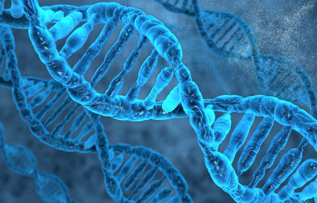
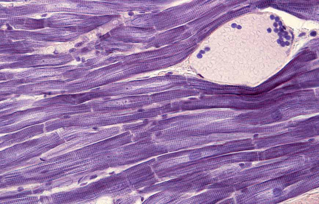
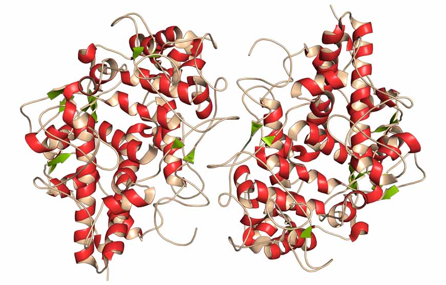
Now, Pier Lorenzo Puri, MD, a professor in the Development, Aging, and Regeneration Program at Sanford Burnham Prebys, has used a cutting-edge technique called Hi-C, which maps millions of interactions between proteins and tightly coiled DNA, called chromatin, to shed light on this mystery. The study, published in Molecular Cell, shows that a specific protein called MyoD—a master regulator of muscle development—reshapes chromatin’s architecture to alter gene expression—revealing fundamental insights into how genetic variations may affect our health. 

“One of the greatest mysteries of medicine is how people with the same mutation can have different symptoms,” says Puri. “Our study indicates that some genetic variations may affect our health by altering how DNA coils and interacts in its 3D shape. This alteration may be helpful or detrimental—and could even explain why some people seem to be naturally athletic.” 

In the study, the scientists used several genomic technologies to map the interactions between MyoD and chromatin as cells turned into skeletal muscle upon MyoD expression. Among other findings, the scientists determined that MyoD rearranged chromatin’s shape during this process—similar to the retying of a tangled shoelace. Importantly, the researchers found that MyoD-driven reconfiguration of 3D chromatin architecture is mediated by interactions between noncoding elements of the genome—where most disease-associated genetic variants occur. These findings demonstrate that the noncoding genome can act as a structural element that defines the chromatin architecture—key information that will help predict the functional outcomes of these variants.

Puri is already applying this insight to help solve other genomic mysteries. He plans to review a worldwide database of gene variations with unknown significance—meaning that scientists are unsure if the change is harmless or a risk factor for disease. Then, he aims to create models that help us better understand the impact of these genetic variations on an individual’s ability to respond to environmental changes and eventually develop disease. 

“It’s possible that many genetic variations alter chromatin folding. Instead of directly causing disease, the changes may increase or decrease our disease risk,” explains Puri. “I hope that my next studies will shed light on these genomic mysteries and help more people get definitive answers about what lies in their DNA.” 


The co-first authors of the study are Alessandra Dall’Agnese, PhD, and Luca Caputo, PhD, of Sanford Burnham Prebys. 

Additional study authors include Chiara Nicoletti, PhD, of Sanford Burnham Prebys and University of Modena and Reggio Emilia; Sole Gatto and Ranjan Perera of Sanford Burnham Prebys; Julia di Iulio, PhD, and Amalio Telenti, MD, PhD, of Scripps Research; Anthony Schmitt, PhD, Yarui Diao, PhD, and Zhen Ye of the Ludwig Institute for Cancer Research; Mattia Forcato, PhD, and Silvio Bicciato, PhD, of University of Modena and Reggio Emilia; and Bing Ren, PhD, of the Ludwig Institute for Cancer Research and UC San Diego School of Medicine. The study’s DOI is 10.1016/j.molcel.2019.07.036. 

Research reported in this article was supported by the U.S. National Institutes of Health (NIH) (R01AR056712, R01AR052779, AR061303), Epigen, Ellison Medical Foundation (AG-NS-0843-11), AFAR (G16294AD), Ludwig Institute for Cancer Research, Human Frontier Program and San Diego Muscle Research Center. The content is solely the responsibility of the authors and does not necessarily represent the official views of the NIH.

Institute News

Cancer’s final frontier: the tumor microenvironment

AuthorMonica May
Date

September 3, 2019

Cancer researchers are setting their sights on a new kind of cancer treatment that targets the tumor’s surrounding environment, called the tumor microenvironment, in contrast to targeting the tumor directly. 

To learn more about this approach, we spoke with cancer experts Jorge Moscat, PhD, director and professor in the Cancer Metabolism and Signaling Networks Program at Sanford Burnham Prebys; and Maria Diaz-Meco, PhD, professor in the Cancer Metabolism and Signaling Networks Program at Sanford Burnham Prebys. Both scientists recently authored a review article centered on a family of cancer-linked proteins that regulate the tumor’s microenvironment. The paper was published in Cancer Cell

What is the tumor microenvironment exactly? 
Moscat: Just like every person is surrounded by a supportive community—their friends, family or teachers—every tumor is surrounded by a microenvironment. This ecosystem includes blood vessels that supply the tumor with nutrients; immune cells that the tumor has inactivated to evade detection; and stroma, glue-like connective tissue that holds the cells together and provides the tumor with nutrients.

Diaz-Meco: These elements are similar to the three legs of a stool. If we remove all three legs, we can deliver a deadly blow to the tumor. FDA-approved drugs exist that target blood vessel growth and reactivate the immune system to destroy the tumor. The final frontier is targeting the stroma.

When did scientists realize it’s important to focus on the tumor’s surroundings—not the tumor itself? 
Diaz-Meco: Scientists have known for more than a century that the tumor’s surroundings are different from normal cells. The tissue surrounding a tumor is inflamed—tumors are often called “wounds that never heal”—and their metabolism is radically different from healthy cells. 

Moscat: The discovery of oncogenes—genes that can lead to cancer—in the 1970s shifted the field’s focus to treatments that target the tumor directly. These targeted treatments work incredibly well, but only for a short time. Cancer researchers are realizing that tumors quickly adapt to this roadblock and become treatment resistant. In addition, many oncogenes are difficult to target, earning the title “undruggable.” As a result, cancer researchers are returning their focus to the tumor microenvironment—especially the stroma. Only a handful of stroma-targeting drugs are in development. None are FDA approved.

Which cancers could benefit most from a stroma-targeting drug? 
Moscat: Pancreatic, colorectal and liver cancers stand to benefit most from a stroma-targeting drug. For example, 90% of a pancreatic tumor consists of stroma—not cancer cells. Combined, these cancers are responsible for more than 20% of all cancer deaths in the U.S. each year. 

What is the focus of your lab’s research? 
Diaz-Meco: Our lab studies the cross talk between tumors and their environment. This conversation is very complex. In addition to “talking” with the tumor, the stroma also “speaks” with the immune system. We are working to map these interactions so we can create drugs that silence this conversation—or change it. For example, we recently showed—in a mouse model that faithfully recapitulates the most aggressive form of human colorectal cancer—that by altering the stroma’s interactions with the immune system, we might make tumors vulnerable to immunotherapy. 

What do new insights into the tumor microenvironment mean for cancer drug development? 
Moscat: It’s likely that the ultimate cancer “cure” won’t be just one drug that kills the tumor cells, but a combination of therapies. I expect this will be a three-part combination treatment that stops blood vessel growth, activates the immune system to attack the tumor and targets the stroma. 

Additionally, this research shows that experimental models of cancer drug development need to take the tumor microenvironment into account. Many current models use mice that lack an immune system—in order to get the tumor to grow—or focus on the tumor in isolation. Based on our knowledge of the tumor microenvironment, this isn’t an accurate representation of human disease. 

Diaz-Meco: In our lab, we have created several animal models of cancers that preserve the immune system and mirror tumor progression. In addition to better modeling human disease, this also allows us to study cancer from its earliest beginnings. This work could lead to early interventions—before the cancer has become large and hard to treat.

Anything else you’d like to add? 
Moscat: We are truly in the golden age of cancer biology. We understand more than we ever have before. New technologies are allowing us to obtain an unprecedented amount of information—we can even map every gene that is “turned on” in a single cancer cell. I am incredibly hopeful for the future. 

Learn more about the future of cancer treatment by attending our next “Conquering Cancer” event at the Fleet Science Center. Details

Institute News

NIH grant aims to boost heart muscle

AuthorMonica May
Date

August 23, 2019

Heart disease is the number one killer of Americans. Now, the National Institutes of Health (NIH) has awarded a four-year grant totaling nearly half a million dollars to Sanford Burnham Prebys to find medicines that could help people repair damaged heart muscle—and potentially reduce the risk of heart attack or other cardiovascular events. 

“Each year we lose far too many loved ones to heart attacks and other heart conditions,” says grant recipient Chris Larson, PhD, adjunct associate professor in the Development, Aging and Regeneration Program at Sanford Burnham Prebys. “Now, we have the opportunity to find medicines that may help more people live long, active lives by strengthening their heart muscles.”

Nearly half of American adults—approximately 120 million people—have cardiovascular disease, according to the American Heart Association and NIH. The condition occurs when blood vessels that supply the heart with oxygen and nutrients become narrowed or blocked, increasing risk of a heart attack, chest pain (angina) or stroke. Current medications for cardiovascular disease can lower blood pressure or thin the blood to minimize risk. Still, five years after a heart attack, 47% of women and 36% of men will die, develop heart failure or experience a stroke. No medicines that repair heart muscle exist. 

To identify drugs that may stimulate heart muscle growth, Larson and his team will screen hundreds of thousands of compounds against human heart muscle cells, called cardiomyocytes. The work will be done in collaboration with Alexandre Colas, PhD, assistant professor in the Development, Aging and Regeneration Program at Sanford Burnham Prebys, who developed the high-throughput screening system that will be employed.

Once the scientists identify drug candidates that promote heart muscle growth, they will study these compounds in additional cellular and animal models of heart disease in the hopes of uncovering insights into the biology behind the repair process. 

“After experiencing a heart attack or other cardiovascular event, many people live in fear that it will happen again,” says Colas. “Today we embark on a journey toward a future where people living with cardiovascular disease don’t have to be afraid of a second heart attack.”

Institute News

Digestion-aiding herbs alter gut microbiome

AuthorMonica May
Date

July 24, 2019

Many medicines used today—including aspirin, penicillin and malaria-fighting quinine—originated from nature. Now, Sanford Burnham Prebys and UC San Diego scientists have turned to ancient digestive herbs to learn about gut health—in the hopes of uncovering new treatments for colon cancer, autoimmune conditions and additional serious diseases.

In a recent study published in Evidence-Based Complementary and Alternative Medicine, the researchers examined how four herbs—turmeric, ginger, long pepper and black pepper—change the gut microbiome. These herbs have been used for more than 5,000 years to aid digestion in Ayurvedic healing, India’s traditional system of medicine. The researchers found that the herbs promoted strong shifts in the gut bacteria that are known to regulate metabolism—providing insights that could help us protect our health. 

“Scientists have long known that these four herbs facilitate digestion and increase bioabsorption of dietary nutrients. However, the effects on the gut microbiome had not been studied,” says Scott Peterson, PhD, senior author of the paper and a professor at Sanford Burnham Prebys. “Our study demonstrates for the first time that these herbs indeed alter the microbiome and produce distinct shifts in microbial populations. This finding is a starting point from which we can begin to decipher how the microbiota may change the gut biochemistry to promote and protect our health.” 

Digestive disorders, including Crohn’s disease, celiac disease and irritable bowel syndrome (IBS), are increasingly prevalent in Western populations. More than 60 million people are affected in the United States alone. Treatments for the disorders are limited.

In the study, the scientists collected stool samples from 12 healthy men and women between the ages of 30 and 60 who ate a vegetarian or vegan diet. The samples were grown in medium (food for bacteria) supplemented with turmeric, ginger, black pepper or long pepper. Genomic sequencing was then used to identify how the abundance of species within the community was altered by the herbal supplement. 

The scientists found that all of the herb-supplemented samples had unique proportions of bacterial families compared to control cultures—indicating the herbs altered the gut microbiome. 

“We are exploring how different herbs produce distinct microbial signatures in the gut,” says Peterson. “It’s clear from this study that each herb works differently. Now the task is to make the connections between the herb profiles and gut health.” 

Next, the researchers plan to test the herbs’ therapeutic potential in a controlled human clinical trial. In parallel, they will work in the lab to dissect the herbs’ molecular components and study how these components influence the gut microbiome and promote digestive health.

“By delving deeper into the beneficial molecules present in these herbs and how microbes may alter those constituents, we may be able to enhance their potential benefit and help people suffering from serious digestive disorders,” explains Peterson.  


The first author of the study is Christine T. Peterson, PhD, of UC San Diego. 

Additional authors include Dmitry A. Rodionov, PhD, of Sanford Burnham Prebys and the Russian Academy of Sciences; Stanislav N. Iablokov of the Russian Academy of Sciences and Yaroslavl State University; Meredith A. Pung, PhD, and Paul J. Mills, PhD, of UC San Diego; Deepak Chopra, MD, of UC San Diego and the Chopra Foundation. Deepak Chopra is the founder of the Chopra Foundation and Chopra Center and a co-owner of the Chopra Center. Mills is the director of research for the Chopra Foundation.

The research was supported by the Samuel Lawrence Foundation, the Chopra Foundation and the Russian Science Foundation (19-14-00305).

Institute News

Antimicrobial protein implicated in Parkinson’s disease

AuthorMonica May
Date

July 17, 2019

An immune system protein that usually protects the body from pathogens is abnormally produced in the brain during Parkinson’s disease, scientists from Sanford Burnham Prebys report. The discovery, published in Free Radical Biology & Medicine, indicates that developing a drug that blocks this protein, called myeloperoxidase (MPO), may help people with Parkinson’s disease.

“Prior to this study we knew that MPO was a powerful oxidizing enzyme found in white blood cells used to protect us from microbial infections,” says Wanda Reynolds, PhD, senior author of the study and adjunct associate professor at Sanford Burnham Prebys. “This is the first time that scientists have found that MPO is produced by neurons in the Parkinson’s disease brain, which opens important new directions for drug development.

Parkinson’s disease occurs when the neurons that control movement are impaired or destroyed. Over time, people with the disease lose mobility. The disorder affects men more than women; most people develop the disease around age 60. Currently available medicines address the disease’s symptoms, not the root cause. There is no cure.

“For this research we compared brain samples from people who had succumbed to Parkinson’s disease to those from normally aged brains,” says Reynolds. “We found that MPO was only expressed in neurons in people who succumbed to Parkinson’s disease—and not the healthy samples. 

“We then created unique mice that modeled Parkinson’s disease and expressed MPO. These mice accumulated toxic, misfolded proteins in the brain. Additionally, the MPO produced in the brain had an altered shape. As a result, instead of being stored inside neurons, MPO is capable of being ejected from the cell and cause further brain damage. We also found that MPO was located preferentially in the memory-associated regions of the brain—the cortex and hippocampus—indicating it plays a role in memory disruption.” 

Reynolds and her team are already working to develop an MPO inhibitor, which they hope will slow the progression of Parkinson’s disease. Based on Reynold’s previous research showing that MPO is abnormally expressed in the Alzheimer’s disease brain, an MPO inhibitor may also hold potential as an Alzheimer’s disease treatment. 


The first author of the study is Richard A. Maki, PhD, of Sanford Burnham Prebys. Additional authors include Michael Holzer, PhD, Gunther Marsche, PhD, and Ernst Malle, PhD, of the Medical University of Graz; Khatereh Motamedchaboki of Sanford Burnham Prebys; and Eliezer Masliah, MD, of the National Institutes of Health (NIH) and University of California, San Diego.

This work was supported by the NIH (ROINS074303, ROIAG017879, and ROI AG040623) and the Austrian National Bank (17600). The content is solely the responsibility of the authors and does not necessarily represent the official views of the NIH.

Institute News

Targeting long-sought EphA2 receptor becomes crystal clear

AuthorMonica May
Date

May 13, 2019

Scientists have long sought to target a cellular receptor called EphA2 because of its known role in many disorders, including cancer, inflammatory conditions, neurological disorders and infectious diseases. However, lack of information about the structure formed when EphA2 links to other molecules—ligands—has hindered drug development. 

Now, scientists at Sanford Burnham Prebys have crystallized EphA2 together with peptide ligands (short proteins) and used the structure to engineer more powerful compounds that activate or inactivate the receptor, paving the way for new therapies. The discovery was published in the Journal of Biological Chemistry.

“EphA2 plays a central role in a plethora of biological and disease processes,” says Elena Pasquale, PhD, professor in the Tumor Initiation and Maintenance Program at Sanford Burnham Prebys. “Our team’s identification of potent, highly selective peptides that regulate the receptor is a key step toward rational design of therapies for the numerous disorders that are driven by EphA2.” 

EphA2 is found in the cells that line the surfaces of our body, including our skin, blood vessels and other organs. The receptor is typically only present at high levels during disease states, making it a promising drug target. Activating the receptor could hinder tumor growth, while inhibiting it could reduce unwanted formation of blood vessels (angiogenesis), treat certain inflammation-driven disorders and block pathogens—such as malaria, chlamydia and the hepatitis C virus—from gaining entry into a cell through the receptor. Because EphA2 travels deep inside of the cell when activated, scientists could also harness it as a Trojan horse by attaching chemotherapies or imaging agents to the peptide ligands, which would subsequently be delivered to the desired cells. 

In the study, the scientists initially crystallized a weakly binding peptide in complex with EphA2, yielding a detailed picture of the binding features and providing clues to the receptor’s “sweet spot” or site of action. The researchers then used this information to repeat this process, engineering increasingly more powerful ligands. This work identified several peptides that strongly clasp the receptor and activate or inactivate it—which can be used to inform drug development.

Further quantitative Förster Resonance Energy Transfer (FRET) microscopy experiments, which measure receptor-receptor interactions, revealed that EphA2 receptors cluster together when activated by a peptide—an effect similar to that caused by its natural ligands—answering an unresolved question in the field. 

“In addition to helping guide therapeutic development paths, these peptides are also valuable research tools for scientists who are working to gain insights into this important receptor,” adds Pasquale. “Our hope is that with this new information, one day we can find targeted therapies to treat cancer, inflammatory disorders and infectious diseases that are regulated by EphA2.”


The co-first authors of the study are Maricel Gomez-Soler, PhD, and Marina Petersen Gehring, PhD, of Sanford Burnham Prebys; and Bernhard C. Lechtenberg, PhD, formerly of Sanford Burnham Prebys and currently of the Walter and Eliza Hall Institute of Medical Research. 

Additional authors include Elmer Zapata-Mercado and Kalina Hristova, PhD, of Johns Hopkins University. The study’s DOI is 10.1074/jbc.RA119.008213. 

This research was supported by the National Institutes of Health (NIH) (R01NS087070, R01GM131374 and P30CA030199). 

Institute News

Drug screen conducted at Sanford Burnham Prebys identifies new therapeutic avenues for Alzheimer’s disease

AuthorMonica May
Date

February 7, 2019

A screen of more than 1,600 Food and Drug Administration (FDA)–approved drugs performed at SBP’s Conrad Prebys Center for Chemical Genomics (Prebys Center) has revealed new therapeutic avenues that could lead to an Alzheimer’s disease treatment. 

The findings come from a collaboration between SBP scientists and researchers at the University of California San Diego School of Medicine, Leiden University Medical Center and Utrecht University in the Netherlands and were published in Cell Stem Cell

The hunt is on for an effective treatment for Alzheimer’s, a memory-robbing disease that is nearing epidemic proportions as the world’s population ages. Nearly six million people in the U.S. are living with Alzheimer’s disease. This number is projected to rise to 14 million by 2060, according to the Centers for Disease Control and Prevention (CDC). 

Scientists have known for many years that a protein called tau accumulates and creates tangles in the brain during Alzheimer’s disease. Additional research is revealing that altered cholesterol metabolism in the brain is associated with Alzheimer’s. But the relationship between these two clues is unknown. 

By testing a library of FDA-approved drugs against induced pluripotent stem cells (iPSC) neurons created from people with Alzheimer’s disease, the scientists were able to identify 42 compounds that reduced the level of phosphorylated tau, a form of tau that contributes to tangle formation. The researchers further refined this group to only include cholesterol-targeting compounds. 

A detailed study of these drugs showed that their effect on tau was mediated by their ability to lower cholesteryl esters, a storage product of excess cholesterol. These results led them to an enzyme called CYP46A1, which normally reduces cholesterol. Activation of this enzyme by the drug efavirenz (brand names Sustiva® and Stocrin®) reduced cholesterol esters and phosphorylated tau in these neurons, making it a promising therapeutic target for Alzheimer’s disease. Further mapping of the enzyme’s action(s) within a cell could reveal even more therapeutic targets. 

“Our Prebys Center is designed to be a comprehensive resource that allows basic research—whether conducted at SBP, academic and nonprofit research institutions or industry—to be translated into medicines for diseases that urgently need better treatments,” says study author Anne Bang, PhD, director of Cell Biology at the Conrad Prebys Center for Chemical Genomics at SBP. “We are proud that the Prebys Centers’ drug discovery technologies helped reveal new paths that could lead to a potential treatment for Alzheimer’s, one of the most devastating diseases of our time.”


The senior author of the study is Lawrence S. B. Goldstein, PhD, distinguished professor at the University of California San Diego (UC San Diego) and scientific director of the Sanford Consortium for Regenerative Medicine. The co-first authors are Vanessa Langness, a PhD graduate student in Goldstein’s lab, and Rik van der Kant, PhD, a senior scientist at Vrije University in Amsterdam and former postdoctoral fellow in Goldstein’s lab. 

Additional study authors include Cheryl M. Herrera, Daniel Williams, Lauren K. Fong and Kevin D. Rynearson, UC San Diego; Yves Leestemaker, Huib Ovaa, Evelyne Steenvoorden and Martin Giera of Leiden University Medical Center; Jos F. Brouwers and J. Bernd Helms; Utrecht University; Steven L. Wagner, UC San Diego and Veterans Affairs San Diego Healthcare System.

Funding for this research came, in part, from the Alzheimer Netherlands Fellowship, ERC Marie Curie International Outgoing Fellowship, the National Institutes of Health (NIH) (5T32AG000216-24, IRF1AG048083-01) and the California Institute for Regenerative Medicine (RB5-07011).

Read more in UC San Diego’s press release.