Immunity Inflammation and Microbiology Archives - Page 2 of 5 - Sanford Burnham Prebys
Institute News

Without this protein, tuberculosis is powerless

AuthorMiles Martin
Date

May 9, 2022

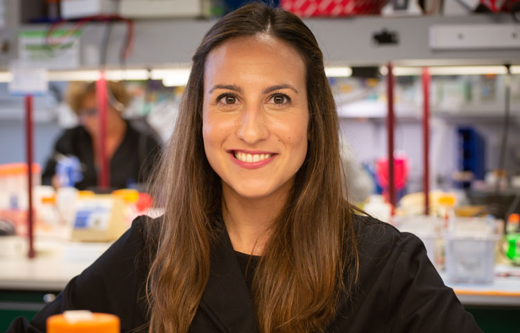
A new study from the lab of Francesca Marassi, PhD could help reveal new treatments for one of the world’s deadliest pathogens.

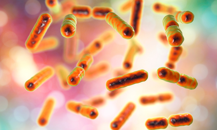
Sanford Burnham Prebys researchers have uncovered the structure of an important protein for the growth of tuberculosis bacteria. The study, published recently in Nature Communications, sheds light on an unusual metabolic system in tuberculosis, which could help yield new treatments for the disease and help make existing therapies more effective.

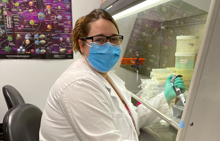
“Molecular discoveries like this give us valuable insight into how these bacteria survive, which is important in terms of finding cures for tuberculosis, and for other areas of health and biology,” says James Kent, a PhD candidate working in Marassi’s lab. “For example, bacteria in this family pose problems in both human health and agriculture, such as leprosy and bovine tuberculosis.”

Tuberculosis caused 1.5 million deaths in 2020 according to the World Health Organization, and this figure is expected to increase in the coming years due to the impact of the COVID-19 pandemic.

Stealing iron has its risks
The new protein, called Rv0455c, is part of a complex transportation system in Mycobacterium tuberculosis. Rv0455C helps the bacteria take up iron from the host cells they infect. This process is essential to their growth and replication.

“They produce these very small molecules called siderophores and send them out of the cell, where they bind to iron and bring it back in,” says Kent. “Rv0455C seems to be essential for secreting these molecules.”

An important step of this iron-uptake process is recycling the siderophores so they can be used again. When this process is interrupted, the leftover molecules can accumulate and poison the cell.

The study found that without Rv0455c, tuberculosis bacteria cannot secrete siderophores, which severely impairs their replication. Bacteria without Rv0455c also experienced poisoning from unrecycled siderophores. 

And while this delicate system can be interrupted by blocking previously known genes, eliminating Rv0455c does it much more efficiently.

“This seems to be the first piece of evidence that there is a single protein in this system that could be targeted by a new class of tuberculosis drugs,” adds Kent.

Structure determines function
Kent’s role in the study was to piece together the structure of the protein, which had posed a significant challenge to the researchers. Revealing the detailed structure of a protein is a critical part of understanding its function.

“The process of figuring out the structure of a protein can be time consuming and requires precise optimization of many conditions,” says Kent. “This protein is small, but it is still a three-dimensional object moving in three-dimensional space, and the way it’s shaped will affect what it does.”

Kent determined that the Rv0455c protein has an unusual “cinched” structure that could help explain its unique function in tuberculosis bacteria. The structure may also help determine whether it’s possible to target the protein with therapeutics. 

Looking ahead
The findings suggest that targeting the recycling of iron-carrying molecules may lead to the development of much-needed drugs to combat one of the world’s deadliest bacterial pathogens.

Kent is also optimistic that the findings could help augment existing treatments for tuberculosis.

“Because treatment cycles are long for tuberculosis, a common problem with is multi-drug resistance,” says Kent. “There’s a very good possibility that there will be implications for this protein in interrupting some of the processes that lead to bacterial resistance.”

Institute News

How our immune system controls gut microbes

AuthorMiles Martin
Date

April 6, 2022

And how this relationship could help fight autoimmune diseases

Sanford Burnham Prebys researchers including Carl Ware, PhD, and John Šedý, PhD, have discovered an immunological process in the gut that could help improve treatment for autoimmune and gastrointestinal diseases. The study, published March 22 in Cell Reports, found that this process regulates the activation of white blood cells in the intestines, which ultimately helps the body control the composition of the gut microbiome. 

“The immune system is like a gardener for our gut bacteria, gently monitoring and responding to their populations and keeping an eye out for unwanted pathogens” says Ware, who directs the Infectious and Inflammatory Diseases Center at Sanford Burnham Prebys. “This ultimately helps the immune system control these microbes.”

This “gardening” relies on a molecule called BTLA, one of several checkpoint proteins used by the body to control the immune system. 

“This is a signaling system we’ve known about for decades, but this is a totally new function for it that we’ve never seen before,” says Šedý, a Sanford Burnham Prebys research assistant professor, who co-led the study with Ware. “I helped discover this system two decades ago, so it’s exciting that we’re still making new discoveries about its function.”  

To explore the role of BTLA in the gut, the team zeroed in on specialized lymph nodes in the intestines called Peyer’s patches, which are full of white blood cells that help monitor and respond to pathogens and other microbes in the gut.

“Gut bacteria are in constant competition, and the populations of specific species can fluctuate,” says Ware. “In a healthy microbiome, there’s a balance, and disrupting that balance can contribute to autoimmune diseases, gastrointestinal disorders and even some brain disorders.”

The team found that BTLA is critical for maintaining this balance because it triggers white blood cells to release antibodies that control the populations of different gut bacteria.

“It’s a finely calibrated system that we’re still only just beginning to understand in detail,” adds Ware.

Immune checkpoints like BTLA are already used in immunotherapy for some cancers, and these results make the researchers confident that this system can be leveraged to treat diseases in the gut, especially those that are also autoimmune disorders, such as Crohn’s disease or ulcerative colitis. 

“The immune system is unimaginably complex, and understanding it gives us the ability to manipulate it, and that can help us treat diseases,” says Šedý. “This discovery is a step forward in that larger narrative.” 

Institute News

New COVID-19 drug passes phase 2 clinical trial

AuthorMiles Martin
Date

January 20, 2022

The new treatment, developed by Avalo Therapeutics with Sanford Burnham Prebys researchers, can mitigate lung damage and improve survival in COVID patients.

In a phase 2 clinical trial conducted by Avalo and supported by Sanford Burnham Prebys, a significantly higher proportion of hospitalized patients with COVID-19 remained alive and free of respiratory failure for 28 days after receiving treatment with the new antibody, called CERC-002. Unlike other experimental COVID therapies, CERC-002 does not target the virus itself, instead targeting the immune response associated with the virus to stop the disease from progressing before it becomes fatal.

“At the beginning of the pandemic we thought vaccines were going to be all we really needed. But with variants like omicron, we’re going to have more people that progress to serious illness even with the vaccine,” says study coauthor Carl F. Ware, PhD, director of the Infectious and Inflammatory Diseases Center at Sanford Burnham Prebys. “We need treatments to stop the progression to severe disease and death.”

The findings were published December 6 in the Journal of Clinical Investigation.

COVID-19: a continuing crisis
In the United States, over 840,000 people have died from COVID-19. A large proportion of these deaths have been among the elderly or those who are immunocompromised due to a preexisting condition. And while three quarters of the population has received at least one dose of the COVID-19 vaccine, many remain unvaccinated.

“A lot of us feel safer now that we’ve gotten our shots,” Ware says, “but the threat of the pandemic has not gone away, even for vaccinated people.”

Most people with COVID-19 experience few to no symptoms. However, elderly individuals, people with a concurrent health condition or those who are immunocompromised are susceptible to a condition called cytokine storm, in which their own immune molecules called cytokines flood the body in higher concentrations than usual.

Rather than helping fight the virus, these extra immune molecules wreak havoc, causing patients to develop the deadly respiratory failure characteristic of severe COVID disease.

“The COVID virus gets the immune system amped up by producing these molecules, which is normally how the immune system fights diseases,” says Ware. “But when there are too many cytokines and they’re not doing their job, it can lead to severe damage.” 

Neutralizing the cytokine storm
The new treatment, CERC-002, is a cytokine neutralizer—an immune molecule that recognizes and deactivates a cytokine known as LIGHT, which is elevated in patients with COVID-19. Cytokine neutralization drugs are currently being tested in the clinic, but they are mainly effective in severely ill patients who are already on a ventilator or other organ support.

“There is a critical need for drugs to stop milder cases from progressing to severe,” says Ware. “This treatment targets the cytokine immune response early enough to stop it in its tracks, which no other treatment does right now.”

83 COVID patients were enrolled in the study, half receiving the treatment, and half receiving a placebo. All patients were hospitalized with mild-to-moderate respiratory distress and were also receiving standard-of-care therapy during the trial.

They found that 83.9% of patients who received a dose of CERC-002 on top of standard of care remained alive and free from respiratory distress for 28 days. For patients receiving placebo, the number was only 64.5%.

Looking ahead
As a phase 2 clinical trial, the purpose of this study was to test whether the compound has therapeutic potential in a small number of patients. Now that the drug has proven successful at a small scale, it can be tested on a larger number of patients to ensure its benefits are consistent across the population.

Additionally, because CEC-002 targets the immune response in COVID cases rather than the virus itself, the compound may have applications that extend beyond COVID.

“Cytokine storm is not unique to COVID. It occurs in other infections—even in autoimmune diseases with no active infection, so this treatment may have some utility in these other diseases as well.” 
While there is more work to be done before CERC-002 becomes widely available, it does offer a glimmer of hope during a pandemic that seems never-ending.

“We have made a lot of progress in controlling the pandemic with vaccines and other new therapies, but it’s not over yet,” says Ware. “Treatments like this may bring physicians an option to protect infected people from severe illness.

Institute News

A strange research ecosystem: Discussing Lyme disease with Victoria Blaho

AuthorMiles Martin
Date

December 22, 2021

As an infectious disease immunologist studying Lyme disease, Victoria Blaho is one of a rare breed.

Sanford Burnham Prebys assistant professor Victoria Blaho, PhD, investigates the biochemical signals of the immune system and how they impact our bodies’ abilities to fight pathogenic infections, a branch of immunology that has become much less popular since the advent of antibiotics in the early 20th century.

Blaho’s disease of choice is Lyme disease, an unusual tick-borne bacterial infection that affects some 476,000 people in America each year, a number that is on the rise.

We caught up with Blaho to talk about why Lyme disease research is important, the progress being made and the work that remains in studying this strange and burdensome disease.

Why is Lyme disease research important?
Blaho: Lyme research is a very small field for a disease that is becoming bigger and bigger every year. Case counts are increasing for Lyme disease all over the world, and people get very sick from it. Some people are infected, take antibiotics and that’s the end of it. But others have chronic symptoms like arthritis or carditis that can last for years and become completely debilitating.

What makes Lyme difficult to study?
Blaho: One reason is that Lyme is an unusual infection from a microbiological standpoint. In the early days of Lyme research, there were studies showing that the bacteria that caused the disease, Borrelia burgdorferi, could change its physical form from a corkscrew shape to dormant blobs—and the blobs could be causing extended disease. This is a problem because scientists haven’t agreed on the true cause of chronic Lyme disease.

To make matters worse, a lot of the medical field still believes Lyme is easily curable with antibiotics, and if people are still having problems, then it’s psychosomatic. This makes it harder to get support for research into the longer-term inflammatory effects of Lyme. These politics make Lyme disease research a strange ecosystem of patients, physicians, researchers and funding agencies, and this is a barrier to learning more about the disease and helping people find relief.

How does your work enter the picture?
Blaho: I’ve been working on Lyme disease for over 15 years, since I was PhD student. It started because Celebrex was hugely popular at the time to treat arthritis, but nobody had ever studied it in the arthritis that emerges in Lyme disease. Celebrex inhibits an enzyme of the immune system that triggers inflammation, so we figured that Celebrex might work just as well in Lyme arthritis as in other types. But research on mice didn’t bear this out.
Inflammation doesn’t just peter out when an infections clears. The immune system has to clean up the mess. We discovered in mice that Celebrex inhibits the resolution of inflammation after Lyme disease has resolved, so the arthritis never went away.

Since then, my career has focused on exploring the signaling molecules that regulate inflammation and its resolution. These molecules affect all parts of the immune system and provide us with a whole host of different potential therapeutic targets for inflammatory diseases like chronic Lyme.

What are the next steps for your Lyme research and for the field at large? 
Blaho: My own immediate next step is to take the work I’ve been doing here at Sanford Burnham Prebys and connect it directly back to my original work with Lyme. My team here is currently working on a signaling molecule called S1P, and while we haven’t studied it in Lyme yet, we think there are connections between it and the immune mediators we first found through those Lyme studies.

Our next steps are to look for the protein that carries S1P in mice with Lyme disease. This protein is associated with disease susceptibility in other inflammatory illnesses like diabetes and cardiovascular disease, and we think it has a role to play in Lyme as well. We’re also planning to partner with the Bay Area Lyme Foundation to see if we can find changes in this protein in their collection of human samples.

More broadly, I think this field is hungry for innovation because there have been a small number of scientists focusing on it. If older ideas about Lyme being simple to treat were the complete picture, we’d already be able to better diagnose and treat patients. But we’re just not there yet.

Lyme may be a lot cleverer than we originally thought, but if we’re able to embrace new technologies and ideas and continue to push forward with new work, we’ll be able to find innovative approaches to fight Lyme and, ultimately, to help people suffering from this horrible disease.

Institute News

Boosting immunotherapy in aggressive brain cancer

AuthorMiles Martin
Date

November 3, 2021

Researchers from Sanford Burnham Prebys have collaborated the University of Pittsburgh Cancer Institute to reveal a new approach to enhance the effects of immunotherapy in glioblastoma, one of the most aggressive and treatment-resistant forms of brain cancer.

The study, published recently in Cancer Discovery, describes a novel method to ‘turn off’ cancer stem cells—the malignant cells that self-renew and sustain tumors—enabling the body’s own defense system to take charge and destroy tumors.

“Tumors are more than just masses of cells—each one is a complex system that relies on a vast network of chemical signals, proteins and different cell types to grow,” says senior author Charles Spruck, PhD, an assistant professor at Sanford Burnham Prebys. “This is part of why cancer is so difficult to treat, but it also presents us with opportunities to develop treatment strategies that target the machinery powering tumor cells rather than trying to destroy them outright.”

Glioblastoma is an extremely aggressive form of cancer that affects the brain and the spinal cord. Occurring more often in older adults and forming about half of all malignant brain tumors, glioblastoma causes worsening headaches, seizures and nausea. And unfortunately for the thousands of people who receive this diagnosis each year, glioblastoma is most often fatal.

“We haven’t been able to cure glioblastoma with existing treatment methods because it’s just too aggressive,” says Spruck. “Most therapies are palliative, more about reducing suffering than destroying the cancer. This is something we hope our work will change.”

Immune checkpoint inhibitors—which help prevent cancer cells from hiding from the immune system—can be effective for certain forms of cancer in the brain, but their results in glioblastoma have been disappointing. The researchers sought a way to improve the effects of these medications.

“Modern cancer treatment rarely relies on just one strategy at a time,” says Spruck. “Sometimes you have to mix and match, using treatments to complement one another.”

The researchers used genomic sequencing to investigate glioblastoma stem cells. These cells are the source of the rapid and consistent regeneration of glioblastoma tumors that make them so difficult to treat.

The team successfully identified a protein complex called YY1-CDK9 as essential to the cells’ ability to express genes and produce proteins. By modifying the activity of this protein complex in the lab, the team was able to improve the effectiveness of immune checkpoint inhibitors in these cells. 

“Knocking out this transcription machinery makes it much more difficult for the cells to multiply” says Spruck. “They start to respond to chemical signals from the immune system that they would otherwise evade, giving immunotherapy a chance to take effect.” 

While the approach will need to be tested in clinical settings, the researchers are optimistic that it may provide a way to improve treatment outcomes for people with glioblastoma. 

“What our results tell us is that these cells are targetable by drugs we already have, so for patients, improving their treatment may just be a matter of adding another medication,” adds Spruck. “For a cancer as treatment-resistant as glioblastoma, this is a great step forward.”

Institute News

How misplaced DNA contributes to chronic illness

AuthorMiles Martin
Date

October 28, 2021

Though DNA is essential for life, it can also wreak havoc on our bodies as we age 

DNA is one of the essential building blocks of life, giving our cells instructions for virtually everything they do, but researchers at Sanford Burnham Prebys are investigating what happens to our cells when DNA ends up in places where it shouldn’t normally be, particularly as we age.

The answer – as described in their recent review in the journal Cell—is disease-causing inflammation. And the researchers hope that targeting this rogue DNA will lead to new therapeutic strategies for a range of age-related illnesses, including cancer, diabetes, rheumatoid arthritis, cardiovascular disease and neurodegenerative disorders.

“Age is the primary risk factor for all of these diseases, but they share another risk factor – chronic inflammation,” says first author Karl Miller, PhD, a postdoctoral researcher in the lab of Peter Adams, PhD, Sanford Burnham Prebys. “We’re trying to understand the underlying processes behind this inflammation so we can potentially treat all these age-related diseases together”

Typically, cells have DNA safely sequestered in their nucleus and in the mitochondria, where the DNA can do its job without interfering with the rest of the cells’ activities. When cells detect DNA in other areas, they unleash a series of biochemical responses designed to protect the cell from invaders. This response is a component of the innate immune system, our body’s first line of defense against infection.

Scientists have known about this system for decades, but until recently it was mostly thought to respond to foreign DNA, such as during a bacterial or viral infection. However, over the last decade, researchers have discovered that pieces of our own DNA, called endogenous cytoplasmic DNA, can escape from the nucleus or mitochondria and trigger this inflammatory response in our own cells, even in the absence of infection. The resulting ‘sterile’ inflammation can accumulate over time, contributing to a range of age-related diseases in all systems of the body.

But this inflammation is not without its upsides. Cytoplasmic DNA is actually an important short-term protective strategy against cancer formation. The inflammation can alert the immune system at the first sign of cancer, preventing its formation. But over the long term, the sterile inflammation caused by cytoplasmic DNA is also thought to contribute to cancer risk. In fact, we’ve only been able to observe the damage associated with sterile inflammation because people are now living long enough to experience it. 

“Systems like this exist because they’re beneficial in youth, but as we age, they break down,” says Miller. “100 years ago, a lot more people died from infectious diseases early in life. Over time, we’ve become better and better at treating these acute infections, and we’re living much longer. It’s in this later period in life that we see chronic diseases emerging that used to be much less common.”

Miller’s review describes four different types of cytoplasmic DNA fragments, classified according to when and how they appear. Some arise from the nucleus during mistakes in cell division. Others emerge because of errors in DNA repair or replication. Some even escape from mitochondria—energy-producing parts of the cell that have their own separate DNA. Others still are of unknown origin.

“They all look similar under a microscope, and they all can cause similar effects. That’s one of the major problems in this field. The benefit of studying how the different types emerge is that it gives us more points to target for therapeutics,” says Miller. 

In the Adams Lab, Miller and his colleagues look specifically at cytoplasmic chromatin fragments, one of the four types of cytoplasmic DNA. These fragments appear in the cell when the membrane surrounding the nucleus is weakened by senescence, a cellular stress response. Senescence is also associated with aging. 

“We’ve shown how this pathway works in mice, and now we’re actually moving forward with therapeutic applications for humans by doing drug screening to find compounds that can target it,” adds Miller. 

And while there is still a lot of work left for the researchers, their progress is encouraging. Adams, senior author on the Cell review, was recently awarded a $13 million grant by the NIH to study the effects of aging, including the role of cytoplasmic DNA, on the progression of liver cancer. 

“We like to call what we’re doing here ‘increasing the healthspan’, as opposed to the lifespan,” says Miller. “We’re hoping to maximize the healthy period of people’s lives.” 

Institute News

How microbes shape human health: an interview with Andrei Osterman

AuthorMiles Martin
Date

October 7, 2021

In his work on the human microbiome, Sanford Burnham Prebys professor Andrei Osterman, PhD, has shown how the organisms living within us can be leveraged to boost human health for a humanitarian cause – the plight of malnourished children. 

Describe your research aimed to improve the gut microbiome in malnourished children.
In infants, it’s been well-established that conditions of severe poverty and food insecurity cause a delayed development of gut microbes, and that this results in stunted growth, numerous syndromes, and even death. My team has been collaborating with researchers at Washington University in St. Louis to develop foods that are designed to enhance the microbiome, and we’ve found that these can actually work to correct some of these pathologies. 

One study involved introducing microbes from undernourished Bangladeshi children into the guts of mice. When these mice were then fed a typical Bangladeshi diet, they exhibited a weaker immune response to the oral cholera vaccine. More importantly, this poor response could be repaired by establishing a more normal gut microbiome in the mice and providing them supplements to boost these microbes’ propagation.

What else can we learn from this research that could be applied more broadly?
From a humanitarian perspective, the progress we’ve made is so valuable that there is no question we will continue the work. But studying the microbiome in infants, regardless of their food security, can also provide us with new insights into the importance of the microbiome in human health. 

In a more recent study with collaborators from University of California San Diego and University of Southern California, we found that adding corn syrup to infant formula can enhance the populations of beneficial microbes they might otherwise have gotten from breast milk. 

We hope this is the first of many studies we work on with this team, because the transition of infants from breast milk or formula to conventional foods is thought to be the most drastic example of how the microbiome changes with diet. Studying infants and their diets at this early point in life could help reveal fundamental truths that we’ll be able to translate to other syndromes related to the microbiome in children and adults worldwide, regardless of food security. 

And this isn’t just speculation. Another study with the team in St. Louis used the same methods as the malnutrition study to develop supplementary foods, called “fiber snacks,” to correct microbiome imbalances in people with obesity. One might think that obesity would be the total opposite of malnutrition, but the microbiome is a key player in both. 

More broadly, gut microbes are already the most well-studied part of the human microbiome, and the list of health associations with these microbes extend well beyond the digestive tract, even into the immune system, affecting the risk for diseases like cancer or diabetes. There’s also a growing body of evidence that suggests that gut microbes can have a direct effect on the brain. For example, the microbiome is being studied closely in connection to autism spectrum disorder, since many people on the spectrum experience concurrent gastrointestinal syndromes.

What would you say is important to know for people not familiar with the subject?
We need to acknowledge that our body and many of its problems have a huge microbiome component. The human body is a complex organism, and we are still learning how the microbiome influences and is influenced by different health conditions. The next step is to incorporate the role of the microbiome into the design of new diagnostics and therapeutics—because this undoubtedly influences their effectiveness. We can’t ignore this aspect of our biology, and the time is ripe to improve our understanding of it and leverage it to our advantage. Moving forward, this is going to help us solve so many problems—from issues we’ve already started looking at like obesity and malnutrition, all the way through to problems we aren’t even aware of yet. 

What are the next steps for you and your team?
What we’re really interested in now is exploring new genomic technologies that are starting to revolutionize the field. The latest development is something called MAG genomics, short for metagenomically assembled genomes. This involves looking at the big picture, sequencing DNA from the whole microbiome at once in a way that is much faster and of much better quality than we’ve ever been capable of before. It’s like the difference between watching a movie on a clunky pixelated monitor from the 80’s and seeing that same movie on an HD monitor. Methods like this are moving us into a new paradigm in biomedical research, one that may be more complex, but also one that has the potential to substantially improve health outcomes for people around the world.

Institute News

Laura Martin-Sancho rises to the challenge of COVID-19

AuthorSusan Gammon
Date

June 29, 2021

The journal Molecular Cell recently asked Laura Martin-Sancho to share her experience working on SARS-CoV-2 during the pandemic

COVID-19 altered our lives and pushed scientific research to operate at breakneck speed, leading to significant breakthroughs in record time. The journal Molecular Cell recently asked experts in the field—including Laura Martin-Sancho—about the challenges they faced in transitioning, rapidly but safely, to working on the virus while navigating the shutdown. Their voices converge on the importance of teamwork, forging new collaborations, and working toward a shared goal.

Here is what Laura had to say:

I remember learning about viral pandemics in university and thinking about the challenges of working with novel viruses. Of course, we virologists contemplate the idea of a global viral pandemic and we discuss this at length in grants and in research articles, but how do you respond when confronted by it? It all started in February 2020 as I was completing my postdoc studying respiratory viruses. The lab had been closely following the worrying news coming from China and decided to drop everything else and work full time on SARS-CoV-2. Once we received the virus in March, it was a race against the clock to get the right conditions, the right cells, and the right reagents to propagate the virus to high enough amounts to start testing small compounds for antiviral activity. With a starting material of barely 100 μl received from BEI Resources, and long lonely hours in the BSL3, I felt a massive relief when I finally saw that the virus was replicating. Soon after, we had optimized experimental conditions and high enough viral yields to begin the essential experiments.

It was a remarkable feeling to have the whole research institute to just the seven of us. It was just me and three other lab members, our PI, Sumit Chanda, and two members of the institute safety department. With nobody around, it felt like we were apocalyptic survivors racing to find a cure. With a non-stop schedule from 8 am to 10 pm in the lab and trying to play catch up with an everyday-evolving literature, we were barely getting any sleep but never felt so energized. I felt so supported and inspired by my family and friends back home in Spain, which was one of the initial pandemic epicenters in Europe. I felt it was our responsibility to keep going and to make discoveries that could have a meaningful contribution. And so we did. In only a few months, we evaluated thousands of small compounds (initially in collaboration with Hong Kong University, then in house), we identified the innate immune sensor for SARS-CoV-2, and we illuminated the cellular antiviral landscape to SARS-CoV-2. Of course, this wouldn’t have been possible without the expertise and assistance from our countless collaborators across the globe. Indeed, a story to tell my grandkids.

Read the stories from other experts in the field in this article published in Molecular Cell: https://doi.org/10.1016/j.molcel.2021.05.021

Institute News

Meet immunologist Jennifer Hope

AuthorMonica May
Date

February 12, 2021

Hope’s research aims to help cancer immunotherapy work for more people

It’s not an overstatement to say that immunotherapy—an approach that uses our own immune system to kill a tumor—has revolutionized the treatment of cancer. Doctors continue to report incredible results, including tough-to-treat tumors seemingly melting away. However, the treatment doesn’t work for everyone, and even if it does work initially, it often stops working as time goes on.  

Jennifer Hope, PhD, a postdoctoral researcher in the Bradley lab at Sanford Burnham Prebys, is working to find ways to make cancer immunotherapy work for more people. We caught up with her as she prepared to take the virtual stage at the Diversity and Science Lecture Series at UC San Diego (DASL) to learn more about what she wishes people knew about science and whom she admires.

Did you always know you wanted to be a scientist?
I always had an interest in science, but at first I wanted to go a totally different route. I was an athlete in high school and college—I played tennis—and really wanted to go into sports medicine. Then I had my first real experience being in a lab in college, and I was hooked. I liked how hands-on it was and how I could keep asking questions. As my family knows, I’ve always been one to ask a lot of questions and always ask why. I found that being in the lab that was my opportunity to keep coming up with new questions, and finding answers that will impact people’s lives.

What do you research, and what is your greatest hope for your work?
I’m trying to understand why the immune system—specifically, T cells—seems to turn a “blind eye” to tumors, which it doesn’t do to other foreign invaders like viruses. My ultimate hope is that we use this information to create better cancer immunotherapies, particularly for skin cancer, which is still really deadly.

What do you wish people knew about science?
That it can be a lot of fun! Most people have this perception of science as being very boring. You see X and you do Y. That part can be true. But there’s a lot of opportunity for creativity and to come up with different ways to ask the same question. Some of the best scientists are incredibly creative people.

How would your coworkers describe you?
Motivated and always willing to try new things.

When you aren’t working, where can you be found?
Reading a book. My family started a book club to stay connected during the pandemic. We just read The Food Explorer by Daniel Evan Stone, which was fascinating. It’s about a botanist who is responsible for transforming what food looked like in the U.S. at the turn of the century. I don’t want to give too much away, but it’s because of him that we have cherry blossoms in Washington D.C., and regulations on importing seeds.

Whom do you admire, and why?
My parents. It sounds cliché, but it’s true. They have always been the biggest supporters of my dreams, whether career or personal.

One example that pops into my head is when I was getting my PhD, and my PI moved from Philadelphia to the Netherlands. I had the opportunity to move, too, if I wished. This was obviously a huge step, and I called my parents to talk it through. Immediately, the conversation was about how this would benefit me—the risks and the advantages—and they said they would support me if I wanted to go or not. That meant, and means, the world to me. Ultimately, I did go, and it was an incredible opportunity that I don’t regret at all.

What do you wish people knew about Sanford Burnham Prebys?
That everyone is willing to help each other. You don’t see that everywhere. It is proof that you can do science at an exceptional level without competing with each other.

Institute News

Hospitals were full. One scientist stepped up.

AuthorMonica May
Date

February 10, 2021

Sanford Burnham Prebys physician-scientist Evan Snyder spent two weeks in a gymnasium-turned-ICU, where he cared for people with severe COVID-19

The novel coronavirus has hit California hard, but one area that has been particularly impacted is Imperial County. Last spring, the rural farming region’s two hospitals became overwhelmed with COVID-19 cases—prompting a college basketball stadium to be converted into a makeshift intensive care unit (ICU). Soon, qualified personal were also needed.

Stem cell scientist Evan Snyder, MD, Ph.D., may not be the first person you would think to call on during such an emergency. But as a physician-scientist who works with critically ill newborns, he knows his way around an ICU. He knows how to run ventilators. And perhaps most importantly, he had an urgent desire to help.

“I had already decided I would study this disease from a scientific perspective,” says Snyder, who is working with UC San Diego’s Sandra Leibel, MD, to use mini lungs” to understand why some people with COVID-19 fare worse than others. “But as I started to see the public health menace it became, I felt like I needed to do more.”

Snyder started to sign up for every volunteer opportunity he could find. However, it wasn’t until the December post-holiday surge in cases when he was deployed to serve in the field. Through the California Medical Assistance Team (CAL-MAT), a group of highly trained medical professionals who provide assistance during disasters, Snyder was deployed to the gym-turned-ICU in Imperial County.

“Although our research examines the impact of the virus on lung cells created from people of many racial and ethnic backgrounds, the degree of disease disparity didn’t hit me at a gut level until this work,” says Snyder. “There’s no question that COVID-19 is unfairly hitting people who are socio-economically challenged and have co-morbidities such as diabetes and hypertension, which are the often the products of a disadvantaged environment.”

“I was like a vampire”

For two weeks Snyder worked through the night, taking down medical histories; giving people oxygen, providing medications such as dexamethasone, remdesivir, anticoagulants and antibiotics; carefully turning people onto their stomachs to ease breathing difficulties or helping individuals walk. He also saw clear patterns emerge.

All of the people he treated had conditions that are linked to poverty. More than 20% of people living in Imperial County live below the poverty line—double the national average. As a result, residents may be more likely to obtain food from food banks and may not have access to regular healthcare—which together can lead to conditions such as diabetes, hypertension or obesity. Many of the people whom Snyder cared for shared that they lived in small quarters with multiple generations, which made quarantining difficult, if not impossible.

“Some people who live in La Jolla and test positive have the luxury of living in a big house. They can afford not to go to work and stay in a separate bedroom while the rest of the family quarantines,” says Snyder. “The people I took care of can’t do that. We need to create places where people who test positive for COVID-19 can quarantine safely away from their families.”

Carrying insights back to the lab

Snyder’s experience has directly informed several new research avenues he plans to pursue.

“We already model real-world COVID-19 infections with ‘mini lungs’ created from different genders and races,” explains Snyder. “But this taught me that we need to better mimic the conditions present in a person who has diabetes or other conditions that create an adverse milieu for their organs and cells.”

This work also imprinted upon him that COVID-19 is more than a lung condition. The risk of blood clots causing strokes, heart attacks or blocking blood flow to the lungs was an ever-present concern.

“It wasn’t just about giving people more oxygen,” says Snyder. “This showed me that we need to focus even more on the vascular and inflammatory components of this disease.”

Lives were saved

Snyder is relieved to report that no lives were lost during those two weeks. He credits the care given—even if relatively primitive—to this success.

“If we weren’t doing what we were doing, about 30% of the people there would have died. And another 30% would have been left with lifelong impairments,” says Snyder. “However, in order to truly tame this virus, we need to find effective drugs, continue to vaccinate as many people as possible and exercise logical public health precautions.”