Immunity Inflammation and Microbiology Archives - Sanford Burnham Prebys
Institute News

Investigating individual immune responses to COVID-19 vaccination and infection

AuthorGreg Calhoun
Date

March 3, 2025

New study analyzes how the set of proteins in blood plasma changes following vaccination and infection, and may contribute to improving vaccine development

While many people have received similar mRNA vaccinations to protect against COVID-19, the strength and duration of the resulting immunity varies. It remains unclear exactly what causes individuals’ immune systems to react differently to the COVID-19 vaccine and other immunizations.

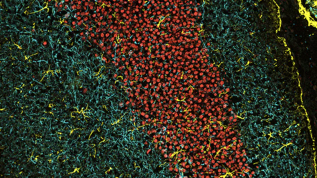
To get a better understanding of this phenomenon, scientists at Sanford Burnham Prebys, Seer Inc. and Federal University of Rio Grande do Sul examined the type and amount of virtually all proteins in the blood plasma of 12 volunteers as they received two doses of the Pfizer-BioNTech mRNA COVID-19 vaccine. The research team published the results of this pilot study in the Journal of Proteome Research on February 4, 2025, detailing the first attempt to comprehensively explore how an mRNA vaccine changes the mix and concentration levels of proteins known as the proteome.

The scientists were able to study a set of more than 3,000 proteins, within which they found a set of proteins that changed following each dose of vaccine. The authors also found a set of proteins that could distinguish between research participants who had or had not tested positive for COVID-19 in the months after receiving the second dose of vaccine.

While more research is needed with larger groups of research volunteers, this pilot study suggests that studying proteome changes can increase our understanding of how individuals’ immune systems react differently to immunization. Future findings from additional experiments may reveal methods for developing more effective vaccines.

Svetlana Maurya, PhD

Svetlana Maurya, PhD, is director of the Sanford Burnham Prebys Proteomics Shared Resource.

Lucélia Santi, PhD, professor adjunto at the Federal University of Rio Grande do Sul, is the senior and corresponding author on the study.

Ting Huang, PhD, a scientist at Seer Inc. focused on data science and machine learning, is first author on the manuscript.

Additional authors include:

  • Alex Rosa Campos, Ramón Díaz and Svetlana Maurya, from Sanford Burnham Prebys
  • Jian Wang, Alexey Stukalov, Khatereh Motamedchaboki, Daniel Hornburg and Serafim Batzoglou, from Seer Inc.
  • Laura R. Saciloto-de-Oliveira, Camila Innocente-Alves, Yohana P. Calegari-Alves and Walter O. Beys-da-Silva, from Federal University of Rio Grande do Sul
Institute News

Bile may be key to immunotherapy effectiveness in liver cancer

AuthorGreg Calhoun
Date

January 17, 2025

Understanding the crucial ingredient in bile may unlock the potential of treatments that help patients’ immune systems eliminate cancer

Hepatocellular carcinoma (HCC) is the most common liver cancer and a growing threat to public health across the globe due to the rising rate of fatty liver disease.

Liver cancer is difficult to treat as it often causes few if any symptoms early on, so it tends to be diagnosed at later, more aggressive stages. While immunotherapies that supercharge patients’ immune systems have proven effective in some cancers, this approach has had limited success in patients suffering from HCC or other forms of the disease.

Scientists are investigating the unique qualities of different tissues that may explain why the effectiveness of immunotherapy varies depending on the location of a tumor. The liver is known to have a flexible immune system capable of defending itself when necessary while not overreacting to a constant flood of foreign materials from digesting food, including metabolic byproducts from bacteria residing in the gut microbiome.

Transplant surgeons see the unique properties of the liver’s immune system firsthand when transplanted livers are typically integrated by recipients with only a low dose of immunosuppressive drugs. This ability to maintain immune tolerance, however, may reduce the ability of the liver’s immune system to find and destroy cancer cells, even when that capability is enhanced by immunotherapy.

In a paper published January 9, 2025, in Science, scientists at Sanford Burnham Prebys, the Salk Institute, the University of California San Diego, Columbia University Irving Medical Center, Memorial Sloan Kettering Cancer Center and the Geisel School of Medicine at Dartmouth, found that a critical ingredient in bile hinders the liver’s immune response against cancer.

Bile is a fluid made by the liver that assists in breaking down fats during digestion. This function is made possible by steroidal acids known as bile acids. The scientists found an increased amount of bile acids in tumor samples from patients with HCC. The team also found that genes involved in creating bile acids were being transcribed to make proteins and enzymes at an abnormally high rate in human samples and in mice genetically modified to develop liver cancer.

The authors went on to remove genes related to bile acid construction to demonstrate that mice without these blueprints developed fewer, smaller tumors. In addition, the liver’s T cells — the primary anti-tumor immune cells — were able to dig deeper into tumors and persist for longer without the immunosuppressive effects of certain bile acids.

“These findings underscore a new appreciation for the influence of bile acids on the liver’s immune system,” said Debanjan Dhar, PhD, associate professor in the Cancer Genome and Epigenetics Program at Sanford Burnham Prebys and coauthor on the study. More research is needed to test the potential use of drugs to directly inhibit certain bile acids or bile acid receptors as a therapeutic strategy to reduce liver cancer growth.

Debanjan Dhar, PhD, headshot outside

Debanjan Dhar, PhD, is an associate professor in the Cancer Genome and Epigenetics Program at Sanford Burnham Prebys.

Peter Adams profile photo in lab

Peter Adams, PhD, is the director of the Cancer Genome and Epigenetics Program at Sanford Burnham Prebys.

It may also be possible to achieve this effect through dietary changes that alter the microbiome and result in modified bile acid production. Based on their findings, the research team suggests that this could be done by using ursodeoxycholic acid, a bile acid that currently is used to treat an autoimmune condition called primary biliary cholangitis. The acid is found at high levels in bear bile, which has served for thousands of years as a treatment in traditional Chinese medicine.

“Given the safety profile of ursodeoxycholic acid and the limited effectiveness of immunotherapy on liver cancer, this study shows significant potential for testing this bile acid as a combination treatment for patients with HCC,” said Peter Adams, PhD, director of the Cancer Genome and Epigenetics Program at Sanford Burnham Prebys and coauthor on the study.


Susan Kaech, PhD, NOMIS Chair, professor and director of the NOMIS Center for Immunobiology and Microbial Pathogenesis at the Salk Institute is the senior and corresponding author on the study.   

Siva Karthik Varanasi, PhD, assistant professor at the UMass Chan Medical School and a former postdoctoral fellow in the Kaech lab at the Salk Institute, is first author on the manuscript. 

Additional authors include:

  • Souradipta Ganguly, Marcos G. Teneche and Aaron Havas, from Sanford Burnham Prebys
  • Dan Chen, Melissa A. Johnson, Kathryn Lande, Michael A. LaPorta, Filipe Araujo Hoffmann, Thomas H. Mann, Eduardo Casillas, Kailash C. Mangalhara, Varsha Mathew, Ming Sun, Yagmur Farsakoglu, Timothy Chen, Bianca Parisi, Shaunak Deota, H. Kay Chung, Satchidananda Panda, April E. Williams and Gerald S. Shadel, from the Salk Institute
  • Yingluo Liu, Cayla M. Miller, Jin Lee and Gen-Sheng Feng, from the University of California San Diego
  • Isaac J. Jensen and Donna L. Farber, from Columbia University Irving Medical Center
  • Andrea Schietinger from Memorial Sloan Kettering Cancer Center
  • Mark S. Sundrud from the Geisel School of Medicine at Dartmouth

Wolfram Goessling, MD, PhD, the Robert H. Ebert Associate Professor of Medicine and associate professor of Health Sciences and Technology at Harvard Medical School, authored a Perspective article on the new study in Science called, “Ena-bile-ing liver cancer growth.”

Institute News

Guglielmi awarded grant to further investigate genetic condition that results in soft, deformed bones and lost teeth

AuthorScott LaFee
Date

January 9, 2025

Hypophosphatasia (HPP) is a rare genetic disorder in which bones and teeth fail to take up sufficient calcium and phosphorus needed to achieve proper hardness and strength. Defective mineralization results in bones that are soft and prone to fracture and deformity, and the loss of teeth. Occasionally, HPP can cause death due to complications.

Prevalence varies by severity and age of onset. It is rarest but most severe at birth (1 in 100,000 live births), with lower prevalence and milder forms in later years. The condition can manifest at any age.

The cause of HPP is a mutation in an enzyme called tissue-nonspecific alkaline phosphatase (TNAP), which plays a critical role in skeletal and dental mineralization. In 2015, an enzyme replacement therapy developed by José Luis Millán, PhD, a professor in the Human Genetics Program at Sanford Burnham Prebys, was approved to treat pediatric onset HPP, dramatically improving patients’ lifespan and quality of life.

But the effects of TNAP deficiency appears to extend beyond faulty mineralization. HPP patients also experience altered immune responses, suggesting TNAP might have a role in immune cells.

Recently, Soft Bones, an advocacy group for HPP patients, awarded Valeria Guglielmi, PhD, a postdoctoral associate in Maximiliano D’Angelo’s lab, with a one-year, $25,000 seed grant to further investigate the involvement of TNAP in inflammatory responses and immune cell functions.

“This study is in line with my broad interest  for immune cells and their contribution to tissue homeostasis and diseases,” said Guglielmi. “I am excited to explore an entirely new area of investigation on HPP.

“Indeed, very little is known about the role of TNAP in the immune system and only a few studies have provided evidence of TNAP involvement in immune cell function. By uncovering how TNAP deficiency affects inflammatory responses, our research represents the first step toward designing interventions to improve immune system dysfunctions in HPP patients.”

Read Soft Bones’ full news release on the award to Guglielmi on Facebook and Instagram.

Institute News

Protein superfamily crucial to the immune system experiences Broadway-style revival

AuthorGreg Calhoun
Date

November 19, 2024

More than 25 years after targeting a member of this superfamily of proteins led to groundbreaking treatments for several autoimmune diseases including rheumatoid arthritis and Crohn’s disease, San Diego scientists note a resurgence of interest in research to find related new drug candidates.

In 1998, the same year “Footloose” debuted on Broadway, REMICADE® (infliximab) was approved by the FDA for the treatment of Crohn’s disease. This was the first monoclonal antibody ever used to treat a chronic condition, and it upended the treatment of Crohn’s disease.

Research published in February 2024 demonstrated better outcomes for patients receiving infliximab or similar drugs right after diagnosis rather than in a “step up” fashion after trying other more conservative treatments such as steroids.

Infliximab and ENBREL® (etanercept) — also approved in 1998 to treat rheumatoid arthritis — were the first FDA-approved tumor necrosis factor-α (TNF) inhibitors. TNF is part of a large family of signaling proteins known to play a key role in developing and coordinating the immune system.

The early success of infliximab and etanercept generated excitement among researchers and within the pharmaceutical industry at the possibility of targeting other members of this protein family. They were interested in finding new protein-based (biologics) drugs to alter inflammation that underlies the destructive processes in autoimmune diseases.

As “Footloose” made it back to Broadway in 2024 for the first time since its initial run, therapies targeting the TNF family are in the midst of their own revival. Carl Ware, PhD, a professor in the Immunity and Pathogenesis Program at Sanford Burnham Prebys, and collaborators at the La Jolla Institute for Immunology and biotechnology company Inhibrx, report in Nature Reviews Drug Discovery that there is a resurgence of interest and investment in these potential treatments.

“Many of these signaling proteins or their associated receptors are now under clinical investigation,” said Ware. “This includes testing the ability to target them to treat autoimmune and inflammatory diseases, as well as cancer.”

Today, there are seven FDA-approved biologics that target TNF family members to treat autoimmune and inflammatory diseases. There also are three biologics and two chimeric antigen receptor (CAR)-T cell-based therapies targeting TNF members for the treatment of cancer. This number is poised to grow as Ware and his colleagues report on the progress of research and many clinical trials to test new drugs in this field and repurpose currently approved drugs for additional diseases.

“The anticipation levels are high as we await the results of the clinical trials of these first-, second- and — in some cases — third-generation biologics,” said Ware.

Ware and his coauthors also weighed in on the challenges that exist as scientists and drug companies develop therapies targeting the TNF family of proteins, as well as opportunities presented by improvements in technology, computational analysis and clinical trial design.

Portrait of Carl Ware

Carl Ware, PhD, is a professor in the Immunity and Pathogenesis Program at Sanford Burnham Prebys.

“There are still many hurdles to get over before we truly realize the potential of these drugs,” noted Ware. “This includes the creation of more complex biologics that can engage several different proteins simultaneously, and the identification of patient subpopulations whose disease is more likely to depend on the respective proteins being targeted.

“It will be important for researchers to use computational analysis of genetics, biomarkers and phenotypic traits, as well as animal models that mimic these variables. This approach will likely lead to a better understanding of disease mechanisms for different subtypes of autoimmune conditions, inflammatory diseases, and cancer, enabling us to design better clinical trials where teams can identify the appropriate patients for each drug.”

Institute News

Gut microbiome repair in children with severe acute malnutrition

AuthorScott LaFee
Date

October 2, 2024

Child malnutrition remains an alarming and appalling scourge.

In 2022, according to the World Health Organization, 148 million children in the world under 5 years were too short for their age (stunting) and another 45 million were too thin for their height (wasting) due to inadequate diet and nutrition.

Researchers around the world, including Andrei L. Osterman, PhD, professor in the Immunity and Pathogenesis Program at Sanford Burnham Prebys, have been investigating potential remedies, in particular some of the consequences of malnutrition, such as disturbed metabolism and immune/gut function.

In a new paper published October 2, 2024 in Science Translational Medicine, the multi-institutional team (including Osterman and colleagues at SBP) describe an interventional diet that essentially repairs the gut microbiome in children with moderate to severe acute malnutrition.

They conducted a three-month randomized controlled trial of a specialized food supplement in 12- to 18-month-old Bangladeshi children living in rural and urban slums with moderate acute malnutrition who had already been treated in hospital for severe acute malnutrition. The supplement, called microbiota-directed complementary food or MDCF-2, contains chickpea flour, peanut flour, soy flour, green banana, sugar, soybean oil and a vitamin-mineral premix, a formulation designed to promote the growth of therapeutic gut bacteria and improve the overall health and balance of the gut microbiome.

They found that MDCF-2 improved weight-for-age better than the traditional ready-to-use supplementary food (RUSF) used by relief agencies and others, which is composed of more traditional ingredients like rice, lentil, sugar, soybean oil and skimmed milk powder mixed with vitamins and minerals.

When excluding children unable to continue study participation due to severe flooding during the trial, the study authors also reported improvement of stunting at a faster rate. They tied these improvements in children’s health to Prevotella copri–associated metabolic changes.

P. copri (recently renamed as Segatella copri) is a bacterium found abundantly in the human gastrointestinal microbiome. Past studies have reported both positive and negative associations with health and disease. In the former, for example, healthy bacterial colonization of the gut has been positively correlated with conditions like inflammation, insulin resistance and diarrhea. It appears to be a major player in regulating dietary metabolism.

The bacterium is more common in non-Westernized populations consuming a diet rich in plants—the bacterium’s source of nutrients. In Western populations, it is associated with consumption of fruits and vegetables.

Genomic reconstruction of the metabolic potential of P. copri strains positively associated with infants’ health improvement confirmed their unique ability to utilize a large repertoire of plant-derived polysaccharides comprising MDCF-2 diet.

“This study can be viewed as a test of the generalizability of the efficacy and mechanism of action of MDCF-2 in acutely malnourished children,” said Osterman. “The main findings include the demonstration of significantly higher efficacy of MDCF-2 vs RUSF with respect to the improvement of (weight) growth.

“The success of the treatment was also manifest by beneficial changes in microbiome composition and by global changes of a range of serum protein biomarkers associated with healthy development.”

The findings, he said, also provide proof-of-concept that improving gut microbial health can be achieved using therapeutic nutrition and offers further guidance on how best to use microbiota-directed complementary foods.

Institute News

Mapping the human body to better treat disease

AuthorGreg Calhoun
Date

August 20, 2024

Scientists build supersized sets of biological data to better treat diseases and reveal the secrets to youth by mapping the body at the single-cell level.

Scientists at Sanford Burnham Prebys are investigating the inner workings of our bodies and the trillions of cells within them at a level of detail that few futurists could have predicted. 

“The scale of the data we can generate and analyze has certainly exploded,” says Yu Xin (Will) Wang, PhD, assistant professor in the Development, Aging and Regeneration Program at Sanford Burnham Prebys. “When I was a graduate student, I would take about a hundred pictures for my experiment and spend weeks manually classifying certain characteristics of the imaged cells.” 

“Now, a single experiment would capture probably hundreds of thousands of images and study the gene and protein expression patterns of millions of individual cells.” 

The Wang lab specializes in advanced spatial multi-omic analyses that capture the location of cells, proteins and other molecules in the body. Wang uses spatial multi-omics to explore how dysfunctional autoimmune responses—when the immune system attacks the body’s own tissues—can interfere with its ability to repair and regenerate. As well as being relevant to disease, autoimmune responses also play a role in “inflammaging,” the low-level, chronic inflammation that occurs with age. Inflammaging is thought to contribute to many of the physical signs of aging.  

“My team thinks about diseases from the perspective of how cells behave in response to changes in the body,” says Wang. “We’re interested in how interactions between the immune and peripheral nervous systems change as people age and make us susceptible to frailty and disease.” 

spectrum of immune cells

A spectrum of immune cells being studied by Will Wang’s lab at Sanford Burnham Prebys. Image courtesy of postdoctoral associate Beatrice Silvestri, PhD.

Yu Xin (Will) Wang, PhD

Yu Xin (Will) Wang, PhD, is an assistant professor in the Development, Aging and Regeneration Program at Sanford Burnham Prebys.

This spatial multi-omics approach is helping scientific teams across the world on projects to understand how the body works at the cellular level. Efforts such as the Human Cell Atlas and the Human BioMolecular Atlas Program seek to develop a cellular map of the human body.  Researchers at Sanford Burnham Prebys are now using these tools to map complex diseases including cancer and degenerative conditions such as muscular dystrophy and ischemic injuries. Wang is also working to map cellular changes in aging through the San Diego Tissue Mapping Center of the Cellular Senescence Network (SenNet), a collaborative effort led by Peter D. Adams, PhD, director of, and professor in, the Institute’s Cancer Genome and Epigenetics Program and Bing Ren, PhD, professor of Cellular and Molecular Medicine at UC San Diego.  

“Integrating multiple types of -omics data can give us a much more comprehensive picture as we study health and disease,” notes Wang. Each additional layer of imaging and sequencing data adds more complexity to how Wang and his peers process and analyze their results. This has driven Wang and his colleagues to develop computational algorithms and AI tools to find patterns and novel translatable targets from these “big data” experiments. 

Wang credits San Diego-based biotechnology company Illumina for playing a major role by creating next-generation sequencing technology that improved the speed and accuracy of genome sequencing. The cost of sequencing steadily declined after Illumina launched the Genome Analyzer platform in 2007, making this research method more accessible to scientists at Sanford Burnham Prebys and around the globe. 

A series of additional technology platforms and research disciplines have followed, allowing scientists to study other parts of biological systems in similarly exhaustive detail. These include epigenomics, transcriptomics, proteomics and metabolomics. Scientists are now able to incorporate more than one of these levels of inquiry into an experiment, which is known as multi-omics.    

Connections in the brain

Connections in the brain photographed during experiments at the Institute.
Image courtesy of postdoctoral associate Sara Ancel, PhD, and Annanya Sethiya, MS, research associate II.

“The amount of information you get back from these sequencing platforms, as well as the application of highly multiplexed biomolecular imaging, has exponentially increased, which really helps us to resolve what we couldn’t before to better understand the genetic regulation of cells and diseases,” says Wang. “The most challenging part is the work to derive the meaning from these massive amounts of information. Thankfully, that’s also the most fun part of what we do.” 


Programming in a Petri Dish, an 8-part series

How artificial intelligence, machine learning and emerging computational technologies are changing biomedical research and the future of health care

  • Part 1 – Using machines to personalize patient care. Artificial intelligence and other computational techniques are aiding scientists and physicians in their quest to prescribe or create treatments for individuals rather than populations.
  • Part 2 – Objective omics. Although the hypothesis is a core concept in science, unbiased omics methods may reduce attachments to incorrect hypotheses that can reduce impartiality and slow progress.
  • Part 3 – Coding clinic. Rapidly evolving computational tools may unlock vast archives of untapped clinical information—and help solve complex challenges confronting health care providers.
  • Part 4 – Scripting their own futures. At Sanford Burnham Prebys Graduate School of Biomedical Sciences, students embrace computational methods to enhance their research careers.
  • Part 5 – Dodging AI and computational biology dangers. Sanford Burnham Prebys scientists say that understanding the potential pitfalls of using AI and other computational tools to guide biomedical research helps maximize benefits while minimizing concerns.
  • Part 6 – Mapping the human body to better treat disease. Scientists synthesize supersized sets of biological and clinical data to make discoveries and find promising treatments.
  • Part 7 – Simulating science or science fiction? By harnessing artificial intelligence and modern computing, scientists are simulating more complex biological, clinical and public health phenomena to accelerate discovery.
  • Part 8 – Acceleration by automation. Increases in the scale and pace of research and drug discovery are being made possible by robotic automation of time-consuming tasks that must be repeated with exhaustive exactness.
Institute News

How a protein component of nuclear pore complexes regulates development of blood cells and may contribute to myeloid disorders

AuthorCommunications
Date

June 5, 2024

Nuclear pore complexes (NPCs) are channels composed of multiple proteins that ferry molecules in and out of the nucleus, regulating many critical cellular functions, such as gene expression, chromatin organization and RNA processes that influence cell survival, proliferation, and differentiation.

In recent years, new studies, including work by Maximiliano D’Angelo, PhD, associate professor in the Cancer Metabolism and Microenvironment Program at Sanford Burnham Prebys, have noted that NPCs in cancer cells are different, but how these alterations contribute to malignancy and tumor development—or even how NPCs function in normal cells—is poorly understood.

In a new paper, published June 5, 2024 in Science Advances, D’Angelo with first author Valeria Guglielmi, PhD, and co-author Davina Lam, uncover Nup358, one of roughly 30 proteins that form the NPCs, as an early player in the development of myeloid cells, blood cells that if not formed or working properly leads to myeloid disorders such as leukemias.

The researchers found that when they eliminated Nup358 in a mouse model, the animals experienced a severe loss of mature myeloid cells, a group of critical immune cells responsible for fighting pathogens that are also responsible for several human diseases including cancer. Notably, Nup358 deficient mice showed an abnormal accumulation of early progenitors of myeloid cells referred as myeloid-primed multipotent progenitors (MPPs).

“MPPs are one of the earliest precursors of blood cells,” said D’Angelo. “They are produced in the bone marrow from hematopoietic stem cells, and they differentiate to generate the different types of blood cells.

Maximiliano D’Angelo and Valeria Guglielmi

“There are different populations of MPPs that are responsible for producing specific blood cells and we found that in the absence of Nup358, the MPPs that generate myeloid cells, which include red blood cells and key components of the immune system, get stuck in the differentiation process.”

Fundamentally, said Gugliemi, Nup358 has a critical function in the early stages of myelopoiesis (the production of myeloid cells). “This is a very important finding because it provides insights into how blood cells develop, and can help to establish how alterations in Nup358 contribute to blood malignancies.”

The findings fit into D’Angelo’s ongoing research to elucidate the critical responsibilities of NPCs in healthy cells and how alterations to them contribute to immune dysfunction and the development and progression of cancer.

“Our long-term goal is to develop novel therapies targeting transport machinery like NPCs,” said D’Angelo, who recently received a two-year, $300,000 Discovery Grant from the American Cancer Society to advance his work.


This research was supported in part by a Research Scholar Grant from the American Cancer Society (RSG-17-148-01), the Department of Defense (grant W81XWH-20-1-0212) and the National Institutes of Health (AI148668).

The study’s DOI is 10.1126/sciadv.adn8963.

Institute News

Perkins Fellow Trains Immune System Against Melanoma

AuthorGreg Calhoun
Date

May 15, 2024

Sanford Burnham Prebys scientist works on new methods to boost the body’s natural defenses against melanoma and other cancers

When she was growing up in India, Sreeja Roy, PhD, looked up to her father as he applied his scientific knowledge to care for the patients in his medical practice.

“He was my inspiration,” says Roy. “I found that I was particularly good at biology, and I liked learning about the mechanisms of how things work. Along the way, I realized I didn’t want to be a medical doctor and began focusing on biomedical research—and I fell in love with it.”

After earning her undergraduate degree in biotechnology from The Australian National University in Canberra, Roy obtained a master’s degree in infection biology from the Universität zu Lübeck in Germany. She returned to The Australian National University for her doctoral degree in immunology with an emphasis on viral vector-based vaccine immunology. After completing graduate school, Roy worked as a postdoctoral researcher at Albany Medical Center in New York before joining Sanford Burnham Prebys in September 2021 as a postdoctoral associate in the Immunity and Pathogenesis Program.

“I had been working on basic science in Albany,” notes Roy. “I really wanted to do translational research so I could work on things that would benefit people much sooner. That is why I chose to move to Sanford Burnham Prebys and focus on cancer immunotherapy.”

Roy’s background in immunology prepared her to enter the emerging field of cancer immunotherapy. This discipline involves developing treatments that enhance the human body’s innate immune response to better locate and dispose of cancer cells. She learned about an opportunity to support her interest in translational immunotherapy through the Jean Perkins Foundation Fellowship.

Roy received one of two prestigious fellowships designed to support postdoctoral researchers in the lab of Carl Ware, PhD, director of the Infectious and Inflammatory Diseases Center and professor in the Immunity and Pathogenesis Program.

“The Jean Perkins Foundation Fellowship has been fantastic,” says Roy. “I can work without the pressure of writing an academic grant, which allows me to focus on the science and be more productive.”

Roy’s project at the Ware lab involves making immunotherapies more effective in treating melanoma, the deadliest form of skin cancer.

“Unfortunately, some tumors never respond to immunotherapy treatments,” explains Roy. “Also, tumors can initially begin to shrink before becoming resistant to a treatment.”

Under Ware’s direction, Roy is testing ways to enhance existing immunotherapies through the lymphotoxin-β receptor, which is found on some types of immune cells.

“When the immune system encounters a foreign substance that may cause an infection, a sample of the invader can be shuttled to the lymph nodes as a way of learning about the threat and generating a better immune response,” explains Roy. “Depending on which tissues are being infected, the lymph nodes cannot always be involved, so an active lymphotoxin-β receptor is able to approximate their effect by organizing immune cells in something akin to training centers so that a better attack can be launched.”

Roy and the Ware lab are developing ways to take advantage of the lymphotoxin-β receptor’s ability to recruit and train immune cells as an approach to making immunotherapies more effective.

“If I can target the lymphotoxin-β receptor signaling against tumors, does that enhance the anti-tumor immunity?” asks Roy. “Do the tumors become more responsive to the treatments now? That is what we’re trying to find out.”

With the help of the Jean Perkins Foundation Fellowship, Roy is determined to continue developing her translational science expertise and find ways to improve the effectiveness of immunotherapies for melanoma and other cancers.

“We’ve made quite a bit of progress,” says Roy. “I look forward to sharing our results and seeing how this project advances from the bench to the bedside.”

Institute News

Seminar Series: extrachromosomal DNA and the metabolic circuits of cancer immune suppression

AuthorScott LaFee
Date

March 25, 2024

The ongoing Sanford Burnham Prebys seminar series will feature a pair of speakers on March 27, from noon to 1p.m., in the Fishman Auditorium. They will be presenting on two topics: extrachromosomal DNA and the tumor microenvironment.

First, Owen Chapman, PhD, a postdoctoral research scientist in the lab of Lukas Chavez, PhD, will discuss clinical and genomic features of circular extrachromosomal DNA (ecDNA) in medulloblastomas, a type of brain tumor.

EcDNA is DNA found off chromosomes, either inside or outside the nucleus of a cell. In a study published last year, Chavez (senior author), Chapman (first author) and colleagues reported that patients with medulloblastomas containing ecDNA are twice as likely to relapse after treatment and three times as likely to die within five years of diagnosis.

The second presentation will be by Kevin Tharp, PhD assistant professor in the Cancer Metabolism and Microenvironment Program. Tharp, who joined Sanford Burnham Prebys in December 2023, studies how tumors manipulate their mitochondria to improve survivability and how those cellular mechanics can be leveraged to create more effective therapies.

Institute News

Sanford Burnham Prebys research plays a key role in developing microbiome-directed complementary food to help save malnourished children

AuthorScott LaFee
Date

January 4, 2024

Among the consequences of childhood malnutrition is the underdevelopment of their gut microbiomes, critical to human health, from innate immunity to appetite and energy metabolism.

Although malnourished children gain some weight and grow better when fed a nutrient-rich diet, they do not catch up to their well-fed counterparts—and their gut microbiomes do not recover.

In a 2021 clinical trial, researchers at Washington University School of Medicine showed how a newly designed therapeutic food—a unique mix of peanuts, bananas and other foods dubbed microbiome-directed complementary food, or MDCF—more effectively nourished healthy gut microbial communities than standard dietary supplements.

Now, with bioinformatics support from Andrei L. Osterman, PhD, professor in the Immunity and Pathogenesis and Cancer Metabolism and Microenvironment programs at Sanford Burnham Prebys  and his colleagues Dmitry Rodionov, PhD, and Alex Arzamasov, the multi-institutional scientific team has published new research that identifies and describes the bioactive elements of microbiome-directed food.

“These are naturally occurring carbohydrate structures that could, in theory, be recovered in large quantities from the by-product streams of food manufacturing and be used to produce prebiotics,” said senior study author Jeffrey I. Gordon, MD, the Dr. Robert J. Glaser Distinguished University Professor at Washington University.

“We also have identified the microbes that process these food components, and in theory, there is potential for the organisms themselves to be part of a therapeutic intervention in children completely lacking these beneficial gut microbes.”

Osterman’s lab contributed bioinformatics analyses of 1,000 new metagenomically assembled genomes, or MAGs, representing the gut microbiomes of healthy Bangladeshi infants. The analyses included genome-based inference of the presence or absence in these MAGs of functional metabolic pathways for 106 major nutrients and intermediary metabolites.

“These predictions enabled the assessment of the microbiome-wide representation or enrichment of dietary carbohydrate utilization capabilities across numerous biospecimens from a randomized, controlled trial of MDCF in Bangladeshi children with moderate acute malnutrition,” said Osterman.

“The analyses helped elucidate glycan components of MDCF metabolized by bacterial taxa that are positively associated with healthy weight growth. The knowledge will help guide the therapeutic use of current MDCF and enable development of new formulations.”

Childhood undernutrition is a global scourge. In 2020, an estimated 149 million children under the age of 5 had stunted growth (low height for age), and 45 million exhibited stunting (low weight for height). More than 30 million children worldwide have moderate, acute malnutrition.

Undernutrition and its consequences are the leading causes of disease and death for children in this age range. An estimated 3 million children die each year due to poor nutrition and hunger.