iPSCs Archives - Sanford Burnham Prebys
Institute News

Using stem cells to study the biochemistry of learning

AuthorMiles Martin
Date

August 18, 2022

A method for studying human neurons could help researchers develop approaches for treating Alzheimer’s, schizophrenia and other neurological diseases

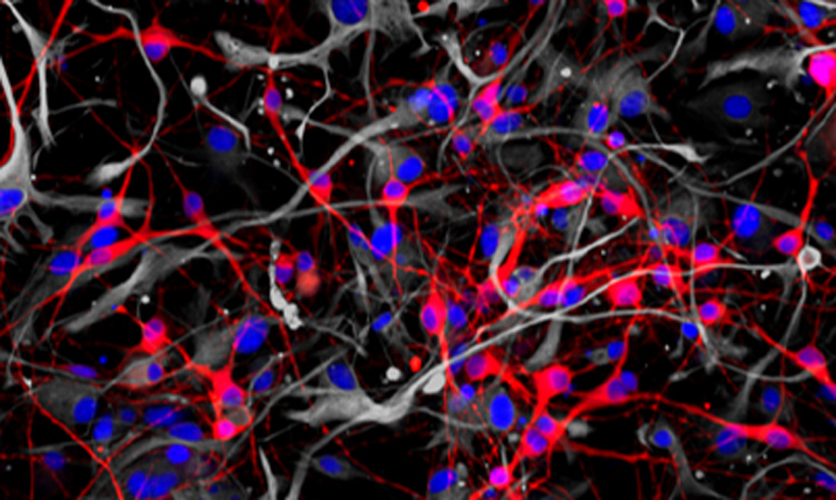
Researchers from the Conrad Prebys Center for Chemical Genomics have developed a procedure to use neurons derived from human stem cells to study the biological processes that control learning and memory. The method, described in Stem Cell Reports, uses electrodes to measure the activity of neuronal networks grown from human-induced pluripotent stem cells (iPSCs). The procedure tracks how synapses—the connections between neurons—strengthen over time, a process called long-term potentiation (LTP).

“Impaired long-term potentiation is thought to be central to many neurological diseases, including Alzheimer’s, addiction and schizophrenia,” says senior author Anne Bang, PhD, director of Cell Biology at the Prebys Center. “We’ve developed an approach to study this process in human cells much more efficiently than current methods, which could help trigger future breakthroughs for researchers working on these diseases.”

LTP helps our brain encode information, which is what makes it so critical for learning and memory. Impairment of LTP is thought to contribute to neurological diseases, but it has proven difficult to verify this hypothesis in human cells.
LTP helps our brain encode information, which is what makes it so critical for learning and memory. Impairment of LTP is thought to contribute to neurological diseases, but it has proven difficult to verify this hypothesis in human cells.

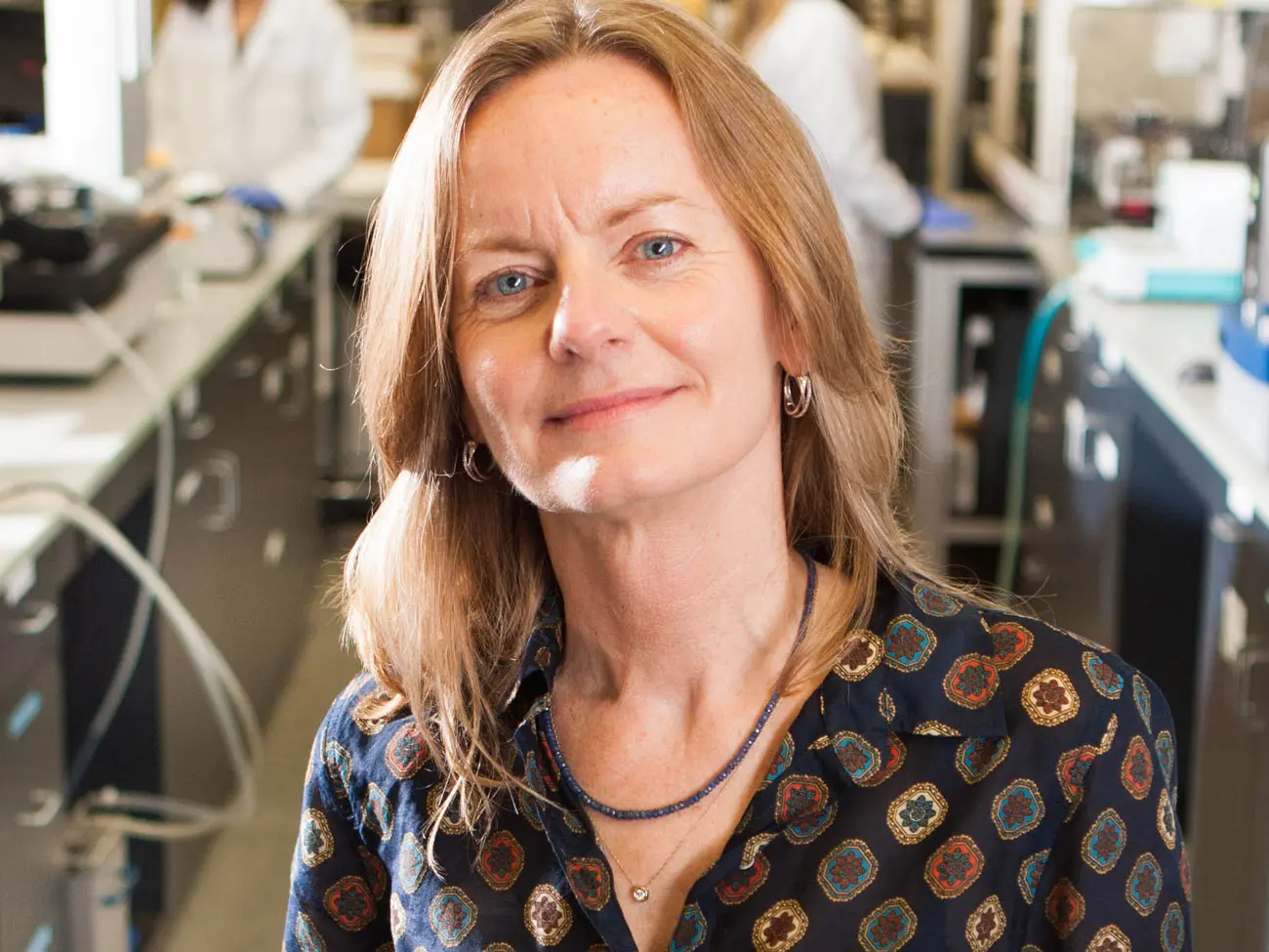
Anne Bang, PhD, director of Cell Biology at the Prebys Center.

“LTP is such a fundamental process,” says Bang. “But the human brain is hard to study directly because it’s so inaccessible. Using neurons derived from human stem cells helps us work around that.”

Although LTP can be studied in animals, these studies can’t easily account for some of the more human nuances of neurological diseases.

“A powerful aspect of human stem cell technology is that it allows us to study neurons produced from patient stem cells. Using human cells with human genetics is important in these types of tests because many neurological diseases have complex genetics underpinning them, and it’s rarely just one or two genes that influence a disease,” adds Bang.

To develop the method, first author and Prebys Center staff scientist Deborah Pré, PhD, grew networks of neurons from healthy human stem cells, added chemicals known to initiate LTP and then used electrodes to monitor changes in neuronal activity that occurred throughout the process.

The method can run 48 tests at once, and neurons continue to exhibit LTP up to 72 hours after the start of the experiment. These are distinct advantages over other approaches, which can often only observe parts of the process and are low throughput, which can make getting results more time consuming.

For this study, the researchers used neurons grown from healthy stem cells to establish a baseline understanding of LTP. The next step is to use the approach on neurons derived from patient-derived stem cells and compare these results to the baseline to see how neurological diseases influence the LTP process.

“This is an efficient method for interrogating human stem cell–derived neurons,” says Bang. “Doing these tests with patient cells could open doors for researchers to discover new ways of treating neurological diseases.”
Institute News

Scientists “turn back time” on cancer using new stem cell reprogramming technique

AuthorMonica May
Date

August 21, 2020

Discovery opens new research avenues that may help catch cancer early and identify potential preventive treatments

Scientists at Sanford Burnham Prebys Medical Discovery Institute have reprogrammed cancer cells back into their pre-cancer identity—opening new doors for studying how cancer develops and how it might be prevented. The research, published in Stem Cell Reports, may lead to tests that identify cancer early on, when it can be more easily treated, and uncover preventive treatments that stop cancer before it starts.

“We believe we have been able to contribute to one of the major goals of modern cancer research: creating next-generation models for studying how cancer develops from its earliest state,” says Evan Snyder, MD PhD, professor and director of the Center for Stem Cells & Regenerative Medicine at Sanford Burnham Prebys and senior author of the study. “We essentially took an adult cancer that has accumulated many mutations and pushed it back to the earliest stages of development, allowing us to emulate a tumor’s premalignant state. Then we watched cancer emerge from normal cells before our eyes.”

Turning back the clock on cancer 

In the study, the scientists set out to transform cells from anaplastic thyroid tumors—an aggressive, fast-growing cancer that is nearly always diagnosed at late stages—into induced pluripotent stem cells (iPSCs). These cells model the embryonic cells that are present at the earliest stages of human development and can become any cell in the body. While iPSCs are used today to create unlimited supplies of cells for research and therapeutic purposes—usually to correct abnormalities—the scientists recognized that tumor-derived iPSCs could be used to study the development of cancer.

However, this feat turned out to be easier said than done. The standard reprogramming method didn’t work, requiring the researchers to hunt for a different method that would induce the cancer cells to reset. Inhibiting a protein called RAS was the key ingredient that coaxed these thyroid cancer cells to become normal iPSC cells.

“We have named the pathway that is critical for making a cancer cell act as if it were a normal cell its ‘reprogram enablement factor,’” explains Snyder. “That factor will likely be different for every cancer and, in fact, may help in defining that cancer type.

“For this cancer type, which we examined in our study as a proof-of-concept, the reprogram enablement factor turned out to be blunting an overactive RAS pathway,” Snyder continues. “Our results suggest that losing control of RAS was the ‘big bang’ for this cancer—the very first event that leads to out-of-control cell growth and development of a tumor.”

The scientists next plan to reprogram additional cancers—including brain and lung cancer—into iPSCs to determine their “reprogram enablement factors.” If successful, they will next map the molecular changes that occur immediately before and after the tumors develop, which could reveal early signals of cancer and new preventive or early treatment measures.

“Unlike other cells, cancer cells are notoriously resistant to reprogramming,” says Snyder. “Our study is the first to successfully reprogram cancer cells into completely normal iPSCs, which opens new doors for cancer research.”

A team effort

The first author of the study is Yanjun Kong of Sanford Burnham Prebys and Shanghai Jiao Tong University. Yang Liu of Sanford Burnham Prebys is a co-corresponding author. Additional study authors include Ryan C. Gimple of UC San Diego; Rachael N. McVicar, Andrew P. Hodges and Jun Yin of Sanford Burnham Prebys; and Weiwei Zhan of Shanghai Jiao Tong University.

This study was funded by the Stem Cell Research Center & Core Facility at Sanford Burnham Prebys and by the China Scholarship Council (201606230202). The study’s DOI is 10.1016/j.stemcr.2020.07.016.

Institute News

First supercentenarian-derived stem cells created

AuthorMonica May
Date

March 19, 2020

Advance primes scientists to unlock the secrets of healthy aging.

People who live more than 110 years, called supercentenarians, are remarkable not only because of their age, but also because of their incredible health. This elite group appears resistant to diseases such as Alzheimer’s, heart disease and cancer that still affect even centenarians. However, we don’t know why some people become supercentenarians and others do not.

Now, for the first time, scientists have reprogrammed cells from a 114-year-old woman into induced pluripotent stem cells (iPSCs). The advance, completed by scientists at Sanford Burnham Prebys and AgeX Therapeutics, a biotechnology company, enables researchers to embark on studies that uncover why supercentenarians live such long and healthy lives. The study was published in Biochemical and Biophysical Research Communications.

“We set out to answer a big question: Can you reprogram cells this old?” says Evan Snyder, MD, PhD, professor and director of the Center for Stem Cells and Regenerative Medicine at Sanford Burnham Prebys, and study author. “Now we have shown it can be done, and we have a valuable tool for finding the genes and other factors that slow down the aging process.”

In the study, the scientists reprogrammed blood cells from three different people—the aforementioned 114-year-old woman, a healthy 43-year-old individual and an 8-year-old child with progeria, a condition that causes rapid aging—into iPSCs. These cells were then transformed into mesenchymal stem cells, a cell type that helps maintain and repair the body’s structural tissues—including bone, cartilage and fat.

The researchers found that supercentenarian cells transformed as easily as the cells from the healthy and progeria samples. As expected, telomeres—protective DNA caps that shrink as we age—were also reset. Remarkably, even the telomeres of the supercentenarian iPSCs were reset to youthful levels, akin to going from age 114 to age zero. However, telomere resetting in supercentenarian iPSCs occurred less frequently compared to other samples—indicating extreme aging may have some lasting effects that need to be overcome for more efficient resetting of cellular aging.

Now that the scientists have overcome a key technological hurdle, studies can begin that determine the “secret sauce” of supercentenarians. For example, comparing muscle cells derived from the healthy iPSCs, supercentenarian iPSCs and progeria iPSCs would reveal genes or molecular processes that are unique to supercentenarians. Drugs could then be developed that either thwart these unique processes or emulate the patterns seen in the supercentenarian cells.

“Why do supercentenarians age so slowly?” says Snyder. “We are now set to answer that question in a way no one has been able to before.”


The senior author of the paper is Dana Larocca, PhD, vice president of Discovery Research at AgeX Therapeutics, a biotechnology company focused on developing therapeutics for human aging and regeneration; and the first author is Jieun Lee, PhD, a scientist at AgeX.

Additional authors include Paola A. Bignone, PhD, of AgeX; L.S. Coles of Gerontology Research Group; and Yang Liu of Sanford Burnham Prebys and LabEaze. The work began at Sanford Burnham Prebys when Larocca, Bignone and Liu were members of the Snyder lab.

The study’s DOI is 10.1016/j.bbrc.2020.02.092.

Institute News

A year in review: Our top 10 discoveries of 2019

AuthorMonica May
Date

December 4, 2019

At Sanford Burnham Prebys, we uncover the origins of disease and launch bold new strategies that lay the foundation for achieving cures. This year our scientists made significant progress—revealing new insights into how we treat some of the deadliest cancers, address neurological disorders such as Parkinson’s and amyotrophic lateral sclerosis (ALS, or Lou Gehrig’s disease) and more.

Read on to learn more about our top 10 discoveries of the year. To receive more frequent updates on our discoveries, subscribe to our monthly newsletter at the bottom of this page.

  1. One-two punch drug combination offers hope for pancreatic cancer therapy. Ze’ev Ronai, PhD, identified a combination of two anti-cancer compounds that shrank pancreatic tumors in mice—supporting the immediate evaluation of the drugs in a clinical trial. The study was published in Nature Cell Biology.
  2. Targeted treatment shrinks deadly pediatric brain tumors. Robert Wechsler-Reya, PhD, reported that a targeted therapy that blocks a protein called LSD1 shrank tumors in mice with a form of pediatric brain cancer known as medulloblastoma. LSD1 inhibitors are currently under evaluation in clinical trials for other cancers, which could speed their potential path to children. The study was published in Nature Communications.
  3. Epigenetic change causes fruit fly babies to inherit diet-induced heart disease. Rolf Bodmer, PhD, showed that reversing an epigenetic modification or over-expressing two genes protected fruit fly children and grandchildren from the negative heart effects of their parents’ fatty diet. These findings help explain how obesity-related heart failure is inherited and uncover potential targets for treatment. The study was published in Nature Communications.
  4. Amyotrophic lateral sclerosis (ALS) research reveals new treatment approach. Huaxi Xu, PhD, extended the survival of mice with ALS-like symptoms by elevating levels of a protein called membralin using a gene therapy approach. The study was published in the Journal of Clinical Investigations.
  5. How prostate cancer becomes treatment resistant. Jorge Moscat, PhD, and Maria Diaz-Meco, PhD, identified how prostate cancer transforms into an aggressive, treatment-resistant subtype called neuroendocrine prostate cancer (NEPC) following treatment with anti-androgen therapy. Their findings uncover new therapeutic avenues that could prevent this transformation from occurring and reveal that an FDA-approved drug holds promise as an NEPC treatment. The study was published in Cancer Cell.
  6. Boosting muscle stem cells to treat muscular dystrophy and aging muscles. Alessandra Sacco, PhD, uncovered a molecular signaling pathway that regulates how muscle stem cells decide whether to self-renew or differentiate—an insight that could lead to muscle-boosting therapeutics for muscular dystrophies or age-related muscle decline. The study was published in Nature Communications.
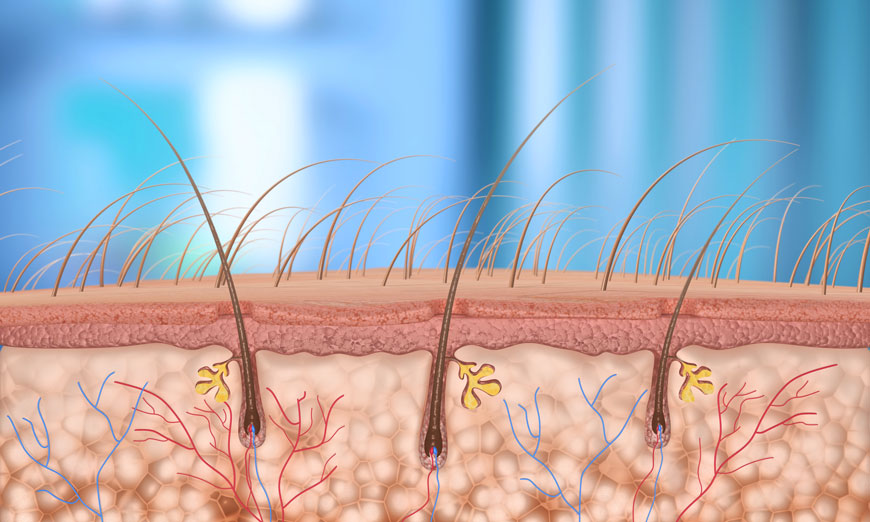
  7. Functional hair follicles grown from stem cells. Alexey Terskikh, PhD, created natural-looking hair that grows through the skin using human induced pluripotent stem cells (iPSCs), a major scientific achievement that could revolutionize the hair growth industry. Stemson Therapeutics has licensed the technology.
  8. Potential targeted treatment for acute myeloid leukemia identified. Ani Deshpande, PhD, showed that a protein called BMI1 is a promising drug target for an AML subtype in which two normally separate genes fuse together. The findings, published in Experimental Hematology, provide a rationale for evaluating a BMl1-inhibiting drug that is currently in clinical development as a potential treatment for this subtype.
  9. Antimicrobial protein implicated in Parkinson’s disease. An immune system protein that usually protects the body from pathogens is abnormally produced in the brain during Parkinson’s disease, Wanda Reynolds, PhD, reported in Free Radical Biology & Medicine. The discovery indicates that developing a drug that blocks this protein, called myeloperoxidase (MPO), may help people with Parkinson’s disease.
  10. Digestion-aiding herbs alter gut microbiome. Scott Peterson, PhD, found that four herbs—turmeric, ginger, long pepper and black pepper—promoted strong shifts in the gut bacteria that are known to regulate metabolism, providing insights that could help us protect our health. The study was published in Evidence-Based Complementary and Alternative Medicine.
Institute News

SBP’s Alexey Terskikh advances hair growth research

AuthorMonica May
Date

August 16, 2018

Three years ago, Alexey Terskikh, PhD, associate professor in Sanford Burnham Prebys Medical Discovery Institute’s (SBP’s) Development, Aging and Regeneration Program, published a groundbreaking study showing that stem cells could be used to grow hair.

This discovery could help more than 80 million men, women and children in the United States experiencing hair loss. Across cultures, personal identity is connected with hair. As a result, hair loss often affects emotional well-being and self-esteem. There is clear interest in the technology: Our 2015 story on this finding remains our blog’s most-read article. 

Since then, Terskikh and his team have been working hard to advance this technology. We caught up with Terskikh to learn about his progress—and how far away the research remains from human studies. 

Alexey Terskikh
     Alexey Terskikh, PhD

Could you fill us in on your work since 2015?

For the past three years, my team and I have been working to overcome several obstacles to the technology’s real-world use. We’ve made progress on multiple fronts, summarized below:

Generating unlimited cells 
Instead of embryonic stem cells, which are difficult to obtain, our method now uses induced pluripotent stem cells (iPSC), which are derived from a simple blood draw or skin sample. iPSCs allow us to create an unlimited supply of cells to grow hair. Not having enough hair is one reason current transplants don’t work, so this is a critical advance.

Creating a natural look
Hair actually grows in a specific direction, so it’s important to control the orientation of hair growth to achieve a natural look. Your hair stylist is familiar with this!

We’ve found a solution—3D biodegradable scaffolds—and partnered with leading scientists in the field to advance our project. The scaffold allows us to control the number of cells transplanted, their direction and where they are placed.

Helping the transplant “take”
The scaffold has a second job of helping seed hair follicles. Skin is a good barrier—that’s its job—so we needed something to help the transplant “take.” The scaffold provides the “soil” from which the hair can grow. 

Hair-generating cells in mouse skin
Hair-inducing human cells (red)
generated from iPSC present within hair
follicles grown in mouse skin. 

I understand you have formed a company based on this research. Can you tell us more? 

Yes, we have formed a company this year and assembled a great team with the expertise needed to move the technology forward. These experts include hair transplantation specialists, experienced entrepreneurs and experts in manufacturing cells at large scale (not a trivial endeavor). 

While hair loss affects people’s self-esteem and self-image, it isn’t life threatening, so it’s not a top priority for many funding agencies. Forming a company gives us a vehicle for raising capital to advance this technology.

Do you know how the stem cell–generated hair will look? Can you control hair color? 
We hope that stem cell–generated hair will look exactly as the original hairs that have been lost. Of course, it will take some time to grow a “perfect” hair, but we believe this should be possible in the long run.

Has anything surprised you during this process? 
I expected to hear from young and older men, but I was surprised by the number of women who reached out to express interest in our research. I received about an equal number of emails from women. Pregnancy, menopause and ovarian conditions may all cause hair loss for women. 

Most heartbreaking were emails from parents of children with alopecia, a condition where a child cannot grow hair. As you can imagine, hair loss at such a young age can affect relationship formation and self-image. All these emails continue to motivate me to keep advancing this research as quickly as possible.

What work needs to be done before you can test this on humans? How far away are we from this product being used on humans?

The good news is that we’ve resolved the biological mystery of hair growth using stem cells. Now, it is mostly an engineering exercise: how to get robust and properly oriented hair growth. 

Before we can discuss human studies with the U.S. Food and Drug Administration (FDA), we need to complete safety and tumorigenicity tests in mice. We are performing these tests very soon. 

Provided we have the proper funding, we expect it will take two years before we can start discussions with the FDA.

Assuming all goes as planned and the FDA approves a first-in-human study, will everyone be eligible for the trial? 

At that point we will work very closely with clinical experts in the field to determine which individuals are most likely to benefit from this research and should be involved in the trial. 

How did you first get started on this research? 

That’s actually a funny story. My father—who is a scientist—wanted to stay more in touch, so we decided to do a joint project. I was researching stem cells, and he was researching skin follicles, so we ended up here! If you look at the paper, you’ll see two authors who have the same last name—him and me. 

Interested in keeping up with SBP’s latest discoveries, upcoming events and more? Subscribe to our monthly newsletter, Discoveries.

Institute News

Measuring heart toxicity of cancer drugs in a dish

AuthorJessica Moore
Date

February 22, 2017

A class of cancer drugs known as tyrosine kinase inhibitors (TKIs) are often damaging to the heart, sometimes to the degree that they can’t be used in patients. These toxic effects are not always predictable using current preclinical methods, so they may not be discovered until they make it to clinical trials.

New research could make it possible to tell which TKIs cause heart toxicity without putting any humans at risk. The collaborative study, involving Wesley McKeithan, a PhD student in the Sanford Burnham Prebys Medical Discovery Institute (SBP) graduate program and Mark Mercola, PhD, adjunct professor at SBP and a professor at Stanford University, used lab-grown heart muscle cells to assess the drugs’ potential to cause damaging effects.

“This new method of screening for cardiotoxicity should help pharma companies focus their efforts on TKIs that will be safe,” says Mercola, who collaborated with Joseph Wu, MD, PhD, also a professor at Stanford, on the study published in Science Translational Medicine. “That could mean better new TKIs will make it to the market, since we will be able to predict whether or not they cause heart problems early in the development process.”

TKIs with tolerable cardiac side effects, which include imatinib (Gleevec) and erlotinib (Tarceva), are widely used to treat multiple types of cancer. Because tumors often become resistant to these drugs, new compounds in this class continue to be developed to provide replacement treatments. Other TKIs can harm the heart in a variety of ways, from altering electrical patterns to causing arrhythmias, reducing pumping capacity, or even increasing risk of heart attacks.

Mercola and Wu’s team used heart muscle cells derived from induced pluripotent stem cells (iPSCs), which can be generated from adult skin or blood cells. After treating heart muscle cells with one of 21 TKIs, they assessed their survival, electrical activity, contractions (beating) and communication with adjacent cells. They used a new method for measuring heart cell contraction developed by the lab of Juan Carlos del Álamo, Ph.D., at UC San Diego to create a ‘cardiac safety index’, which correlates in vitro assay results with the drugs’ serum concentrations in humans. Importantly, the safety index values matched nicely with clinical reports on the cardiotoxicity of currently used TKIs.

The study also identified a possible way to protect heart muscle cells from impairment caused by TKIs—treating them with insulin or insulin-like growth factor. Although more research is needed, the findings suggest that it may be possible to alleviate some of the heart damage in patients receiving these chemotherapies.

Mercola adds, “By using cells derived from a broader group of individuals, this screening strategy could easily be adopted by the pharma industry to predict cardiotoxicity.”

This story is based in part on a press release from Stanford University School of Medicine.

Institute News

Consortium awarded $15 million to unravel bipolar disorder and schizophrenia

AuthorSusan Gammon
Date

August 31, 2016

Sanford Burnham Prebys Medical Discovery Institute (SBP), the Johns Hopkins University School of Medicine, the Salk Institute for Biological Studies, and the University of Michigan will embark on a $15.4 million effort to develop new systems for quickly screening libraries of drugs for potential effectiveness against schizophrenia and bipolar disorder, the National Institute of Mental Health (NIMH) has announced. The consortium, which includes two industry partners, will be led by Hongjun Song, PhD, of Johns Hopkins and Rusty Gage, PhD, of Salk.

Bipolar disorder affects more than 5 million Americans, and treatments often help only the depressive swings or the opposing manic swings, not both. And though schizophrenia is a devastating disease that affects about 3 million Americans and many more worldwide, scientists still know very little about its underlying causes — which cells in the brain are affected and how — and existing treatments target symptoms only.

With the recent advance of induced pluripotent stem cell (iPSC) technology, researchers are able to use donated cells, such as skin cells, from a patient and convert them into any other cell type, such as neurons. Generating human neurons in a dish that are genetically similar to patients offers researchers a potent tool for studying these diseases and developing much-needed new therapies.

“IPSCs are a powerful platform for studying the underlying mechanisms of disease,” says Gage, a professor of genetics at Salk. “Collaborations that bring together academic and industry partners, such as this one enabled by NIMH, will greatly facilitate the improvement of iPSC approaches for high-throughput diagnostic and drug discovery.”

A major aim of this collaboration is to improve the quality of iPSC technology, which has been limited in the past by a lack of standards in the field and inconsistent practices among different laboratories. “There has been a bottleneck in stem cell research,” says Gage, a professor of genetics at Salk. “Every lab uses different protocols and cells from different patients, so it’s really hard to compare results. This collaboration gathers the resources needed to create robust, reproducible tests that can be used to develop new drugs for mental health disorders.”

The teams will use iPSCs generated from more than 50 patients with schizophrenia or bipolar disorder so that a wide range of genetic differences is taken into account. By coaxing iPSCs to become four different types of brain cells, the teams will be able to see which types are most affected by specific genetic differences and when those effects may occur during development.

First the researchers must figure out, at the cellular level, what features characterize a given illness in a given brain cell type. To do that, they will assess the cells’ shapes, connections, energy use, division and other properties. They will then develop a way of measuring those characteristics that works on a large scale, such as recording the activity of cells under hundreds of different conditions simultaneously.

“SBP’s Conrad Prebys Center for Chemical Genomics will play a key role in this initiative,” says Anne Bang, PhD, a director at the Center. “We will be developing assays and testing prototype drug compounds to see if they induce the desired response in iPSC disease models from the consortium. Our goal is to establish assays suitable for high throughput drug screening, ultimately leading to discovery of drugs for preclinical and clinical studies.”

Once a reliable, scalable and reproducible test system has been developed, the industry partners will have the opportunity to use it to identify or develop drugs that might combat mental illness. “This exciting new research has great potential to expedite drug discovery by using human cells from individuals who suffer from these devastating illnesses. Starting with a deeper understanding of each disorder should enable the biopharmaceutical industry to design drug discovery strategies that are focused on molecular pathology,” says Husseini K. Manji, MD, F.R.C.P.C., global therapeutic area head of neuroscience for Janssen Research & Development.

The researchers also expect to develop a large body of data that will shed light on the molecular and genetic differences between bipolar disorder and schizophrenia. And, since other mental health disorders share some of the genetic variations found in schizophrenia and bipolar disorder, the data will likely inform the study of many illnesses.

The National Cooperative Reprogrammed Cell Research Groups program, which is funding the research, was introduced by the National Institute of Mental Health in 2013 to overcome barriers to collaboration by creating precompetitive agreements that harness the unique strengths of academic and industry research.