NIH funding Archives - Page 3 of 7 - Sanford Burnham Prebys
Institute News

How misplaced DNA contributes to chronic illness

AuthorMiles Martin
Date

October 28, 2021

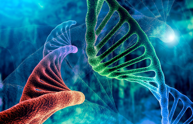
Though DNA is essential for life, it can also wreak havoc on our bodies as we age 

DNA is one of the essential building blocks of life, giving our cells instructions for virtually everything they do, but researchers at Sanford Burnham Prebys are investigating what happens to our cells when DNA ends up in places where it shouldn’t normally be, particularly as we age.

The answer – as described in their recent review in the journal Cell—is disease-causing inflammation. And the researchers hope that targeting this rogue DNA will lead to new therapeutic strategies for a range of age-related illnesses, including cancer, diabetes, rheumatoid arthritis, cardiovascular disease and neurodegenerative disorders.

“Age is the primary risk factor for all of these diseases, but they share another risk factor – chronic inflammation,” says first author Karl Miller, PhD, a postdoctoral researcher in the lab of Peter Adams, PhD, Sanford Burnham Prebys. “We’re trying to understand the underlying processes behind this inflammation so we can potentially treat all these age-related diseases together”

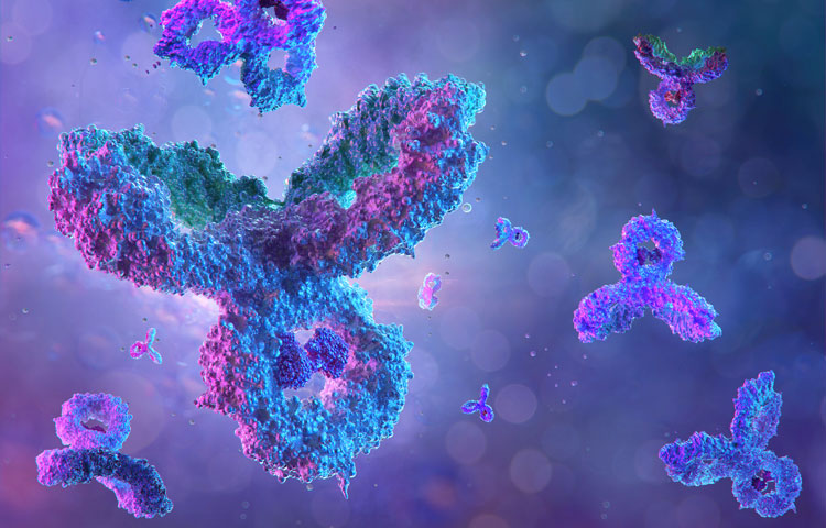
Typically, cells have DNA safely sequestered in their nucleus and in the mitochondria, where the DNA can do its job without interfering with the rest of the cells’ activities. When cells detect DNA in other areas, they unleash a series of biochemical responses designed to protect the cell from invaders. This response is a component of the innate immune system, our body’s first line of defense against infection.

Scientists have known about this system for decades, but until recently it was mostly thought to respond to foreign DNA, such as during a bacterial or viral infection. However, over the last decade, researchers have discovered that pieces of our own DNA, called endogenous cytoplasmic DNA, can escape from the nucleus or mitochondria and trigger this inflammatory response in our own cells, even in the absence of infection. The resulting ‘sterile’ inflammation can accumulate over time, contributing to a range of age-related diseases in all systems of the body.

But this inflammation is not without its upsides. Cytoplasmic DNA is actually an important short-term protective strategy against cancer formation. The inflammation can alert the immune system at the first sign of cancer, preventing its formation. But over the long term, the sterile inflammation caused by cytoplasmic DNA is also thought to contribute to cancer risk. In fact, we’ve only been able to observe the damage associated with sterile inflammation because people are now living long enough to experience it. 

“Systems like this exist because they’re beneficial in youth, but as we age, they break down,” says Miller. “100 years ago, a lot more people died from infectious diseases early in life. Over time, we’ve become better and better at treating these acute infections, and we’re living much longer. It’s in this later period in life that we see chronic diseases emerging that used to be much less common.”

Miller’s review describes four different types of cytoplasmic DNA fragments, classified according to when and how they appear. Some arise from the nucleus during mistakes in cell division. Others emerge because of errors in DNA repair or replication. Some even escape from mitochondria—energy-producing parts of the cell that have their own separate DNA. Others still are of unknown origin.

“They all look similar under a microscope, and they all can cause similar effects. That’s one of the major problems in this field. The benefit of studying how the different types emerge is that it gives us more points to target for therapeutics,” says Miller. 

In the Adams Lab, Miller and his colleagues look specifically at cytoplasmic chromatin fragments, one of the four types of cytoplasmic DNA. These fragments appear in the cell when the membrane surrounding the nucleus is weakened by senescence, a cellular stress response. Senescence is also associated with aging. 

“We’ve shown how this pathway works in mice, and now we’re actually moving forward with therapeutic applications for humans by doing drug screening to find compounds that can target it,” adds Miller. 

And while there is still a lot of work left for the researchers, their progress is encouraging. Adams, senior author on the Cell review, was recently awarded a $13 million grant by the NIH to study the effects of aging, including the role of cytoplasmic DNA, on the progression of liver cancer. 

“We like to call what we’re doing here ‘increasing the healthspan’, as opposed to the lifespan,” says Miller. “We’re hoping to maximize the healthy period of people’s lives.” 

Institute News

This enzyme is one of the hardest working proteins in the body

AuthorMiles Martin
Date

October 21, 2021

Researchers from Sanford Burnham Prebys have shown that a protein they identified plays a major role in the breakdown of hyaluronic acid, a compound found in the scaffolding between our cells. The findings, published recently in the Journal of Biological Chemistry, could have implications for epilepsy, cancer and other human diseases associated with hyaluronic acid and similar compounds.

They also shed light on one of the most active biochemical processes in the body. 

“Our body turns over hyaluronic acid at an extremely rapid rate, far faster than the other compounds surrounding our cells,” says senior author Yu Yamaguchi, MD, PhD, a professor in the Human Genetics Program at Sanford Burnham Prebys.

Hyaluronic acid, a common ingredient in cosmetic anti-aging products, is a one of several large sugar molecules known as glycosaminoglycans (GAGs). These are found naturally in the extracellular matrix, the complex network of organic compounds surrounding our cells that gives structure to our tissues. In addition to its structural role, the extracellular matrix is involved in regulating the immune system and is critical in the early development of connective tissues like cartilage, bone and skin.

“The extracellular matrix is found in every organ and tissue of the body, and malfunctions in its biochemistry can trigger or contribute to a variety of diseases, some of which we don’t even know about yet,” says Yamaguchi. His team studies how GAGs affect childhood diseases including congenital deafness, epilepsy and multiple hereditary exostoses, a rare genetic disorder that causes debilitating cartilage growths on the skeleton.

Hyaluronic acid is also known to be correlated with several health conditions, depending on its concentration in certain tissues. Reduced levels of hyaluronic acid in the skin caused by aging contribute to loss of skin elasticity and reduced capacity to heal without scarring. Levels of hyaluronic acid in the blood dramatically increase in alcoholic liver disease, fatty liver and liver fibrosis. In addition, hyaluronic acid levels have been correlated with increased tumor growth in certain cancers.

“These compounds are literally everywhere in the body, and we continue to learn about how GAG’s influence disease, but there’s also a lot we still don’t know about how these molecules are processed,” says Yamaguchi, “Research like this is about understanding what’s happening at the molecular level so we can later translate that into treatments for disease.” 

For this study, the team focused on a protein called TMEM2, which they had previously found to break down hyaluronic acid by cutting the longer molecule into manageable pieces for other enzymes to process further. Using mice as a research model, they selectively shut off the gene that codes for TMEM2 and were able to successfully measure precisely how much the absence of TMEM2 affects the overall levels of hyaluronic acid.

The answer: a lot.

“We saw up to a 40-fold increase in the amount of hyaluronic acid in the study mice compared to our controls,” says Yamaguchi. “This tells us that TMEM2 is one of the key players in the process of degrading this compound, and its dysfunction may be a key player in driving human diseases.” 

The team further confirmed this role of the TMEM2 protein by using fluorescent compounds that detect hyaluronic acid to determine where the TMEM2 protein is most active. They found the most activity on the surface of cells lining blood vessels in the liver and lymph nodes, which are known to be the main sites of hyaluronic acid degradation. 

“These findings refine our understanding of this critical biochemical process and set us up to explore it further in the interest of developing treatments for human diseases,” says Yamaguchi. “Hyaluronic acid is so much a part of our tissues that there could be any number of diseases out there waiting to benefit from discoveries like these.”

Institute News

How Sanford Burnham Prebys is helping map the brain

AuthorMiles Martin
Date

October 11, 2021

By joining forces with hundreds of researchers across the country, a team from the Chun Lab at Sanford Burnham Prebys are working to create a comprehensive map of the human brain, in the hopes of leveraging that knowledge to better treat brain disorders.

Researchers in the lab of Sanford Burnham Prebys professor Jerold Chun, MD, PhD, have helped the NIH create a cellular atlas of the motor cortex – the area of the brain responsible for movement. Their work, published recently in the journal Nature, is the flagship paper for the NIH’s BRAIN initiative, a massive multi-institution project to unravel the mysteries of the human brain.

“There are hundreds of billions of cells in the brain, and identifying and classifying all the different types of brain cells is just too big a job for any single lab,” says Chun, who is a coauthor on the study. “Similar to efforts in particle physics, hundreds of neuroscientists have now come together and it’s really exciting for us to be part of this major effort.”

The NIH Brain Research through Advancing Innovative Neurotechnologies® (BRAIN) Initiative aims to revolutionize our understanding of the human brain to more effectively diagnose, treat and prevent neurological diseases and disorders. Since its launch in 2013, the BRAIN Initiative has awarded more than 900 grants to research institutions across the country, totaling $1.8 billion. 

Chun is one of the principal investigators of the BRAIN Initiative Cell Census Network (BICCN), a subset of the Initiative that aims to develop a database of all the brain cell types in humans, mice, and non-human primates.

“While the project is about exploring the brain, what we’re really interested in over the long term is the clinical applications,” says study coauthor Carter Palmer. “Understanding the nuances of the brain and how the trillions of neural connections really work is going to lead us to new targets for therapies and diagnostics so we can help people heal.” Palmer is a graduate student in Chun’s lab, alongside fellow co-authors Christine Liu and William Romanow.

There are over 150 billion cells in the average human brain and well over a thousand different cell types, depending on how you characterize them. With such a vast landscape to track, many different types of data are needed to develop a comprehensive atlas of the brain.

For their part, the Chun Lab provided single-cell transcriptomes for human brain cells, focusing on the motor cortex. Single cell transcriptomes provide a measure of how hundreds to thousands of genes are expressed in individual cells and can provide hints as to what functions those cells are serving. This process also provides a molecular definition of cell types, making it easier for researchers to identify and classify them.

“Looking at how genes are expressed gives us a wealth of information on what cells are doing, how they develop and how they’re interacting with other cells,” says Palmer. “And when our data feed into the data from other teams, we start to get a much clearer picture of what’s happening in the brain than has ever been possible.” 

Their flagship Nature paper is one of seventeen in a special edition of the journal, chronicling recent advances by hundreds of BICCN researchers. The team also contributed to a second paper in the issue, which expands on the first by comparing the motor cortex cells of humans, mice, and marmosets. These publications speak not only to the expertise of Chun and his colleagues, but to the power of collaborative, interdisciplinary work to achieve previously unheard-of research goals.

“Fifty years ago, a project like this would have been impossible, because we just didn’t have the technology or even basic knowledge to collaborate on such a large scale,” says Chun. “Huge initiatives like BRAIN are an important part of the future of scientific research, and we’re thrilled we were able to contribute to this milestone in neuroscience.” 

Institute News

Sanford Burnham Prebys drug enters Phase 1 study for the treatment of tobacco use disorder

AuthorSusan Gammon
Date

August 26, 2021

A drug discovered in the lab of Nicholas Cosford, PhD, professor and deputy director of the NCI-designated Cancer Center at Sanford Burnham Prebys, has entered a Phase 1 clinical study.

The compound, SBP-9330, targets a neuronal signaling pathway underlying addictive behaviors and would be a first-in-class oral therapeutic to help people quit smoking. 

The study is being funded by the National Institute on Drug Abuse (NIDA) at the National Institutes of Health (NIH) through a grant awarded to Sanford Burnham Prebys, the Department of Psychiatry, University of California San Diego, School of Medicine, and Camino Pharma, LLC, who will oversee activities related to the Phase 1 study.  

“Smoking continues to be the leading cause of preventable death in the US. Nearly 70% of adult smokers try to quit smoking, but only succeed less than 30% of the time, and often relapse after quitting,” says Cosford, who is also co-founder of Camino Pharma. “It has been 15 years since the U.S. Food and Drug Administration (FDA) last approved a therapeutic for this indication. We hope that SBP-9330 ultimately becomes a viable therapeutic option for smokers to quit for good.”

As a novel selective positive allosteric modulator of the metabotropic glutamate receptor 2 (mGlu2), SBP-9330 is designed to reduce levels of glutamate, a neurotransmitter linked to addiction and relapse behavior. Preclinical studies of SBP-9330, supported by a previous NIDA grant awarded to the same three institutions, demonstrated that the drug candidate reduces nicotine self-administration in animal models and is safe and well tolerated in preclinical safety and toxicology studies.

“We are excited to initiate the first-in-human study of SBP-9330 and are grateful for the investment the NIDA has made in the treatment of tobacco use disorder,” says Gonul Velicelebi, PhD, CEO and co-founder of Camino Pharma. “In the future, we also hope to broaden the indication of SBP-9330 to other types of addiction, such as cocaine, opioid, or methamphetamine use disorders. This is supported by preclinical data in other models of substance abuse as well as the mechanism of action of SBP-9330.”

The randomized, placebo-controlled, double-blind, single-ascending and multiple-ascending dose study is being conducted at a single site in the United States under an Investigational New Drug (IND) application recently allowed by the FDA and will enroll up to 80 healthy volunteers through multiple cohorts. The goal of the study is to determine the safety, tolerability and pharmacokinetic profile of SBP-9330 in humans and to determine a safe dose range for further clinical development SBP-9330 for the treatment of people with tobacco use disorder. 

“We are excited about collaborating in the development of SBP-9330 to treat tobacco use disorder. Each year in the United States, roughly half a million people die from tobacco-related diseases. It is critical to have more therapeutic options if we want to reduce the number of deaths and illnesses related to smoking,” says Robert Anthenelli, MD, UC San Diego professor of psychiatry and one of the co-principal investigators on the NIDA project.


Dr. Cosford has an equity interest in Camino Pharma, LLC. Dr. Cosford’s relationship with Camino Pharma, LLC has been reviewed and approved by Sanford Burnham Prebys in accordance with its conflict-of-interest policies.

Institute News

Laura Martin-Sancho rises to the challenge of COVID-19

AuthorSusan Gammon
Date

June 29, 2021

The journal Molecular Cell recently asked Laura Martin-Sancho to share her experience working on SARS-CoV-2 during the pandemic

COVID-19 altered our lives and pushed scientific research to operate at breakneck speed, leading to significant breakthroughs in record time. The journal Molecular Cell recently asked experts in the field—including Laura Martin-Sancho—about the challenges they faced in transitioning, rapidly but safely, to working on the virus while navigating the shutdown. Their voices converge on the importance of teamwork, forging new collaborations, and working toward a shared goal.

Here is what Laura had to say:

I remember learning about viral pandemics in university and thinking about the challenges of working with novel viruses. Of course, we virologists contemplate the idea of a global viral pandemic and we discuss this at length in grants and in research articles, but how do you respond when confronted by it? It all started in February 2020 as I was completing my postdoc studying respiratory viruses. The lab had been closely following the worrying news coming from China and decided to drop everything else and work full time on SARS-CoV-2. Once we received the virus in March, it was a race against the clock to get the right conditions, the right cells, and the right reagents to propagate the virus to high enough amounts to start testing small compounds for antiviral activity. With a starting material of barely 100 μl received from BEI Resources, and long lonely hours in the BSL3, I felt a massive relief when I finally saw that the virus was replicating. Soon after, we had optimized experimental conditions and high enough viral yields to begin the essential experiments.

It was a remarkable feeling to have the whole research institute to just the seven of us. It was just me and three other lab members, our PI, Sumit Chanda, and two members of the institute safety department. With nobody around, it felt like we were apocalyptic survivors racing to find a cure. With a non-stop schedule from 8 am to 10 pm in the lab and trying to play catch up with an everyday-evolving literature, we were barely getting any sleep but never felt so energized. I felt so supported and inspired by my family and friends back home in Spain, which was one of the initial pandemic epicenters in Europe. I felt it was our responsibility to keep going and to make discoveries that could have a meaningful contribution. And so we did. In only a few months, we evaluated thousands of small compounds (initially in collaboration with Hong Kong University, then in house), we identified the innate immune sensor for SARS-CoV-2, and we illuminated the cellular antiviral landscape to SARS-CoV-2. Of course, this wouldn’t have been possible without the expertise and assistance from our countless collaborators across the globe. Indeed, a story to tell my grandkids.

Read the stories from other experts in the field in this article published in Molecular Cell: https://doi.org/10.1016/j.molcel.2021.05.021

Institute News

Enzyme therapy helps rebuild teeth

AuthorBrandon Levy
Date

May 28, 2021

Study in mice suggests a new approach to treating periodontal disease

Our teeth are extremely tough, but neglectful oral hygiene practices and certain genetic disorders can still massively damage them. If this deterioration becomes bad enough, teeth can be permanently lost. In a recent study co-authored by José Luis Millán, PhD, professor at Sanford Burnham Prebys, researchers identified a promising new strategy for helping the body regenerate a part of the tooth that is particularly difficult to repair.

When it comes to taking care of our teeth, the enamel coating surrounding the upper portion of teeth tends to get most of the attention. It is, after all, the most visible part of our teeth and the hardest substance in the human body. However, a substance called cementum that surrounds the roots of our teeth is also incredibly important. The cementum helps our teeth remain in our mouths by attaching them to periodontal ligaments connected to surrounding jaw bone.

“The cementum around the tooth root is one of the tissues that has to be repaired to restore the tooth’s function after periodontal disease,” explains National Institutes of Health (NIH) senior investigator Martha J. Somerman, D.D.S, PhD, the new study’s senior author. “A lot of scientists have been focusing on promoting bone regrowth, but if you do that without considering the need for a healthy cementum, you will not restore proper function.”

Unfortunately, damaged cementum doesn’t regenerate very quickly when left to its own devices, if it heals at all, and current approaches to rebuilding it have not proven to be very effective. In the new study, Somerman’s team investigated whether an enzyme naturally found in the human body called alkaline phosphatase (ALP) could help repair damaged cementum by boosting the process that builds teeth and bone, known as mineralization. Prior research had shown that ALP transforms a chemical called pyrophosphate, which inhibits mineralization, into another molecule called phosphate, which promotes mineralization.

The new study utilized a mouse model of periodontal disease that lacks the gene for an important bone- and cementum-building protein called bone sialoprotein. The researchers began by giving five-day-old mice a ‘systemic’ therapy that quadrupled their blood’s level of tissue-nonspecific alkaline phosphatase (TNAP), a form of ALP found in bones. At two months of age, the mice that received the therapy had cementum that was more than twice as thick as the cementum of their untreated counterparts, and they also showed greater growth of the jaw bone that surrounds the cementum. Moreover, their teeth were just as well-attached to the periodontal ligament as the teeth of untreated, genetically normal mice.

“Bone sialoprotein is thought to be a critical molecule for mineralization,” Somerman explains, “so this is a perfect proof-of-principle model to examine whether you can regenerate cementum.”

Next, using five-week-old mice with the same genetic defect, the research team investigated the effects of delivering TNAP directly to the area where the degraded periodontal tissue was rather than raising TNAP levels in the entire body. The treated animals showed similar beneficial effects to the mice that had received the systemic TNAP-boosting therapy. What’s more, the locally delivered TNAP treatment also promoted growth of the cementum and surrounding jaw bone in genetically normal mice.

A final set of experiments in cells showed that ALP corrected mineralization deficiencies in cementum-producing cells, called cementoblasts, that had the same genetic defect as the mice. However, treating those cells with a chemical that disrupts the transport of phosphate into cells diminished the ALP’s beneficial effects, strongly suggesting that the TNAP treatment given to the mice promoted regeneration of the cementum and surrounding bone by increasing the amount of phosphate available for cementoblasts to use for the rebuilding process.

Moving forward, the scientists will continue refining their TNAP treatment and working to move therapies based on their findings into clinical trials. Based on the original research of Millán, TNAP is now FDA-approved for use in humans to treat hypophosphatasia—a debilitating genetic condition that causes soft-brittle bones. This could hasten its adoption as a treatment to help rebuild the cementum and jaw bone of people with severe periodontal disease. Importantly, delivering TNAP directly into the damaged area, as the researchers did in their new study, would likely have fewer side effects than introducing it throughout the body.

“I’m incredibly excited that our past and ongoing research on TNAP is proving to be valuable for conditions that weren’t even on our radar when we started,” says Millán. “We knew that TNAP was very important for the proper mineralization of teeth but employing it as a therapy for periodontal disease is a very novel and promising strategy.

This piece was originally published on the NIH’s “I Am Intramural” blog.  

Institute News

Alumni adventures: Catching up with student intern Daniel Million

AuthorMonica May
Date

March 23, 2021

Million reflects on how a summer internship at Sanford Burnham Prebys accelerated his scientific career.

Daniel Million was always fascinated by biology. But, like most high school students, he wasn’t sure what life as a scientist was really like. That all changed when he had the opportunity to complete a summer internship at Sanford Burnham Prebys.

“We thought it would be a strict environment where we were all very serious, with nobody talking,” says Million. “But my mentors both made the work in the research lab exciting. They taught me early on that you can have fun while doing great science.”

For six weeks during the summer of 2013, he and nine other classmates from the Preuss School at UC San Diego—a charter school for students who would be the first in their families to graduate from college—gained valuable laboratory skills while working directly with cancer researchers.

“Now that I’ve had the opportunity to do biological research in college and grad school, I look back and am amazed at what we were able to accomplish while in high school,” says Million. “We were doing PCRs, gel electrophoresis—techniques you usually don’t get to experience until college. They gave us a great preview of what it’s like to work in science.”

Million believes that this experience gave him a leg up that led to his acceptance to the University of Southern California, and to his receipt of a prestigious GATES Millennium scholarship, which covered all of his college costs through graduation. The benefits also extended to when he arrived on campus to start his degree.

“When you go into a research lab, that can be an intimidating place,” says Million. “If I didn’t get the chance to build my confidence in the research setting, I don’t feel that I would have performed as well when I got to college.”

Today, Million is wrapping up his master’s degree in infectious disease at Keck Graduate Institute. Whatever his future holds—perhaps medical school, or a master’s degree in public health—he remains a supporter of the internship program.

“This experience not only changed my life but changed a lot of students’ lives at Preuss,” says Million. “For a student who is going to be a first-generation college student, and who is already going to have a lot of barriers entering higher education, this is the extra push and extra knowledge they need to be successful.”

This internship was funded by the National Cancer Institute’s Continuing Umbrella of Research Experiences (CURE) program, which supports training and career-development opportunities from middle school through junior investigator levels with the goal of increasing diversity in the cancer research workforce.

Institute News

Scientists design potential drug for triple-negative breast cancer

AuthorMonica May
Date

February 16, 2021

Drug candidate blocks autophagy, a cellular recycling process that cancer cells hijack as a way to resist treatment

Scientists at Sanford Burnham Prebys Medical Discovery Institute have designed a next-generation drug, called SBP-7455, which holds promise as a treatment for triple-negative breast cancer—an aggressive cancer with limited treatment options. The drug blocks a cellular recycling process called autophagy, which cancer cells hijack as a way to resist treatment. The proof-of-concept study was published in the Journal of Medicinal Chemistry.

“Scientists are now learning that autophagy is one of the main ways that cancer cells are able to survive, even in the presence of growth-blocking treatments,” says Huiyu Ren, a graduate student in the laboratory of Nicholas Cosford, PhD, at Sanford Burnham Prebys, and first author of the study. “If all goes well, we hope this compound will stop cancer cells from turning on autophagy and allow people with triple-negative breast cancer to benefit from their treatment for as long as possible.”

Cells normally use autophagy as a way to recycle waste products. However, when cancer cells’ survival is threatened by a growth-blocking treatment, this process is often “revved up” so the cancer cell can continue to receive nutrients and keep growing. Certain cancers are more likely to rely on the autophagy process for survival, including breast, pancreatic, prostate and lung cancers.

“While this study focused on triple-negative breast cancer, an area of great unmet need, we are actively testing this drug’s potential against more cancer types,” says Cosford, professor and deputy director in the National Cancer Institute (NCI)-designated Cancer Center at Sanford Burnham Prebys and senior author of the study. “An autophagy-inhibiting drug that stops treatment resistance from taking hold would be a great addition to an oncologist’s toolbox.”

About 15% to 20% of all breast cancers are triple negative, which means they do not respond to hormonal therapy or targeted treatments. The cancer is currently treated with surgery, chemotherapy and radiation, and is deadlier than other breast cancer types. If the tumor returns, other treatments such as PARP inhibitors or immunotherapy are considered. People under the age of 50 are more likely to have triple-negative breast cancer, as well as women who are Black, Hispanic, and/or have an inherited BRCA mutation.

An optimized drug

In this study, the scientists optimized a first-generation drug they created in 2015. The result is a compound called SBP-7455 that blocks two autophagy proteins, ULK1 and ULK2. SBP-7455 exhibits promising bioavailability in mice and reduces autophagy levels in triple-negative breast cancer cells, resulting in cell death. Importantly, combining the drug with PARP inhibitors, which are currently used to treat people with recurrent triple-negative breast cancer, makes the drug even more effective.

“We are hopeful that we have found a new potential therapy for people living with triple-negative breast cancer,” says Reuben Shaw, PhD, a study author and professor in the Molecular and Cell Biology Laboratory and director of the NCI-designated Cancer Center at the Salk Institute. “We envision this drug being used in combination with targeted therapies, such as PARP inhibitors, to prevent cancer cells from becoming treatment resistant.”

Next, the scientists plan to test the drug in mouse models of triple-negative breast cancer to confirm that the compound can stop tumor growth in an animal model. In parallel, they will continue optimization efforts to ensure the drug has the greatest chance of clinical success.

“Triple-negative breast cancer is one of the hardest cancers to treat today,” says Ren. “I hope that our research marks the start of a path to successful treatment that helps more people survive this aggressive cancer.”


Additional study authors include Nicole A. Bakas, Mitchell Vamos, Allison S. Limpert, Carina D. Wimer, Lester J. Lambert, Lutz Tautz, Maria Celeridad and Douglas J. Sheffler of Sanford Burnham Prebys; Apirat Chaikuad and Stefan Knapp of the Buchmann Institute for Molecular Life Sciences and Goethe-University Frankfurt; and Sonja N. Brun of the Salk Institute.

This work was supported by the National Institutes of Health (P30CA030199, T32CA211036), Epstein Family Foundation, Larry L. Hillblom Foundation (2019-A-005-NET), Pancreatic Cancer Action Network (19-65-COSF), SGC—a registered charity that receives funds from AbbVie, Bayer Pharma AG, Boehringer Ingelheim, Canada Foundation for Innovation, Eshelman Institute for Innovation, Genome Canada through Ontario Genomics Institute [OGI-196], EU/EFPIA/OICR/McGill/KTH/Diamond, Innovative Medicines Initiative 2 Joint Undertaking (875510), Janssen, Merck KGaA, Merck & Co, Pfizer, São Paulo Research Foundation-FAPESP, Takeda, and Wellcome.

The study’s DOI is 0.1021/acs.jmedchem.0c00873.

Institute News

Mining “junk DNA” reveals a new way to kill cancer cells

AuthorMonica May
Date

February 11, 2021

Scientists unearth a previously unknown vulnerability for cancer and a promising drug candidate that leverages the approach

Scientists at Sanford Burnham Prebys have uncovered a drug candidate, called F5446, that exposes ancient viruses buried in “junk DNA” to selectively kill cancer cells. Published in the journal Cell, the proof-of-concept study reveals a previously unknown Achilles’ heel for cancer that could lead to treatments for deadly breast, brain, colon and lung cancers.

“We found within ‘junk DNA’ a mechanism to stimulate an immune response to cancer cells, while also causing tumor-specific DNA damage and cell death,” says Charles Spruck, PhD, assistant professor in the National Cancer Institute (NCI)-designated Cancer Center and senior author of the study. “This is a very new field of research, with only a handful of papers published, but this has the potential to be a game-changer in terms of how we treat cancer.”

Since the human genome was fully sequenced in 2003, scientists have learned that our DNA is filled with some very strange stuff—including mysterious, noncoding regions dubbed “junk DNA.” These regions are silenced for a reason—they contain the genomes of ancient viruses and other destabilizing elements. An emerging area of cancer research called “viral mimicry” aims to activate these noncoding regions and expose the ancient viruses to make it appear that a cancer cell is infected. The hypothesis is that the immune system will then be triggered to destroy the tumor.

A one-two punch to cancer

In the study, Spruck and his team set out to find the molecular machinery that silences “junk DNA” in cancer cells. Using sophisticated molecular biology techniques, they found that a protein called FBXO44 is key to this process. Blocking this protein caused the noncoding sections of DNA to unwind—but not for long.

“When we revealed noncoding regions, which aren’t meant to be expressed, this caused DNA breakage. This told the cell that something is deeply wrong, and it committed suicide,” explains Spruck. “At the same time, the DNA of the ancient virus was exposed, so the immune system was recruited to the area and caused more cell death. So, we really delivered a one-two punch to cancer.”

The scientists then showed that a drug that targets the FBXO44 pathway, called F5446, shrank tumors in mice with breast cancer. The drug also improved the survival of mice with breast cancer that were resistant to anti-PD-1 treatment, an immunotherapy that is highly effective but often stops working over time. Additional studies in cells grown in a lab dish showed that the drug stops the growth of other tumors, including brain, colon and lung cancers.

The scientists also conducted many experiments to show that this silencing mechanism only occurs in cancer cells, not regular cells. Analysis of patient tumor databases confirmed that FBXO44 is overproduced in many cancers and correlated with worse outcomes—further indicating that a drug that inhibits this protein would be beneficial.

Moving the research toward people

As a next step, the scientists are working with the Conrad Prebys Center for Chemical Genomics to design an FBXO44 pathway-inhibiting drug that is more potent and selective than F5446. This state-of-the-art drug discovery facility is located at Sanford Burnham Prebys.

“Now that we have a compound that works, medicinal chemists can make modifications to the drug so we have a greater chance of success when we test it in people,” says Jia Zack Shen, PhD, staff scientist at Sanford Burnham Prebys and co-first author of the study. “Our greatest hope is that this approach will be a safe and effective pan-cancer drug, which maybe one day could even replace toxic chemotherapy.”


Institute News

Scientists uncover protein that empowers antibodies

AuthorMonica May
Date

January 11, 2021

Discovery may lead to better vaccine strategies and improve treatments for cancer and autoimmune disorders

Antibodies are the heroes of our immune system. They protect us from viruses, like SARS-CoV-2 (which can lead to COVID-19), as well as bacteria and other pathogens. They can provide lifelong protection from future infections—if they are strong enough. But, like any hero, they are fallible, and certain cancers or autoimmune disorders can arise when things go wrong.

Now, Sanford Burnham Prebys scientists have revealed that a protein called cyclin D3 tells antibody-producing B cells to start dividing—opening new research avenues that could improve vaccine development or the treatment of B cell lymphoma and autoimmune disorders. The discovery was published in Cell Reports.

Antibodies get their power from a complicated process. When an “intruder” is detected in the body, B cells—which produce antibodies—are activated. Each B cell is unique—they contain slight genetic variations to produce a diverse set of antibodies to attack the “intruder.” Later, they undergo optimization through a “survival of the fittest” process to identify the most protective versions.

“Our findings reveal that cyclin D3 is the ‘go’ signal for B cells to start rapidly dividing and producing a set of diverse antibodies,” says Parham Ramezani-Rad, PhD, a postdoctoral researcher in the Tumor Microenvironment and Cancer Immunology Program at Sanford Burnham Prebys and the lead author of the study. “This information might help scientists create better vaccine strategies in the future. On the flip side, researchers may be able to develop better weapons against B cell lymphoma and autoimmune disorders by removing malignant B cells.”

Parham Ramezani-Rad, PhD

​Parham Ramezani-Rad, a postdoctoral researcher at Sanford Burnham Prebys and lead author of the study.

Diving into the “dark zone”

After infection, B cells grow and divide in special structures called germinal centers that form in our spleen and lymph nodes. In this structure, a “dark zone”—referring to what scientists saw under the microscope in the 1930s—and a “light zone” are visible. Now researchers know the dark zone is where B cells are rapidly expanding, and this cell density appeared darker in the original microscope studies. After proliferating in the dark zone, B cells head to the light zone where the best potential antibody options are selected—while less desirable options are eliminated.

Cell Reports cover

Parham Ramezani-Rad designed the image that was featured on the cover of Cell Reports. The image is an artistic impression of the dynamics occurring inside of the germinal center, where antibody-producing B cells undergo a “survival of the fittest” selection process.

​Ramezani-Rad made the discovery when studying B cell lymphoma, a blood cancer that often contains a mutation that leads to hyper-stable cyclin D3. Using mice and sophisticated CRISPR gene editing technology, he discovered that cyclin D3 regulates the expansion or contraction of B cells specifically in the dark zone of germinal centers—and not the light zone. He also identified other regulatory aspects involved in this process that scientists might be able to harness for the benefit of human health.

“B cell lymphoma is often treated with an intensive chemo and immunotherapy combination. The side effects of this treatment can be immense, and relapses may occur,” says Ramezani-Rad. “Our findings about cyclin D3 could form the basis for a more tailored medicine that targets exactly what goes wrong during B cell lymphoma, and is potentially less toxic and more effective.”

Ramezani-Rad also designed the image that was selected for the journal cover, which is his artistic impression of the dynamics occurring inside the germinal center. He finds many parallels between scientists and artists.

“As a scientist, I see myself describing what already exists in nature,” explains Ramezani-Rad. “Musicians and painters are also describing the world. They are just using instruments or paint strokes to express emotions, whereas scientists use data to express knowledge.”