proteins Archives - Sanford Burnham Prebys
Institute News

Two Sanford Burnham Prebys scientists selected for American Cancer Society postdoctoral fellowships

AuthorGreg Calhoun
Date

October 18, 2024

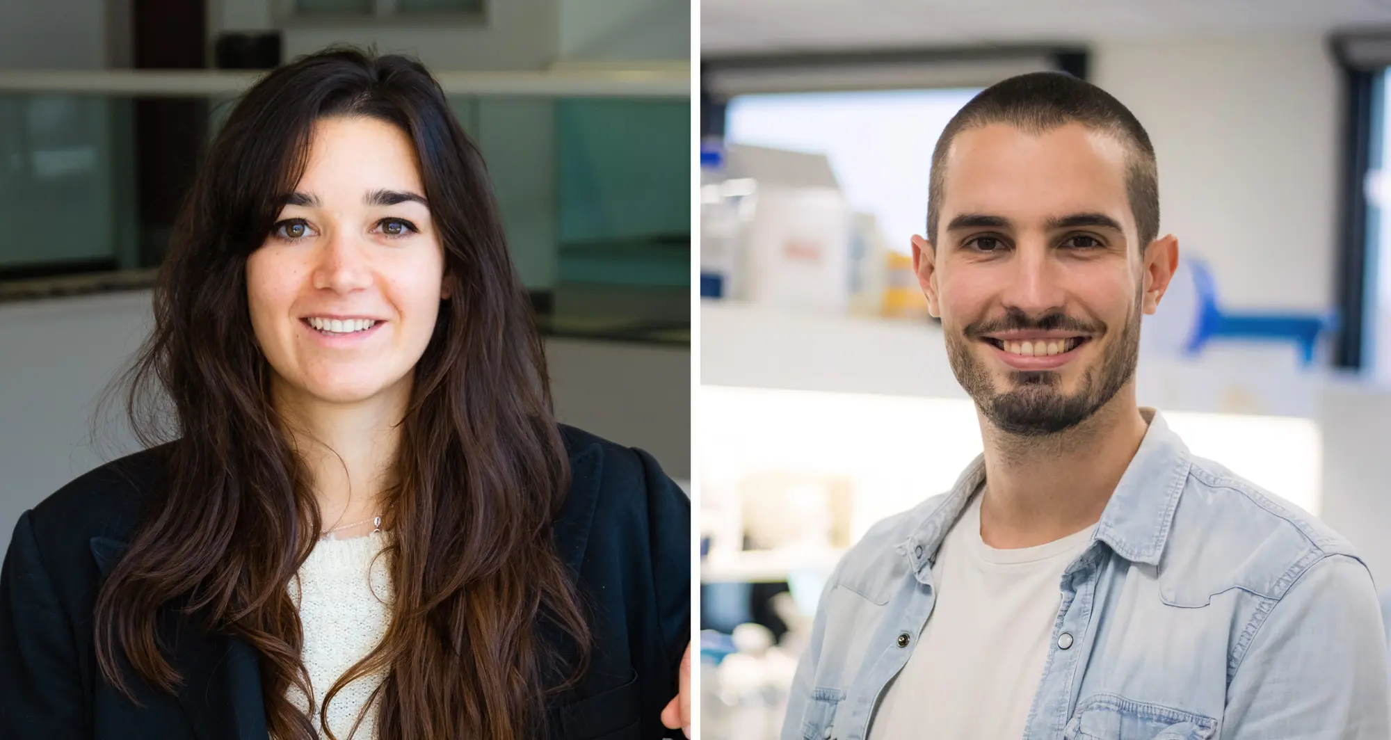
Funds will support Alicia Llorente Lope and Ambroise Manceau who study breast and pancreatic cancer

Alicia Llorente Lope, PhD, and Ambroise Manceau, PhD, were awarded 2024 Postdoctoral Fellowships from the American Cancer Society (ACS). These prestigious awards provide more than $65,000 per year for up to three years to support early career scientists studying cancer.

“I was so excited when I heard the news,” said Llorente. “It is a privilege to have this award, and it feels very validating to know that someone saw enough potential in my research to deem it worthy of funding.”

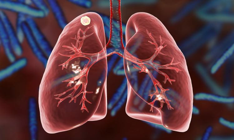
Tackling treatment-resistant breast cancer

Llorente joined the lab of Brooke Emerling, PhD, director of the Cancer Metabolism and Microenvironment Program at Sanford Burnham Prebys, nearly three years ago after beginning her breast cancer research career as a doctoral student.

“I was first interested in breast cancer because my grandmother died of the disease, and I wanted to contribute to finding new therapeutic opportunities for cancer patients,” said Llorente. Llorente’s ACS-funded research project focuses on HER2-positive (HER2+) breast cancer.

Roughly one in five breast cancer tumors have elevated levels of the HER2 protein. While these tumors tend to grow quickly, drugs targeting the HER2 protein are usually effective at first. However, HER2+ tumors often are able to adapt and develop resistance to these drugs over time, leaving patients with few if any remaining treatments options.

Llorente has found evidence that a form of the protein phosphatidylinositol-5-phosphate 4-kinase (PI5P4K) plays a role in breast cancer tumors becoming resistant to HER2 drugs.

Brooke Emerling

Brooke Emerling, PhD

“We’ve revealed a strong connection between elevated levels of PI5P4K gamma and reduced survival rates in patients with HER2+ breast cancer,” explained Llorente. “I plan to explore whether targeting both HER2 and PI5P4K gamma in breast cancer cells may provide a path to overcoming treatment resistance.” Llorente also will study the functions of PI5P4K gamma in breast cancer cells to see why these cells cease to respond to HER2-targeting drugs.

“I am incredibly proud of Alicia for spearheading this groundbreaking project targeting the lipid kinase PI5P4K gamma,” said Emerling. “Her insightful analysis of breast cancer datasets, which uncovered a correlation between elevated expression of PI5P4K gamma and worse outcomes in HER2+ patients, has set the stage for vital research aimed at overcoming the significant challenge of resistance to targeted therapies in HER2+ tumors.”

Cosimo Commisso headshot

Cosimo Commisso, PhD

Powering down pancreatic cancer

Manceau is in the second year of his postdoctoral training in the lab of Cosimo Commisso, PhD, interim director and deputy director of the institute’s NCI-Designated Cancer Center. During his doctoral program, Manceau studied how abnormal cells die in a programmed series of steps called apoptosis, a process known to go awry in cancer and neurodegenerative diseases.

“It began as a basic science project about the molecular processes around cell death, and over time it led to possible therapeutic implications,” said Manceau. “I learned that I like to study fundamental biology and then try to find an application for it, and I saw in the Commisso lab an opportunity to do just that in pancreatic cancer.”

Manceau’s fellowship project focuses on pancreatic ductal adenocarcinoma (PDAC) — the most common form of pancreatic cancer with only a 13% five-year survival rate — and its ravenous pursuit of energy. Because of PDAC cells’ constant need for fuel to sustain their rampant growth, they adapt by reshaping the surface of their cells to snatch extra nutrients from the jelly-like substance between cells.

Commisso and others have shown that cutting off the extra power supplied by this process — known as macropinocytosis — reduces tumor growth. Manceau has studied the contents taken by contorted pancreatic cell surfaces in pockets called macropinosomes. By analyzing every single protein in this scooped goop, he found that calcium transporter proteins present in macropinosomes also are required for macropinocytosis.

“During the fellowship, I will work to understand how these transporter proteins affect macropinocytosis,” said Manceau. “These proteins have never been targeted before in pancreatic cancer, so our long-term goal is to use this strategy to cut the nutrient supply to tumors and see if we can inhibit tumor growth.”

“By disrupting the cancer cells’ ability to feed themselves through macropinocytosis, we can potentially starve tumors and inhibit their growth,” added Commisso. “Ambroise’s research aims to target key proteins involved in this process, opening up new possibilities for treatments that could significantly improve outcomes for patients battling pancreatic cancer.”

Institute News

Simulating science or science fiction? 

AuthorGreg Calhoun
Date

August 27, 2024

By harnessing artificial intelligence and modern computing, scientists are simulating more complex biological, clinical and public health phenomena to accelerate discovery.

While scientists have always employed a vast set of methods to observe the immense worlds among and beyond our solar system, in our planet’s many ecosystems, and within the biology of Earth’s inhabitants, the public’s perception tends to reduce this mosaic to a single portrait.

A Google image search will reaffirm that the classic image of the scientist remains a person in a white coat staring intently at a microscope or sample in a beaker or petri dish. Many biomedical researchers do still use their fair share of glassware and plates while running experiments. These scientists, however, now often need advanced computational techniques to analyze the results of their studies, expanding the array of tools researchers must master to push knowledge forward. For every scientist pictured pipetting, we should imagine others writing code or sending instructions to a supercomputer.

In some cases, scientists are testing whether computers can be used to simulate the experiments themselves. Computational tools such as generative artificial intelligence (AI) may be able to help scientists improve data inputs, create scenarios and generate synthetic data by simulating biological processes, clinical outcomes and public health campaigns. Advances in simulation one day might help scientists more quickly narrow in on promising results that can be confirmed more efficiently through real-world experiments.

“There are many different types of simulation in the life sciences,” says Kevin Yip, PhD, professor in the Cancer Genome and Epigenetics Program at Sanford Burnham Prebys and director of the Bioinformatics Shared Resource. “Molecular simulators, for example, have been used for a long time to show how certain molecules will change their shape and interact with other molecules.”

“One of the most successful examples is in structural biology with the program AlphaFold, which is used to predict protein structures and interactions,” adds Yip. “This program was built on a very solid foundation of actual experiments determining the structures of many proteins. This is something that other fields of science can work to emulate, but in most other cases simulation continues to be a work in progress rather than a trusted technique.”

In the Sanford Burnham Prebys Conrad Prebys Center for Chemical Genomics (Prebys Center), scientists are using simulation-based techniques to more effectively and efficiently find new potential drugs.

Click to Play VideoNanome Virtual Reality demonstration

To expedite their drug discovery and optimization efforts, the Prebys Center team uses a suite of computing tools to run simulations that model the fit between proteins and potential drugs, how long it will take for drugs to break down in the body, and the likelihood of certain harmful side effects, among other properties.

“In my group, we know what the proteins of interest look like, so we can simulate how certain small molecules would fit into those proteins to try and design ones that fit really well,” says Steven Olson, PhD, executive director of Medicinal Chemistry at the Prebys Center. In addition to fit, Olson and team look for drugs that won’t be broken down too quickly after being taken.

“That can be the difference between a once-a-day drug and one you have to take multiple times a day, and we know that patients are less likely to take the optimal prescribed dose when it is more than once per day,” notes Olson. 

Steven Olson, PhD, profile photo

Steven Olson, PhD, is the executive director of Medicinal Chemistry at the Prebys Center.

“We can use computers now to design drugs that stick around and achieve concentrations that are pharmacologically effective and active. What the computers produce are just predictions that still need to be confirmed with actual experiments, but it is still incredibly useful.”

In one example, Olson is working with a neurobiologist at the University of California Santa Barbara and an x-ray crystallographer at the University of California San Diego on new potential drugs for Alzheimer’s disease and other forms of dementia.

“This protein called farnesyltransferase was a big target for cancer drug discovery in the 1990s,” explains Olson. “While targeting it never showed promise in cancer, my collaborator showed that a farnesyltransferase inhibitor stopped proteins from aggregating in the brains of mice and creating tangles, which are a pathological hallmark of Alzheimer’s.”

“We’re working together to make drugs that would be safe enough and penetrate far enough into the brain to be potentially used in human clinical trials. We’ve made really good progress and we’re excited about where we’re headed.”

To expedite their drug discovery and optimization efforts, Olson’s team uses a suite of computing tools to run simulations that model the fit between proteins and potential drugs, how long it will take for drugs to break down in the body, and the likelihood of certain harmful side effects, among other properties. The Molecular Operating Environment program is one commercially available application that enables the team to visualize candidate drugs’ 3D structures and simulate interactions with proteins. Olson and his collaborators can manipulate the models of their compounds even more directly in virtual reality by using another software application known as Nanome. DeepMirror is an AI tool that helps predict the potency of new drugs while screening for side effects, while StarDrop uses learning models to enable the team to design drugs that aren’t metabolized too quickly or too slowly.

Steven Olson et al using VR in Prebys Center

The Prebys Center team demonstrates how the software application known as Nanome allows scientists to manipulate the models of potential drug compounds directly in virtual reality.

“In addition, there are certain interactions that can only be understood by modeling with quantum mechanics,” Olson notes. “We use a program called Gaussian for that, and it is so computationally intense that we have to run it over the weekend and wait for the results.”

“We use these tools to help us visualize the drugs, make better plans and give us inspiration on what we should make. They also can help explain the results of our experiments. And as AI improves, it’s helping us to predict side effects, metabolism and all sorts of other properties that previously you would have to learn by trial and error.”

While simulation is playing an active and growing role in drug discovery, Olson continues to see it as complementary to the human expertise required to synthesize new drugs and put predictions to the test with actual experiments.

“The idea that we’re getting to a place where we can simulate the entire drug design process, that’s science fiction,” says Olson. “Things are evolving really fast right now, but I think in the future you’re still going to need a blend of human brainpower and computational brainpower to design drugs.”


Programming in a Petri Dish, an 8-part series

How artificial intelligence, machine learning and emerging computational technologies are changing biomedical research and the future of health care

  • Part 1 – Using machines to personalize patient care. Artificial intelligence and other computational techniques are aiding scientists and physicians in their quest to prescribe or create treatments for individuals rather than populations.
  • Part 2 – Objective omics. Although the hypothesis is a core concept in science, unbiased omics methods may reduce attachments to incorrect hypotheses that can reduce impartiality and slow progress.
  • Part 3 – Coding clinic. Rapidly evolving computational tools may unlock vast archives of untapped clinical information—and help solve complex challenges confronting health care providers.
  • Part 4 – Scripting their own futures. At Sanford Burnham Prebys Graduate School of Biomedical Sciences, students embrace computational methods to enhance their research careers.
  • Part 5 – Dodging AI and computational biology dangers. Sanford Burnham Prebys scientists say that understanding the potential pitfalls of using AI and other computational tools to guide biomedical research helps maximize benefits while minimizing concerns.
  • Part 6 – Mapping the human body to better treat disease. Scientists synthesize supersized sets of biological and clinical data to make discoveries and find promising treatments.
  • Part 7 – Simulating science or science fiction? By harnessing artificial intelligence and modern computing, scientists are simulating more complex biological, clinical and public health phenomena to accelerate discovery.
  • Part 8 – Acceleration by automation. Increases in the scale and pace of research and drug discovery are being made possible by robotic automation of time-consuming tasks that must be repeated with exhaustive exactness.
Institute News

How a protein component of nuclear pore complexes regulates development of blood cells and may contribute to myeloid disorders

AuthorCommunications
Date

June 5, 2024

Nuclear pore complexes (NPCs) are channels composed of multiple proteins that ferry molecules in and out of the nucleus, regulating many critical cellular functions, such as gene expression, chromatin organization and RNA processes that influence cell survival, proliferation, and differentiation.

In recent years, new studies, including work by Maximiliano D’Angelo, PhD, associate professor in the Cancer Metabolism and Microenvironment Program at Sanford Burnham Prebys, have noted that NPCs in cancer cells are different, but how these alterations contribute to malignancy and tumor development—or even how NPCs function in normal cells—is poorly understood.

In a new paper, published June 5, 2024 in Science Advances, D’Angelo with first author Valeria Guglielmi, PhD, and co-author Davina Lam, uncover Nup358, one of roughly 30 proteins that form the NPCs, as an early player in the development of myeloid cells, blood cells that if not formed or working properly leads to myeloid disorders such as leukemias.

The researchers found that when they eliminated Nup358 in a mouse model, the animals experienced a severe loss of mature myeloid cells, a group of critical immune cells responsible for fighting pathogens that are also responsible for several human diseases including cancer. Notably, Nup358 deficient mice showed an abnormal accumulation of early progenitors of myeloid cells referred as myeloid-primed multipotent progenitors (MPPs).

“MPPs are one of the earliest precursors of blood cells,” said D’Angelo. “They are produced in the bone marrow from hematopoietic stem cells, and they differentiate to generate the different types of blood cells.

Maximiliano D’Angelo and Valeria Guglielmi

“There are different populations of MPPs that are responsible for producing specific blood cells and we found that in the absence of Nup358, the MPPs that generate myeloid cells, which include red blood cells and key components of the immune system, get stuck in the differentiation process.”

Fundamentally, said Gugliemi, Nup358 has a critical function in the early stages of myelopoiesis (the production of myeloid cells). “This is a very important finding because it provides insights into how blood cells develop, and can help to establish how alterations in Nup358 contribute to blood malignancies.”

The findings fit into D’Angelo’s ongoing research to elucidate the critical responsibilities of NPCs in healthy cells and how alterations to them contribute to immune dysfunction and the development and progression of cancer.

“Our long-term goal is to develop novel therapies targeting transport machinery like NPCs,” said D’Angelo, who recently received a two-year, $300,000 Discovery Grant from the American Cancer Society to advance his work.


This research was supported in part by a Research Scholar Grant from the American Cancer Society (RSG-17-148-01), the Department of Defense (grant W81XWH-20-1-0212) and the National Institutes of Health (AI148668).

The study’s DOI is 10.1126/sciadv.adn8963.

Institute News

Without this protein, tuberculosis is powerless

AuthorMiles Martin
Date

May 9, 2022

A new study from the lab of Francesca Marassi, PhD could help reveal new treatments for one of the world’s deadliest pathogens.

Sanford Burnham Prebys researchers have uncovered the structure of an important protein for the growth of tuberculosis bacteria. The study, published recently in Nature Communications, sheds light on an unusual metabolic system in tuberculosis, which could help yield new treatments for the disease and help make existing therapies more effective.

“Molecular discoveries like this give us valuable insight into how these bacteria survive, which is important in terms of finding cures for tuberculosis, and for other areas of health and biology,” says James Kent, a PhD candidate working in Marassi’s lab. “For example, bacteria in this family pose problems in both human health and agriculture, such as leprosy and bovine tuberculosis.”

Tuberculosis caused 1.5 million deaths in 2020 according to the World Health Organization, and this figure is expected to increase in the coming years due to the impact of the COVID-19 pandemic.

Stealing iron has its risks
The new protein, called Rv0455c, is part of a complex transportation system in Mycobacterium tuberculosis. Rv0455C helps the bacteria take up iron from the host cells they infect. This process is essential to their growth and replication.

“They produce these very small molecules called siderophores and send them out of the cell, where they bind to iron and bring it back in,” says Kent. “Rv0455C seems to be essential for secreting these molecules.”

An important step of this iron-uptake process is recycling the siderophores so they can be used again. When this process is interrupted, the leftover molecules can accumulate and poison the cell.

The study found that without Rv0455c, tuberculosis bacteria cannot secrete siderophores, which severely impairs their replication. Bacteria without Rv0455c also experienced poisoning from unrecycled siderophores. 

And while this delicate system can be interrupted by blocking previously known genes, eliminating Rv0455c does it much more efficiently.

“This seems to be the first piece of evidence that there is a single protein in this system that could be targeted by a new class of tuberculosis drugs,” adds Kent.

Structure determines function
Kent’s role in the study was to piece together the structure of the protein, which had posed a significant challenge to the researchers. Revealing the detailed structure of a protein is a critical part of understanding its function.

“The process of figuring out the structure of a protein can be time consuming and requires precise optimization of many conditions,” says Kent. “This protein is small, but it is still a three-dimensional object moving in three-dimensional space, and the way it’s shaped will affect what it does.”

Kent determined that the Rv0455c protein has an unusual “cinched” structure that could help explain its unique function in tuberculosis bacteria. The structure may also help determine whether it’s possible to target the protein with therapeutics. 

Looking ahead
The findings suggest that targeting the recycling of iron-carrying molecules may lead to the development of much-needed drugs to combat one of the world’s deadliest bacterial pathogens.

Kent is also optimistic that the findings could help augment existing treatments for tuberculosis.

“Because treatment cycles are long for tuberculosis, a common problem with is multi-drug resistance,” says Kent. “There’s a very good possibility that there will be implications for this protein in interrupting some of the processes that lead to bacterial resistance.”

Institute News

This enzyme is one of the hardest working proteins in the body

AuthorMiles Martin
Date

October 21, 2021

Researchers from Sanford Burnham Prebys have shown that a protein they identified plays a major role in the breakdown of hyaluronic acid, a compound found in the scaffolding between our cells. The findings, published recently in the Journal of Biological Chemistry, could have implications for epilepsy, cancer and other human diseases associated with hyaluronic acid and similar compounds.

They also shed light on one of the most active biochemical processes in the body. 

“Our body turns over hyaluronic acid at an extremely rapid rate, far faster than the other compounds surrounding our cells,” says senior author Yu Yamaguchi, MD, PhD, a professor in the Human Genetics Program at Sanford Burnham Prebys.

Hyaluronic acid, a common ingredient in cosmetic anti-aging products, is a one of several large sugar molecules known as glycosaminoglycans (GAGs). These are found naturally in the extracellular matrix, the complex network of organic compounds surrounding our cells that gives structure to our tissues. In addition to its structural role, the extracellular matrix is involved in regulating the immune system and is critical in the early development of connective tissues like cartilage, bone and skin.

“The extracellular matrix is found in every organ and tissue of the body, and malfunctions in its biochemistry can trigger or contribute to a variety of diseases, some of which we don’t even know about yet,” says Yamaguchi. His team studies how GAGs affect childhood diseases including congenital deafness, epilepsy and multiple hereditary exostoses, a rare genetic disorder that causes debilitating cartilage growths on the skeleton.

Hyaluronic acid is also known to be correlated with several health conditions, depending on its concentration in certain tissues. Reduced levels of hyaluronic acid in the skin caused by aging contribute to loss of skin elasticity and reduced capacity to heal without scarring. Levels of hyaluronic acid in the blood dramatically increase in alcoholic liver disease, fatty liver and liver fibrosis. In addition, hyaluronic acid levels have been correlated with increased tumor growth in certain cancers.

“These compounds are literally everywhere in the body, and we continue to learn about how GAG’s influence disease, but there’s also a lot we still don’t know about how these molecules are processed,” says Yamaguchi, “Research like this is about understanding what’s happening at the molecular level so we can later translate that into treatments for disease.” 

For this study, the team focused on a protein called TMEM2, which they had previously found to break down hyaluronic acid by cutting the longer molecule into manageable pieces for other enzymes to process further. Using mice as a research model, they selectively shut off the gene that codes for TMEM2 and were able to successfully measure precisely how much the absence of TMEM2 affects the overall levels of hyaluronic acid.

The answer: a lot.

“We saw up to a 40-fold increase in the amount of hyaluronic acid in the study mice compared to our controls,” says Yamaguchi. “This tells us that TMEM2 is one of the key players in the process of degrading this compound, and its dysfunction may be a key player in driving human diseases.” 

The team further confirmed this role of the TMEM2 protein by using fluorescent compounds that detect hyaluronic acid to determine where the TMEM2 protein is most active. They found the most activity on the surface of cells lining blood vessels in the liver and lymph nodes, which are known to be the main sites of hyaluronic acid degradation. 

“These findings refine our understanding of this critical biochemical process and set us up to explore it further in the interest of developing treatments for human diseases,” says Yamaguchi. “Hyaluronic acid is so much a part of our tissues that there could be any number of diseases out there waiting to benefit from discoveries like these.”

Institute News

Ze’ev Ronai wins Lifetime Achievement Award from the Society for Melanoma Research

AuthorJessica Moore
Date

November 10, 2016

Ze’ev Ronai, PhD, chief scientific advisor at Sanford Burnham Prebys Medical Research Institute (SBP) and professor in its NCI-designated Cancer Center, is the 2016 recipient of the Society for Melanoma Research’s Lifetime Achievement Award. The award honors “an individual who has made major and impactful contributions to melanoma research throughout their career.”

Ronai is being recognized for his significant contributions to melanoma research that are advancing understanding of this deadly form of skin cancer and could lead to new treatments. His studies on ultraviolet (UV) irradiation-induced changes that promote melanoma showed how they rewire signaling networks. A major discovery from those inquiries was that one player in that rewiring, a protein called ATF2, can switch from its usual tumor-preventive function to become a tumor promoter. Work by the Ronai lab also mapped how ATF2 contributes to melanoma development, and identified specific factors involved in melanoma response to therapy and metastatic potential.

In mapping the landscape of melanoma signaling, Ronai’s lab also uncovered the important role the enzyme PDK1 plays in melanoma development and metastasis. More recently, Ronai’s studies identified a mechanism underlying resistance of melanoma to BRAF inhibitor therapy, paving the road for a new clinical trial. Integral to Ronai’s research are translational initiatives, including the development of SBI-756, a small molecule that disrupts the complex that initiates protein synthesis and prevents melanoma resistance when combined with BRAF inhibition.

Ronai and his team also study how cancer cells thrive under harsh conditions, such as lack of oxygen or nutrients. That line of research has produced important insights into cancer heterogeneity and its capacity to drive the survival of the select few cancer cells that are resistant to therapy and able to metastasize. Ronai’s studies of proteins that control stress responses, such as Siah and RNF5, have furthered understanding of these processes and identified new targets for future therapies.

Ronai’s record of scientific accomplishments was recognized by the National Cancer Institute with an Outstanding Investigator Award, a seven-year grant that allows recipients to pursue projects of unusual potential. Ronai’s unique focus on how gene activity changes in cancer promises to continue establishing new paradigms for how cancers develop and respond to therapy.

About the Society for Melanoma Research

The Society for Melanoma Research (SMR) is an all-volunteer group of scientists dedicated to finding the mechanisms responsible for melanoma and, consequently, new therapies for this cancer. SMR contributes to advances in melanoma research by catalyzing collaborations among basic, translational, and clinical researchers, carrying new technology-based discoveries from bench to bedside and back.

About melanoma

The incidence of melanoma, the most lethal form of skin cancer, is rising at one of the fastest rates of all cancers in the U.S. Melanoma can strike people of all ages and is the most common form of cancer among young adults ages 25 to 29.

Institute News

How cholesterol-lowering drugs ameliorate fatty liver disease

AuthorJessica Moore
Date

October 27, 2016

Nonalcoholic fatty liver disease (NAFLD) is quietly becoming an epidemic alongside obesity—up to 20% of people in Western countries have it. Though NAFLD, the mildest of a spectrum of liver diseases characterized by excess fat in liver cells, has no symptoms at first, it increases risk for liver cancer and can worsen to nonalcoholic steatohepatitis (NASH) or even liver failure.

There are no specific treatments for NAFLD, but cholesterol-lowering drugs called statins appear to slow its progression to more serious liver inflammation and fibrosis/scarring, characteristics of NASH. However, they haven’t been widely adopted, in part because of concerns about statins’ potential liver toxicity, though recent analyses suggest that severe toxicity is rare.

Now, a study co-led by Timothy Osborne, PhD, professor and director of the Integrative Metabolism Program, and published in Scientific Reports, outlines the molecular pathway through which statins break down fat stores in the liver.

“We show directly that these drugs reduce the amount of fat molecules and cholesterol in the liver in an animal model of NAFLD,” said Osborne. “Our results provide support for using statins to treat NAFLD itself, even if patients’ serum cholesterol isn’t dangerously high.”

The experiments were initiated by Young-Kyo Seo, PhD, now a professor at the Ulsan National Institute of Science and Technology, while he was a postdoc in Osborne’s laboratory. The study was based on previous work that found statins activate a protein called SREBP-2, a transcription factor that activates genes to regulate cholesterol balance.

To figure out how statins work on liver cells, the team searched SREBP-2’s target genes for enzymes that break down fat molecules and found PNPLA8, which splits certain fat molecules into pieces that regulate cell signaling. Further experiments showed that PNPLA8 helps liver cells break down stored fat molecules.

The new study provides some hints as to PNPLA8’s mechanism. Statins are known to enhance a cellular recycling process called autophagy, which breaks down cell parts—such as lipid droplets, the site of fat storage—for energy and re-use. The new results suggest that this may depend on PNPLA8’s ability to target the autophagy machinery directly to lipid droplets.

“This is the first time PNPLA8 has been implicated in freeing fat from liver cells,” Osborne commented. “Looking in more detail at how it mobilizes fat stores will give us an idea of whether it might be a good drug target.”

Institute News

New therapeutic target for Crohn’s disease

AuthorJessica Moore
Date

September 20, 2016

Research from the Sanford Burnham Prebys Medical Discovery Institute (SBP) identifies a promising new target for future drugs to treat inflammatory bowel disease (IBD). The study, published in Cell Reports, also indicates that another protein, protein kinase C (PKC) λ/ι, may serve as a biomarker of IBD severity.

“The intestine is protected by specialized cells, called Paneth cells, that secrete antimicrobial peptides,” said Jorge Moscat, PhD, deputy director and professor in the NCI-designated Cancer Center and senior author of the paper. “We found that maintaining normal numbers of Paneth cells requires PKC λ/ι, and that the amount of PKC λ/ι decreases as IBD gets worse. We also discovered a way to prevent Paneth cell loss—inhibiting a protein called EZH2, which could be a new therapeutic strategy for IBD.”

IBD, which includes Crohn’s disease and ulcerative colitis, affects 1.4 million people in the U.S. These chronic conditions are often debilitating, as they cause unpredictable abdominal pain and diarrhea. Because current medications only help control symptoms and not the underlying disease, 70% of Crohn’s patients and 30% of those with colitis must eventually undergo surgery. In addition, IBD increases risk of intestinal cancer by as much as 60%.

“We also examined the effect of PKC λ/ι on tumor formation,” said Maria Diaz-Meco, PhD, also a professor in the Cancer Center and co-author of the paper. “In contrast to some previous studies indicating that it might promote cancer development, we demonstrate that in the intestine, PKC λ/ι is protective.”

“We inactivated the PKC λ/ι gene in the intestine of mice, which caused them to have very few Paneth cells,” added Diaz-Meco. “Without Paneth cells, the intestine is more susceptible to bacterial infiltration, which leads to inflammation. Since inflammation favors cancer, it makes sense that PKC λ/ι is a tumor suppressor in this setting.”

To find a way to boost Paneth cell numbers and possibly treat IBD, the team looked for what drives the deficit in these protector cells. The key link was overactive EZH2, which turns off genes needed to generate Paneth cells.

“We used an in vitro model—‘mini guts’ in a dish—to show that blocking EZH2 helps return the number of Paneth cells to normal,” said Yuki Nakanishi, MD, a postdoctoral fellow in the Moscat/ Diaz-Meco lab and lead author of the work. “This demonstrates that inhibiting EZH2 could be a new way to slow the progression of IBD.”

Importantly, the team verified the relevance of their findings in intestinal biopsy samples from 30 patients with Crohn’s disease. Disease progression correlated with lower levels of PKC λ/ι.

“EZH2 inhibitors are currently being developed by the pharmaceutical industry to treat other cancers, so they could be tested for IBD relatively soon,” said Moscat. “But first, we need to do preclinical studies to test whether they block progression of the disease.”

The paper is available online here.

Institute News

Super-oncogenic protein that promotes development of melanoma

AuthorJessica Moore
Date

May 19, 2016

An international collaborative study led by scientists at the Sanford Burnham Prebys Medical Discovery Institute (SBP) has identified a malicious form of a protein that drives the formation of melanoma. The findings, published in Cell Reports, reveal unexpected insight into how this lethal skin cancer develops and progresses, and may help understand and develop novel therapies against these aggressive tumors.

“We found that an inactive version of a protein called activating transcription factor 2 (ATF2) elicits a tumor-promoting effect in a way not seen before,” said Ze’ev Ronai, PhD, chief scientific advisor of SBP and professor of its NCI-designated Cancer Center. “We have known for years that the active version of ATF2 promotes melanoma, but this result was a surprise because we thought ATF2 transcriptional activity was essential to activate cancer-related genes.”

Ronai’s team has been studying ATF2’s role in melanoma for two decades. Their past work led to the view that it’s dangerous when it’s in the nucleus because it controls cancer-enabling genes, but benign when it’s not.

In the current study, researchers looked at the oncogenic potential of a ‘dead’ form of ATF2 in mice with mutations in BRAF, a kinase that transmits signals promoting cell division and is often mutated in pigmented skin cells. The same mutation is found in about half of all human melanomas.

“Inactive ATF2, in mice with mutant BRAF, resulted in the formation of pigmented lesions and later, melanoma tumors,” said Ronai, senior author of the study.

“What makes this discovery relevant to human melanoma is that we identified a structurally similar form of inactive ATF2 in human melanoma samples that has the same effects on cancer cells,” added Ronai. “Inactive ATF2 could be an indicator of tumor aggressiveness in patients with BRAF mutations, and maybe other types of cancer as well.”

“Unlike models with more complex genetic changes, like the inactivation of PTEN and p16 combined with BRAF mutations that result in rapid tumorigenesis (within a few weeks), the inactive ATF2 caused BRAF mutant mice to develop melanoma much slower, more similar to the timescale seen in patients,” commented Ronai. “This improves our ability to monitor the development of melanoma and efficacy of possible interventions.”

“We’re now investigating why inactive ATF2 so potently promotes BRAF-mutant melanoma, and looking for other types of cancer where it acts the same way,” Ronai said. “Our findings may guide precision therapies for tumors with mutant ATF2.”

The paper is available online here.

Institute News

How proteins age

Authorsgammon
Date

October 19, 2015

SBP researchers and colleagues discover a mechanism that regulates the aging and abundance of secreted proteins.

Physiological processes in the body are in large part determined by the composition of secreted proteins found in the circulatory systems, including the blood. Each of the hundreds of proteins in the blood has a specific life span that determines its unique range of abundance. In fact, measurements of their quantities and activities contribute to many clinical diagnoses. However, the way in which normal protein concentrations in the blood are determined and maintained has been a mystery for decades.

Biomedical scientists at Sanford Burnham Prebys Medical Discovery Institute (SBP) and UC Santa Barbara (UCSB) have now discovered a mechanism by which secreted proteins age and turnover at the end of their life spans. Their findings, which shed light on a crucial aspect of health and disease, appear today in the Proceedings of the National Academy of Sciences (PNAS).

“This is a fundamental advance that is broadly applicable and provides an understanding of how secreted proteins, which are involved in many important physiological processes, normally undergo molecular aging and turnover,” said senior author Jamey Marth, PhD, professor in SBP’s NCI-designated Cancer Center.

“When a secreted protein is made, it has a useful life span and then it must be degraded — the components are then basically recycled,” added Marth, also director of UCSB’s Center for Nanomedicine and a professor in the campus’s Department of Molecular, Cellular, and Developmental Biology. “We can now see how the regulation and alteration of secreted protein aging and turnover is able to change the composition of the circulatory system and thereby maintain health as well as contribute to various diseases.”

This newly discovered mechanism encompasses multiple factors, including circulating enzymes called glycosidases. These enzymes progressively remodel N-glycans, which are complex structures of monosaccharide sugars linked together and attached to virtually all secreted proteins.

It is the N-glycan structure itself that identifies the protein as nearing the end of its life span. Subsequently, multiple receptors known as lectins — carbohydrate-binding proteins — recognize these aged proteins and eliminate them from circulation.

Marth and colleagues identified more than 600 proteins in the bloodstream that exhibit molecular signs of undergoing this aging and turnover process. Many of these proteins are regulators of proteolysis (the breakdown of proteins), blood coagulation and immunity.

Honing in on individual examples, the researchers were able to track each of them through time and watch the process unfold. “In these studies we further saw that the different life spans of distinct proteins are accounted for by the different rates of aging due to N-glycan remodeling,” said lead author Won Ho Yang, PhD, a postdoctoral associate at SBP and at UCSB’s Center for Nanomedicine.

“Altering this aging and turnover mechanism is the fastest way to change the abundance of a secreted protein, which we increasingly note is occurring at the interface of health and disease,” Marth explained. “In retrospect from published literature and from studies in progress, we can now see how sepsis, diabetes and inflammatory bowel disorders can arise by the targeted acceleration or deceleration of secreted protein aging and turnover.”

“The discovery of this mechanism provides a unique window into disease origins and progression,” Marth added. “It has been known that circulating glycosidase enzyme levels are altered in diseases such as sepsis, diabetes, cancer and various inflammatory conditions. The resulting changes in the composition and function of the circulatory systems, including the blood and lymphatic systems, can now be identified and studied. We are beginning to see previously unknown molecular pathways and connections in the onset and progression of disease.”