chromosomes Archives - Sanford Burnham Prebys
Institute News

Coding clinic

AuthorGreg Calhoun
Date

August 6, 2024

Rapidly evolving computational tools may unlock vast archives of untapped clinical information—and help solve complex challenges confronting healthcare providers

The wealth of data stored in electronic medical records has long been considered a veritable treasure trove for scientists able to properly plumb its depths.  

Emerging computational techniques and data management technologies are making this more possible, while also addressing complicated clinical research challenges, such as optimizing the design of clinical trials and quickly matching eligible patients most likely to benefit.  

Scientists are also using new methods to find meaning in previously published studies and creating even larger, more accessible datasets.  

“While we are deep in the hype cycle of artificial intelligence [AI] right now, the more important topic is data,” says Sanju Sinha, PhD, an assistant professor in the Cancer Molecular Therapeutics Program at Sanford Burnham Prebys. “Integrating data together in a clear, structured format and making it accessible to everyone is crucial to new discoveries in basic and clinical biomedical research.” 

Sinha is referring to resources such as the  St. Jude-Washington University Pediatric Cancer Genome Project, which makes available to scientists whole genome sequencing data from cancerous and normal cells for more than 800 patients.

Medulloblastoma tumor cells with hundreds of circular DNA pieces

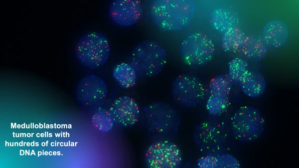
The Chavez lab uses fluorescent markers to observe circular extra-chromosomal DNA elements floating in cancer cells. Research has shown that these fragments of DNA are abundant in solid pediatric tumors and associated with poor clinical outcomes. Image courtesy of Lukas Chavez.

The Children’s Brain Tumor Network is another important repository for researchers studying pediatric brain cancer, such as Lukas Chavez, PhD, an assistant professor in the Cancer Genome and Epigenetics Program at Sanford Burnham Prebys. 

“We have analyzed thousands of whole genome sequencing datasets that we were able to access in these invaluable collections and have identified all kinds of structural rearrangements and mutations,” says Chavez. “Our focus is on a very specific type of structural rearrangement called circular extra-chromosomal DNA elements.” 

Circular extra-chromosomal DNA elements (ecDNA) are pieces of DNA that have broken off normal chromosomes and then been stitched together by DNA repair mechanisms. This phenomenon leads to circular DNA elements floating around in a cancer cell.  

Sanju Sinha, PhD

Sanju Sinha, PhD, is an assistant professor in the Cancer Molecular
Therapeutics Program at  Sanford Burnham Prebys.

“We have shown that they are much more abundant in solid pediatric tumors than we previously thought,” adds Chavez. “And we have also shown that they are associated with very poor outcomes.” 

To help translate this discovery for clinicians and their patients, Chavez is testing the use of deep learning AI algorithms to identify tumors with ecDNA by analyzing the biopsy slides that are routinely created by pathologists to diagnose brain cancer. 

“We have already done the genomic analysis, and we are now turning our attention to the histopathological images to see how much of the genomic information can be predicted from these images,” says Chavez. “Our hope is that we can identify tumors that have ecDNA by evaluating the images without having to go through the genomic sequencing process.”  

Currently, this approach serves only as a clinical biomarker of a challenging prognosis, but Chavez believes it can also be a diagnostic tool—and a game changer for patients.  

“I’m optimistic that in the future we will have drugs that target these DNA circles and improve the therapeutic outcome of patients,” says Chavez.  

“Once medicine catches up, we need to be able to find the patients and match them to the right medicine,” says Chavez. “We’re not there yet, but that’s the goal.” 

Chavez is also advancing his work as scientific director of the Pediatric Neuro-Oncology Molecular Tumor Board at Rady Children’s Hospital in San Diego.  

“Recently, it has been shown that new sequencing technologies coupled with machine learning tools make it possible to compress the time it takes to sequence and classify types of tumors from days or weeks to about 70 minutes,” says Chavez. “This is quick enough to take that technology into the operating room and use a surgical biopsy to classify a tumor.  

“Then we could get feedback to the surgeon in real time so that more or less tissue can be removed depending on if it is a high- or low-grade tumor—and this could dramatically affect patient outcomes.  

“When I talk to neurosurgeons, they are always in a pickle between trying to be aggressive to reduce recurrence risk or being conservative to preserve as much cognitive function and memory as possible for these patients.  

“If the surgeon knows during surgery that it’s a tumor type that’s resistant to treatment versus one that responds very favorably to chemotherapy, radiation or other therapies, that will help in determining how to strike that surgical balance.” 

Lukas Chavez, PhD

Lukas Chavez, PhD, is an assistant professor in the Cancer Genome and Epigenetics Program at Sanford Burnham Prebys.

Artist’s rendering X-shaped chromosomes floating in a cell

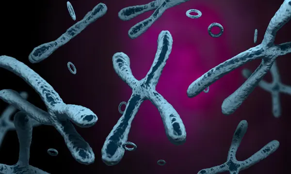
Artist’s rendering of X-shaped chromosomes floating in a cell alongside circular extra-chromosomal DNA elements.

Rady Children’s Hospital has also contributed to the future of genomic and computational medicine through BeginNGS, a pilot project to complement traditional newborn health screening with genomic sequencing that screens for approximately 400 genetic conditions. 

“The idea is that if there is a newborn baby with a rare disease, their family often faces a very long odyssey before ever reaching a diagnosis,” says Chavez. “By sequencing newborns, this program has generated success stories, such as identifying genetic variants that have allowed the placement of a child on a specific diet to treat a metabolic disorder, and a child to receive a gene therapy to restore a functional immune system.”


Programming in a Petri Dish, an 8-part series

How artificial intelligence, machine learning and emerging computational technologies are changing biomedical research and the future of health care

  • Part 1 – Using machines to personalize patient care. Artificial intelligence and other computational techniques are aiding scientists and physicians in their quest to prescribe or create treatments for individuals rather than populations.
  • Part 2 – Objective omics. Although the hypothesis is a core concept in science, unbiased omics methods may reduce attachments to incorrect hypotheses that can reduce impartiality and slow progress.
  • Part 3 – Coding clinic. Rapidly evolving computational tools may unlock vast archives of untapped clinical information—and help solve complex challenges confronting health care providers.
  • Part 4 – Scripting their own futures. At Sanford Burnham Prebys Graduate School of Biomedical Sciences, students embrace computational methods to enhance their research careers.
  • Part 5 – Dodging AI and computational biology dangers. Sanford Burnham Prebys scientists say that understanding the potential pitfalls of using AI and other computational tools to guide biomedical research helps maximize benefits while minimizing concerns.
  • Part 6 – Mapping the human body to better treat disease. Scientists synthesize supersized sets of biological and clinical data to make discoveries and find promising treatments.
  • Part 7 – Simulating science or science fiction? By harnessing artificial intelligence and modern computing, scientists are simulating more complex biological, clinical and public health phenomena to accelerate discovery.
  • Part 8 – Acceleration by automation. Increases in the scale and pace of research and drug discovery are being made possible by robotic automation of time-consuming tasks that must be repeated with exhaustive exactness.
Institute News

Seminar Series: extrachromosomal DNA and the metabolic circuits of cancer immune suppression

AuthorScott LaFee
Date

March 25, 2024

The ongoing Sanford Burnham Prebys seminar series will feature a pair of speakers on March 27, from noon to 1p.m., in the Fishman Auditorium. They will be presenting on two topics: extrachromosomal DNA and the tumor microenvironment.

First, Owen Chapman, PhD, a postdoctoral research scientist in the lab of Lukas Chavez, PhD, will discuss clinical and genomic features of circular extrachromosomal DNA (ecDNA) in medulloblastomas, a type of brain tumor.

EcDNA is DNA found off chromosomes, either inside or outside the nucleus of a cell. In a study published last year, Chavez (senior author), Chapman (first author) and colleagues reported that patients with medulloblastomas containing ecDNA are twice as likely to relapse after treatment and three times as likely to die within five years of diagnosis.

The second presentation will be by Kevin Tharp, PhD assistant professor in the Cancer Metabolism and Microenvironment Program. Tharp, who joined Sanford Burnham Prebys in December 2023, studies how tumors manipulate their mitochondria to improve survivability and how those cellular mechanics can be leveraged to create more effective therapies.

Institute News

Brain map connects brain diseases to specific cell types

AuthorSusan Gammon
Date

January 8, 2018

Researchers have developed new single-cell sequencing methods that could be used to map the cell origins of various brain disorders, including Alzheimer’s, Parkinson’s, schizophrenia and bipolar disorder.

By analyzing individual nuclei of cells from adult human brains, Jerold Chun, MD, PhD, professor at SBP, in collaboration with researchers at UC San Diego and Harvard Medical School, have identified 35 different subtypes of neurons and glial cells and discovered which of these subtypes are most susceptible to common risk factors for different brain diseases.

There are multiple theories regarding the roots of brain diseases. The new study, published in Nature Biotechnology, allows scientists to narrow down and rank the cell types in the brain that carry the most genetic risk for developing brain disorders. The information can guide researchers to pick the best drug-targets for future therapies.

The work builds off of a previous study by the authors that identified 16 subtypes of neurons in the cerebral cortex. That study was the first large-scale mapping of gene activity in the human brain and provided a basis for understanding the diversity of individual brain cells.

In the new study, researchers developed a new generation of single-cell sequencing methods that enabled them to identify additional neuronal subtypes in the cerebral cortex as well as the cerebellum, and even further divide previously identified neuronal subtypes into different classes. The new methods also enabled researchers to identify different subtypes of glial cells, which wasn’t possible in the previous study due to the smaller size of glial cells.

“These data confirm and significantly expand our prior work, further highlighting the enormous transcriptional diversity among brain cell types, especially neurons,” says Chun. “This diversity, which continues to emerge from our single-cell analytical approach, will provide a foundation for better understanding the normal and diseased brain.” The advance was made possible by combining next-generation RNA sequencing with chromatin mapping—mapping of DNA and proteins in the nucleus that combine to form chromosomes—for more than 60,000 individual neurons and glial cells.

“While the analysis of RNA can tell us how cell types differ in their activity, the chromatin accessibility can reveal the regulatory mechanisms driving the distinctions between different cells”, notes Peter Kharchenko, PhD, an assistant professor of biomedical informatics at Harvard Medical School who co-led the study.

Using the information from RNA sequencing and chromatin mapping methods, researchers were able to map which cell types in the brain were affected by common risk alleles—snippets in DNA that occur more often in people with common genetic diseases. Researchers could then rank which subtypes of neurons or glial cells are more genetically susceptible to different brain diseases. For example, they found that two subtypes of glial cells, microglia and oligodendrocytes, were the first and second most at risk, respectively, for Alzheimer’s disease. They also identified microglia as most at risk for bipolar disorder, and a subtype of excitatory neurons as most at risk for schizophrenia.

“Now we can locate where the disease likely starts,” says Kun Zhang, PhD, professor of bioengineering at the UC San Diego Jacobs School of Engineering and co-senior author of the study. “However, we are only mapping the genetic risk. We don’t know the precise mechanism of how these specific cells actually trigger the disease.”

One caveat of this study, explains Zhang, is that it primarily analyzed data from adult brains (ages 20 to 50), so the findings do not represent younger or older populations. In order to better understand brain disorders that manifest early on, for example in infants, like autism spectrum disorder, the study would need to analyze cells from younger brains, he said.

The team also plans to expand their studies to map additional regions of the brain.

Authors of the study are Blue B. Lake*, Song Chen*, Brandon C. Sos*, Thu E. Duong, Derek Gao and Kun Zhang of UC San Diego; Jean Fan* and Peter V. Kharchenko of Harvard Medical School; and Gwendolyn Kaeser, Yun C. Yung and Jerold Chun of Sanford Burnham Prebys Medical Discovery Institute.

*These authors contributed equally to this work.

This story is based on a UC San Diego press release written by Liezel Labios.

Institute News

Slowing down the “aging clock”

AuthorJessica Moore
Date

April 14, 2017

What if it were possible to slow down the clock on aging? There may indeed be such a clock in all your cells. New research from the laboratory of Peter Adams, PhD, professor at Sanford Burnham Prebys Medical Discovery Institute (SBP), provides further evidence that the epigenome—the pattern of chemical tags across our chromosomes that help determine which genes can be read—is the key to aging.

“We found that conditions or treatments that extend lifespan make the epigenome of an old animal look like that of a much younger one,” says Adams, senior author of one of a pair of studies in Genome Biology. “In other words, the ‘epigenetic clock’ can be slowed. That suggests that to help people stay healthy longer and lower their risk of diseases like Alzheimer’s, cancer, and atherosclerosis, we should find molecules that do the same thing.”

Adams’ studies, performed in collaboration with the lab of Trey Ideker, PhD, professor at UC San Diego, build on previous findings in humans. Ideker and subsequently other teams of scientists had identified the genomic sites at which the presence or absence of a chemical tag correlates with age, and created an algorithm to tell a person’s age within two or three years by analyzing all those sites. This epigenetic clock speeds up in people with diseases that lead to earlier onset of aging-associated problems, such as obesity or HIV infection, or who have survived severe psychological stress in childhood.

Adams’ and Ideker’s teams showed that longevity-conferring interventions have the opposite effect—they put a brake on age-associated epigenetic changes. To make that discovery, they compared the epigenomes of normal mice to those of mice in which aging was slowed by three strategies that are all well known to extend the mouse lifespan: a longevity mutation (Prop1df/df, which also causes dwarfism), caloric restriction (reducing dietary intake significantly, but not enough to harm the mice), and rapamycin, a drug with multiple effects on metabolism and the immune system.

“To show that longer life correlates with slower epigenetic aging, we first had to delineate the mouse epigenetic clock,” adds Adams. “That provides us with a very useful tool. Now we can do experiments to find out whether epigenetic changes actually drive aging. For example, we can compare animals with slow and fast epigenetic clocks to see if the ones that age slower stay healthier as they age.

“And we can investigate how the epigenetic clock “ticks”—what cellular processes cause these changes over time? The answers to that question could identify targets for anti-aging medicines.”

Institute News

How arsenic cures leukemia

Authorsgammon
Date

June 18, 2015

For the first time, Sanford-Burnham researchers have shown how the reversible interactions of the small protein SUMO work to facilitate treatment of acute promyelocytic leukemia (APL). The study, published recently in the journal Science Signaling, explains how the on-off associations of SUMO are required to destroy the APL causing oncoprotein and pave the way for an arsenic-based cure. Continue reading “How arsenic cures leukemia”