drug resistance Archives - Sanford Burnham Prebys
Institute News

Objective omics

AuthorGreg Calhoun
Date

August 1, 2024

Although the hypothesis is a core concept in science, unbiased omics methods may reduce attachments to incorrect hypotheses that can reduce impartiality and slow progress

Biological techniques that study the entire landscape of a sample’s genes or proteins—genomics or proteomics, respectively—help scientists discover new results without becoming too narrowly focused on what they predicted would happen. Although some scientists pursuing studies with this wider lens have been accused of going on “fishing expeditions,” many researchers counter that they now are able to investigate their hypotheses without missing other important results.

“I am a major proponent of omics, and especially unbiased omics,” says Sanju Sinha, PhD, an assistant professor in the Cancer Molecular Therapeutics Program at Sanford Burnham Prebys. “If someone now doesn’t show me unbiased results, it deeply bothers me. If every experiment only shows results from one pathway, it’s concerning and increases my skepticism about the study.”

An omics approach differs from traditional hypothesis-driven research in that it includes a comprehensive perspective about the phenomenon a scientist is studying and what might be causing it.

Sinha Lab

The Sinha Lab

“Unbiased omics look at the global picture of how everything is changing,” explains Sinha. “If you’re looking at genetic factors, you present all 20,000 genes and how they change, rather than just one pathway and maybe 10 genes.”

Sanju Sinha, PhD

Sanju Sinha, PhD, is an assistant professor in the Cancer Molecular
Therapeutics Program at  Sanford Burnham Prebys.

This method reflects the dynamic nature of biomedical research.

“Biomedical research is currently experiencing a period of accelerating and metamorphic discoveries fueled by unparalleled technologies that generate enormous amounts of data that, in turn, spur and spawn avenues of new inquiry and questions previously unimagined,” says David A. Brenner, MD, president and CEO of Sanford Burnham Prebys.

“An effective and successful biomedical researcher in the 21st century requires input from different disciplines that previously were not part of standard practice or the scientific method.”

Sinha agrees. “People used to work in small silos. They could work on the same biological pathway for 30 years.” The new model, he said, is quickly shifting to more multidisciplinary, team-based science where experts from many fields collaborate to make the most of new technology and the rich data it can provide.

Some teams employing these omics approaches have been criticized for conducting aimless studies due to the lack of traditional hypotheses. Sinha is quick to defend against these claims.

“I don’t mind these so-called fishing expeditions. I like to say that there are only two kinds of science: applied science and not-yet-applied science. Fishing expeditions are valuable if the data is made available and other scientists can make discoveries with it for years to come.”

“We should remember that fishing expeditions in biomedical research have done a great service to humanity.”

The hypothesis is not an endangered species destined to be replaced by unbiased omics approaches. On the contrary, omics experiments can often be kick-starters that help scientists generate new hypotheses to explore.

A team of scientists at Sanford Burnham Prebys and their collaborators are using an omics technique called resistomics to develop a new class of antibiotics effective against a drug-resistant pathogen.

In a paper published on January 3, 2024 in Nature, a multi-institutional team including  Andrei Osterman, PhD, a professor in the Immunity and Pathogenesis Program at Sanford Burnham Prebys, with colleagues at  Roche—the Swiss-based pharmaceutical/healthcare company—and others, describe a novel class of small-molecule-tethered macrocyclic peptide (MCP) antibiotics with potent antibacterial activity against carbapenem-resistant  Acinetobacter baumannii  (CRAB).

The World Health Organization and the Centers for Disease Control and Prevention have both categorized multidrug-resistant  A. baumannii as a top-priority pathogen and public health threat.

In the study, Osterman and colleagues applied an experimental evolution approach to help identify the drug target (the LPS transporter complex) of a new class of antibiotics—a macrocyclic peptide called Zosurabalpin—and elucidate the dynamics and mechanisms of acquired drug resistance in four distinct strains of A. baumannii. 

Andrei Osterman, PhD

Andrei Osterman, PhD, is a professor in the Immunity and Pathogenesis program at Sanford Burnham Prebys.

They used an integrative workflow that employs continuous bacterial culturing in an “evolution machine” (morbidostat) followed by time-resolved, whole-genome sequencing and bioinformatics analysis to map resistance-inducing mutations. 

“This comprehensive mapping of the drug-resistance landscape yields valuable insights for a variety of practical applications,” says Osterman, “from therapy optimization via genomics-based assessment of drug resistance/susceptibility of bacterial pathogens to a rational development of novel drugs with minimized resistibility potential.”


Programming in a Petri Dish, an 8-part series

How artificial intelligence, machine learning and emerging computational technologies are changing biomedical research and the future of health care

  • Part 1 – Using machines to personalize patient care. Artificial intelligence and other computational techniques are aiding scientists and physicians in their quest to prescribe or create treatments for individuals rather than populations.
  • Part 2 – Objective omics. Although the hypothesis is a core concept in science, unbiased omics methods may reduce attachments to incorrect hypotheses that can reduce impartiality and slow progress.
  • Part 3 – Coding clinic. Rapidly evolving computational tools may unlock vast archives of untapped clinical information—and help solve complex challenges confronting health care providers.
  • Part 4 – Scripting their own futures. At Sanford Burnham Prebys Graduate School of Biomedical Sciences, students embrace computational methods to enhance their research careers.
  • Part 5 – Dodging AI and computational biology dangers. Sanford Burnham Prebys scientists say that understanding the potential pitfalls of using AI and other computational tools to guide biomedical research helps maximize benefits while minimizing concerns.
  • Part 6 – Mapping the human body to better treat disease. Scientists synthesize supersized sets of biological and clinical data to make discoveries and find promising treatments.
  • Part 7 – Simulating science or science fiction? By harnessing artificial intelligence and modern computing, scientists are simulating more complex biological, clinical and public health phenomena to accelerate discovery.
  • Part 8 – Acceleration by automation. Increases in the scale and pace of research and drug discovery are being made possible by robotic automation of time-consuming tasks that must be repeated with exhaustive exactness.
Institute News

A potential new weapon against a deadly, drug-resistant bacterial pathogen

AuthorScott LaFee
Date

January 8, 2024

Carbapenems are a class of highly effective antibiotics that are often used to treat severe bacterial infections. They are usually reserved for known or suspected bacterial infections resistant to other drugs.

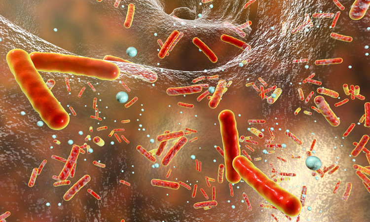
Carbapenem-resistant Acinetobacter baumannii (CRAB) is, as the name suggests, impervious to carbapenems; and it has become a major global pathogen, particularly in hospital settings and conflict zones. No new antibiotic chemical class with activity against A. baumannii has successfully emerged in more than 50 years.

In a paper published January 3, 2024, in Nature, a multi-institutional team including Andrei Osterman, PhD, at Sanford Burnham Prebys, with colleagues at Roche—the Swiss-based pharmaceutical/healthcare company—and others, describe a novel class of small-molecule tethered macrocyclic peptide (MCP) antibiotics with potent antibacterial activity against CRAB. Osterman’s lab provided critical data and discoveries related to the drug target and mapping of drug-resistant mutations.

Developing a new class of antibiotics effective against CRAB is critical. The bacterium is resistant to nearly all antibiotics and is difficult to remove from the environment. It poses a particular health threat to hospitalized patients and nursing home residents, with an estimated mortality rate in invasive cases of 40–60%.

The World Health Organization and the Centers for Disease Control (CDC) have both categorized multidrug-resistant A. baumannii as a top-priority pathogen and public health threat.

In the new study, Osterman and colleagues applied an experimental evolution approach to help identify the drug target (the LPS transporter complex) of a new class of antibiotics—a macrocyclic peptide called Zosurabalpin—and elucidate the dynamics and mechanisms of acquired drug resistance in four distinct strains of A. baumannii.

They used an integrative workflow that employs continuous bacterial culturing in an “evolution machine” (morbidostat) followed by time-resolved, whole-genome sequencing and bioinformatics analysis to map resistance-inducing mutations.

In addition to a mechanistic understanding (crucial from a regulatory perspective), the new information also helped reveal the drug-binding site. A related paper in the same issue experimentally verified the findings.

“This comprehensive mapping of the drug-resistance landscape yields valuable insights for a variety of practical applications,” says Osterman, “from therapy optimization via genomics-based assessment of drug resistance/susceptibility of bacterial pathogens to a rational development of novel drugs with minimized resistibility potential.”

A commentary in Nature said the research was “cause for cautious celebration” and urged further development.

Institute News

The “Eph” system may pave the way for novel cancer therapies

AuthorSusan Gammon
Date

November 27, 2023

Over the past three decades, researchers have been investigating an important cell communication system called the “Eph system,” and the evidence implicating the system in cancer is staggering.

The Eph system is comprised of multiple Eph receptors and their ligands—ephrins—and are involved in contact-dependent communication between cells. They play essential roles in regulating various cellular processes.

Modern studies have shed light on the Eph system’s role in tumor expansion, invasiveness, metastasis, cancer stem cell maintenance and therapy resistance.

This month, Elena Pasquale, PhD, published a review in Nature Reviews Cancer that summarizes the current state of research on the Eph system and its links to cancer progression and drug resistance.

“The Eph system has many critical functions during the development of tissues and organs, but it also has the capacity to either promote or suppress cancer progression and malignancy” says Pasquale. “In cancer, the activities of the Eph system can differ depending on the circumstances—for example, which Eph receptors and ligands are present in a tumor cell, the types of tumor cells in which they function, and the characteristics of these cells.”

“It’s this remarkable versatility that makes the Eph system a compelling but also challenging target for potential therapies,” says Pasquale.

“The aims of this review were to comprehensively survey the large body of data regarding various aspects related to Eph signaling in tumors and to highlight potential strategies for therapeutic targeting,” says Pasquale. “Overall, while significant progress has been made in deciphering the Eph system in cancer, there is much more to learn.

“Gaining a deeper understanding of how the Eph system functions in cancer is challenging but will be essential for the development of targeted therapies and personalized treatment approaches for patients.”

Institute News

Sanford Burnham Prebys and Roche fight back against antibiotic resistance

AuthorMiles Martin
Date

December 8, 2021

Researchers from Sanford Burnham Prebys have teamed up with prominent drug developer Roche Pharma to learn how bacteria develop antibiotic resistance.

Their new results, published in the journal mBio, are one piece of a long-standing collaboration between the two organizations, the goal of which is to mitigate the growing threat of antibiotic resistance by developing more “irresistible” drugs and by helping improve antibiotic prescribing practices.

“The emergence of antibiotic resistance is inevitable for any single drug, new or old. It’s only a question of time,” says senior author Andrei Osterman, PhD, a professor at Sanford Burnham Prebys. “But the precise time is different for every drug and every microbe, so studying when and how resistance to antibiotics evolves gives us powerful information for improving antibiotic treatment.”

Antibiotic resistance develops rapidly

When a patient is treated with antibiotics, most individual bacteria die, but a few cells will survive, usually as a lucky consequence of a random genetic mutation. These survivors go on to multiply into a whole new population of antibiotic-resistant bacteria.

“The development of antibiotic resistance is a strictly Darwinian process, very similar to evolution in larger organisms,” says Osterman. “The difference is that in bacteria, it happens much more rapidly, which makes antibiotic resistance one of the most pressing challenges facing medicine today.” 

Although the speed at which evolution occurs in bacteria makes antibiotic resistance a threat, the researchers were also able to take advantage of this speed to study its development. The team cultured three species of bacteria in a morbidostat, a device that allows bacteria to grow continuously over multiple generations while being dosed with antibiotics. Although theirs was not the first morbidostat device, the team designed a new, more effective version of the system for their experiments.

“It’s like an evolution machine, letting us watch the development of antibiotic resistance in real time and in an environment that more accurately models what happens to bacteria in a clinical setting than other approaches,” says Osterman. “This gives us a clearer and more comprehensive view of resistance than we’ve ever had before.”

Different bacteria develop resistance differently

By observing the bacteria’s evolution in the morbidostat and sequencing their genomes as they evolved, the researchers found that all three species had a similar pattern of resistance development. However, they also found subtle differences in the ways certain genes were expressed, particularly those that help bacteria remove toxins, a critical process in developing resistance.

“It’s like three remakes of the same movie by three different directors, and their comparison gives us a wealth of information to guide the development and use of antibiotics,” says Osterman. 

Understanding resistance is critical to reducing its harm

Working with Roche, the team has completed similar studies on several other classes of antibiotic drugs, which is helping Roche identify promising candidates for antibiotics that are less prone to resistance.

And because antibiotic resistance is often not assessed in drug candidates until years into the process, using resistance to screen for drug candidates this way could save the biomedical industry millions of dollars and help patients benefit from effective drugs sooner.

“A completely ‘irresistible’ drug is a holy grail, something we can never truly achieve,” says Osterman. “But some drugs are less resistible than others, and our methods allow us to figure out which is which in a systematic way.”

In addition to helping develop new drugs, the researchers claim that their findings are easily translatable to the clinic, where doctors can use detailed knowledge of resistance to select optimal drug combinations with less likelihood of failure due to resistance.

“We are moving away from trial-and-error approaches in medicine and moving toward being able to predict exactly what drugs will work best for each patient,” says Osterman. “It is going to take time, effort and money to make this happen, but it will all be worth it if we’re able to alleviate the threat of antibiotic resistance and help save lives, which I’m confident can be done.”

Institute News

Battling infectious diseases with 3D structures

AuthorSusan Gammon, PhD
Date

April 25, 2017

Sanford Burnham Prebys Medical Discovery Institute (SBP) scientists are part of an international team led by Northwestern University Feinberg School of Medicine that has determined the 3D atomic structure of more than 1,000 proteins that are potential drug and vaccine targets to combat some of the world’s most dangerous emerging and re-emerging infectious diseases.

These experimentally determined structures have been deposited into the World-Wide Protein Data Bank, an archive supported by the National Institutes of Health (NIH), and are freely available to the scientific community. The 3D structures help expedite drug and vaccine research and advance the understanding of pathogens and organisms causing infectious disease.

“Almost 50 percent of the structures that we have deposited in the Protein Data Bank are proteins that were requested by scientific investigators from around the world,” said Feinberg’s Wayne Anderson, PhD, director of the project. “The NIH has also requested us to work on proteins for potential drug targets or vaccine candidates for many diseases, such as the Ebola virus, the Zika virus and antibiotic-resistant bacteria. We have determined several key structures from these priority organisms and published the results in high-impact journals such as Nature and Cell.

Teamwork with an international consortium

This milestone effort, funded by two five-year contracts from the National Institute of Allergy and Infectious Diseases (NIAID), totaling a budget of $57.7 million, represents a decade of work by the Center for Structural Genomics of Infectious Diseases (CSGID) at Feinberg, led by Anderson in partnership with these institutions:

  • University of Chicago
  • University of Virginia School of Medicine
  • University of Calgary
  • University of Toronto
  • Washington University School of Medicine in St. Louis
  • UT Southwestern Medical Center
  • J. Craig Venter Institute
  • Sanford Burnham Prebys Medical Discovery Institute
  • University College London

How the 3D structures are made

Before work begins on a targeted protein, a board appointed by the NIH examines each request. Once approved, the protein must be cloned, expressed and crystallized, and then X-ray diffraction data is collected at the Advanced Photon Source at Argonne National Laboratory. This data defines the location of each of the hundreds or even thousands of atoms to generate 3-D models of the structures that can be analyzed with graphics software. Each institution in the Center has an area of expertise it contributes to the project, working in parallel on many requests at once.

The bioinformatics group SBP, led by Adam Godzik, PhD, focuses on steps that have to be taken before the experimental work starts. Every protein suggested by the research community as a target for experimental structure determination is analyzed and an optimal procedure for its experimental determination is mapped out.

Experimental structure determination used to have a very high failure rate and the money and time spent on failed attempts is a major contributor to the total expense and time needed to solve protein structures. Both can be significantly improved using “Big Data” approaches, as researchers learn from thousands of successful and failed experiments in structural biology. The SBP bioinformatics group uses these approaches to improve success rates at CSGID, allowing our center to solve more structures at lower costs.

Until recently the process of determining the 3D structure of a protein took many months or even years to complete, but advances in technology, such as the Advanced Photon Source, and upgrades to computational hardware and software has dramatically accelerated the process. The Seattle Structural Genomics Center for Infectious Disease, a similar center funded by NIAID, is also on track to complete 1,000 3-D protein structures soon. Browse all of the structures deposited by the CSGID.

Anyone in the scientific community interested in requesting the determination of structures of proteins from pathogens in the NIAID Category A-C priority lists or organisms causing emerging and re-emerging infectious diseases, can submit requests to the Center’s web portal. As part of the services offered to the scientific community, the CSGID can also provide expression clones and purified proteins, free of charge.

This project has been supported by federal funds from the NIAID, NIH,  Department of Health and Human Services, under contract numbers HHSN272200700058C and HHSN272201200026C.

Institute News

Sanford-Burnham researchers identify a new target for treating drug-resistant melanoma

Authorsgammon
Date

May 28, 2015

A new collaborative study led by researchers at Sanford-Burnham, published today in Cell Reports, provides new insight into the molecular changes that lead to resistance to a commonly prescribed group of drugs called BRAF inhibitors. The findings suggest that targeting newly discovered pathways could be an effective approach to improving the clinical outcome of patients with BRAF inhibitor-resistant melanoma tumors. Continue reading “Sanford-Burnham researchers identify a new target for treating drug-resistant melanoma”