NIH funding Archives - Page 4 of 7 - Sanford Burnham Prebys
Institute News

What scientists are learning about COVID-19 and the brain

AuthorMonica May
Date

December 8, 2020

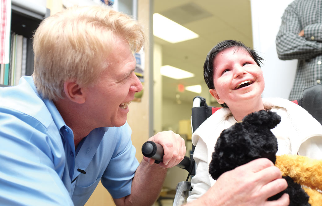
We caught up with cell biologist Anne Bang, who recently teamed up with her husband to study how SARS-CoV-2 affects the brain

Brain fog. Memory loss. Dizziness and confusion. Although COVID-19 is primarily thought of as a lung disease, survivors continue to report lingering and highly concerning neurological effects—severe enough to impact their ability to work and live normal lives. Doctors are also seeing a worrisome increase in strokes in younger patients, among other observations.

To learn what scientists know so far about COVID-19 and its effect on the brain, we caught up with Anne Bang, PhD, director of Cell Biology at Sanford Burnham Prebys’ Conrad Prebys Center for Chemical Genomics. Bang recently teamed up with scientists at Penn Medicine and a virologist at Scripps Research—who also happens to be her husband—to investigate whether SARS-CoV-2 infects brain cells. Their findings were published in Cell Stem Cell.

What do scientists know about the brain and COVID-19 so far?

Unfortunately, information is still very limited. There are reports of viral replication in the brain and spinal cord fluid of people with COVID-19 who have neurological symptoms. But as you can imagine, taking brain biopsies from someone who has COVID-19 is not realistic. So we really don’t know a lot yet. For this reason, scientists are turning to systems that can model the human brain, such as brain cells created from induced pluripotent stem cells (iPSCs) and brain organoids, to study SARS-CoV-2’s impact on the brain.

What did you find in your study?

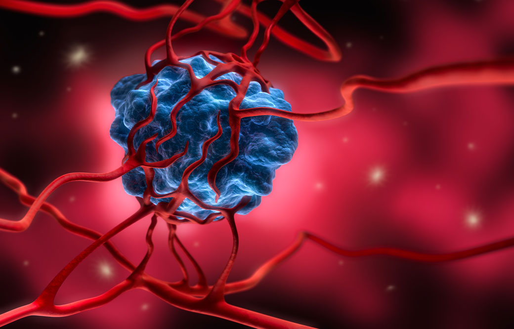
We created several types of brain cells using iPSCs and brain organoids, which we then infected with SARS-CoV-2. We found that SARS-CoV-2 primarily infects a brain cell type called choroid plexus cells—largely bypassing neurons and astrocytes. The choroid plexus is a specialized part of the blood-brain barrier, which controls what can enter your brain and produces cerebral spinal fluid. More research emerges every day, but so far, the consensus in the field seems to align with our findings.

SARS_CoV2_ Infected human choroid plexus cells a type of brain cell

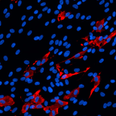
The scientists found that SARS-CoV-2 (red) primarily infects brain cells called choroid plexus cells (blue), which are part of the brain’s protective blood-brain barrier.

How might this finding translate to what we’re seeing in patients?

We know that choroid plexus cells produce high levels of ACE2, which is the receptor that SARS-CoV-2 uses to enter and infect cells. Because the choroid plexus is the “gatekeeper” to the brain, it’s possible that the virus enters the brain by infecting these cells. However, much more research is needed before we can give a definitive answer to this question.

We have more questions than answers right now about COVID-19. What is one question you wish we had the answer to?

How does the virus get from the nose and mouth and spread to other parts of the body? This is a big question for me and the scientific field. Once we know how the virus travels throughout the body, we can potentially stop its spread and control the dangerous symptoms.

What was it like working with your husband? Was this your first time working together?

It was really fun. I found out that he is great to work with. We’ve been together for 30 years, and incredibly, this was the first time we worked together.

Institute News

Top San Diego researchers receive $5 million to study cellular aging

Authorsgammon
Date

September 22, 2020

Professors Peter Adams, PhD, and Malene Hansen, PhD, of Sanford Burnham Prebys will lead key research and development cores

Sanford Burnham Prebys researchers are joining forces with University of California San Diego (UC San Diego) and the Salk Institute to form a world-class San Diego Nathan Shock Center (SD-NSC), a consortium established to study cellular and tissue aging in humans. The center will be funded by the National Institute on Aging (NIA), part of the National Institutes of Health, and is expected to receive $5 million over the next two years.

Professors Peter Adams, Ph.D., and Malene Hansen, PhD, of Sanford Burnham Prebys will lead key research and development cores, along with Professors Rusty Gage, PhD, Martin Hetzer, PhD, and Tatyana Sharpee, PhD, of Salk; and Anthony Molina, PhD, of UC San Diego. Salk Professor Gerald Shadel, PhD, will be the director of the SD-NSC.

“This is a special opportunity for San Diego’s aging research community to share our ideas, skills and technologies to drive innovative research in the basic biology of aging,” says Adams. “We are grateful for this support and will work to create the strongest environment possible to achieve meaningful breakthroughs that will benefit human health.”

Aging is the most significant risk factor for human disease. Human cells and tissues age at different rates depending on their intrinsic properties, where they are in the body and environment exposures. Yet, scientists do not fully understand this variability (“heterogeneity”) and how it contributes to overall human aging, risk for disease or therapeutic responses.

To explore the complex heterogeneity of human aging, the SD-NSC will deploy three cutting-edge Research Resource Cores, including the Human Cell Models of Aging Core, to be led by Gage and Molina; the Heterogeneity of Aging Core, to be led by Hetzer and Adams; and the Integrative Models of Aging Core, to be led by Sharpee.

The cores will allow detailed analysis of human cells and organoids (artificially grown clusters of cells that model tissues), derived from a unique aging cohort at UC San Diego that is annotated for multiple measures of the actual biological age of individuals. In addition, the cores will provide scientific services to the Nathan Shock Centers network and the aging research community at large, including the dissemination of samples, protocols and computational tools to facilitate the study of heterogeneity in aging.

A Research Development Core headed by Hansen will also be established to encourage and support new investigators to enter the field of aging research. Through this core, the SD-NSC will provide pilot research grants, workshops and customized mentoring programs to promote the research and development of young investigators, as well as in-person and virtual trainings to spur collaboration and the sharing of knowledge related to the basic biology of aging.

“For years, my colleagues and I have been organizing successful symposia such as the annual La Jolla Aging Meeting (LJAM), where we share new aging research and discuss opportunities for collaboration,” says Hansen, who also hold the positions of associate dean for Student Affairs and faculty adviser for Postdoctoral Training at Sanford Burnham Prebys. “The San Diego Nathan Shock Center will enable us to broaden the reach and impact of LJAM, as well as take training and mentoring of the next generation of researchers to a new level.”

The SD-NSC builds on Sanford Burnham Prebys’ strengths in fundamental aging research, renowned for its use of model organisms to unravel cell changes associated with normal development and aging. By building, analyzing and probing models of disease, scientists in the Institute’s Aging, Development and Regeneration Program are providing new tools to uncover novel therapeutic targets for heart disease, neurodegeneration, muscle disorders, diabetes, cancer and other debilitating diseases.

The SD-NSC will be one of a network of eight Nathan Shock Centers nationwide, and is funded by NIA grant number P30AG068635.

Institute News

Scientists discover an early sign of type 2 diabetes: Misfolded proinsulin

AuthorMonica May
Date

March 19, 2020

The findings could lead to tests or treatments that help prevent type 2 diabetes.

Misfolded proinsulin—a protein the body normally processes into insulin—is an early sign of type 2 diabetes, according to a study by scientists at Sanford Burnham Prebys and the University of Michigan Medical School. The discovery, published in eLife, could lead to tests or treatments that help prevent people from developing type 2 diabetes.

“Understanding the molecular events that occur as prediabetes progresses to diabetes opens new avenues for us to detect or interrupt these processes,” says Randal Kaufman, PhD, director and professor in the Degenerative Diseases Program at Sanford Burnham Prebys and co-corresponding author of the study. “With this information, we can start to find interventions that might spare millions of people from a serious, lifelong condition.”

More than one in three Americans, or approximately 88 million people, have prediabetes—which is characterized by elevated blood sugar. If left untreated, within four years nearly 40% of people with prediabetes develop type 2 diabetes, which occurs when the body doesn’t use insulin properly. In 2017, the cost of treating diabetes exceeded $327 billion, according to the American Diabetes Association. Due to increasing obesity rates, the number of people with the condition—particularly children—is on the rise.

Identifying the molecular events that occur during progression from prediabetes to full-blown diabetes remains one of the most perplexing problems in diabetes research. In the study, the scientists set out to answer this question by tracking proinsulin folding in the beta cells of humans and mice that are healthy, prediabetic and diabetic.

These studies revealed that instead of undergoing its normal folding process, proinsulin proteins were abnormally linked to each other. Levels of the abnormal proinsulin accumulated as prediabetes progressed to type 2 diabetes. Obese mice in the earliest stages of diabetes had the highest levels of abnormal proinsulin in their beta cells.

“Proinsulin misfolding is the earliest known event that may contribute to the progression from prediabetes to diabetes,” says Kaufman. “Together, these studies show that abnormally linked proinsulin holds promise as a potential measure of how close someone may be to developing type 2 diabetes.”

Now, the researchers are set to uncover more details about this process, such as the proteins that interact with the misfolded proinsulin.

“Understanding the fundamental molecular events that lead to type 2 diabetes is critical as the number of people with prediabetes continues to rise,” says Kaufman. “If we don’t find preventive measures, we will soon have a diabetes epidemic.”


The study’s first author is Anoop Arunagiri, PhD; and the study’s senior author is Peter Arvan, both of the University of Michigan Medical School.

Additional authors include Leena Haataja and Fawnnie Pamenan of the University of Michigan Medical School; Ming Liu of the University of Michigan Medical School and Tianjin Medical University in China; Anita Pottekat and Pamela Itkin-Ansari of Sanford Burnham Prebys; Soohyun Kim of Konkuk University in South Korea; Lori M. Zeltser of Columbia University; Adrienne W. Paton and James C. Paton of the University of Adelaide in Australia; and Billy Tsai of the University of Michigan.

The study’s DOI is 10.7554/eLife.44532.

This work was supported by the National Institutes of Health (R01DK111174, R24DK110973 and R01DK48280) and the Juvenile Diabetes Research Foundation International (2-SRA-2018-539-A-B).

Institute News

Scientists discover new survival strategy for oxygen-starved pancreatic cancer cells

AuthorMonica May
Date

October 23, 2019

Oxygen is essential to life. When fast-growing tumor cells run out of oxygen, they quickly sprout new blood vessels to keep growing, a process called angiogenesis. 

By blocking pancreatic cancer’s oxygen-sensing machinery—the same field of research studied by the winners of the 2019 Nobel Prize in Medicine—Sanford Burnham Prebys scientists have uncovered a new way that tumors turn on angiogenesis in an animal model. The discovery, published in Cancer Research, could lead to a treatment that is given with an anti-angiogenetic medicine, thereby overcoming drug resistance. 

“Treatment resistance is a major challenge for cancer treatments that block blood vessel growth,” says Garth Powis, D.Phil., professor and director of Sanford Burnham Prebys’ National Cancer Institute (NCI)-designated Cancer Center and senior author of the study. “Our research identifies a new way angiogenesis is activated, opening new opportunities to find medicines that might make existing cancer treatments more effective.” 

Many cancer treatments work by blocking angiogenesis, which rarely occurs in healthy tissues. However, these medicines eventually stop working, and the cancer returns, sometimes in as little as two months. Scientists have been researching why this treatment resistance occurs so it can be stopped.

In this study, the scientists focused on pancreatic cancer, which is notoriously desperate for oxygen and also difficult to treat. Fewer than 10% of people diagnosed with pancreatic cancer are alive five years later. 

To see how a pancreatic tumor responds to a disruption in its oxygen supply, the Sanford Burnham Prebys researchers used a mouse model to block an oxygen-sensing protein called HIF1A—which should cripple the tumor’s growth. Instead of dying, however, after about a month the cells multiplied—indicating they had developed a new way to obtain oxygen. 

Further work revealed that the cancer cells were clear and swollen with the nutrient glycogen (a characteristic also seen in some ovarian and kidney cancers). In response to the excess glycogen, special immune system cells were summoned to the tumor, resulting in blood vessel formation and tumor survival. Each of these responses represents a new way scientists could stop pancreatic tumors from evolving resistance to treatment.

“Our team’s next step is to test tumor samples from people with pancreatic cancer to confirm this escape mechanism occurs in a clinical setting,” says Powis. “One day, perhaps we can create a second medicine that keeps anti-angiogenic drugs working and helps more people survive pancreatic cancer.”


Research reported in this press release was supported by the U.S. National Institutes of Health (NIH) (5F31CA203286, CA216424 and P30CA030199). The content is solely the responsibility of the authors and does not necessarily represent the official views of the NIH. The study’s DOI is 10.1158/0008-5472.CAN-18-2994. 

Institute News

Sanford Burnham Prebys welcomes U.S. Congressman Mike Levin

AuthorMonica May
Date

October 22, 2019

On October 1, 2019, U.S. Representative Mike Levin (D-CA) toured Sanford Burnham Prebys and met with several faculty members to learn more about the innovative biomedical research taking place in his backyard. Levin represents California’s 49th Congressional District, which includes North County San Diego, South Orange County and neighbors our La Jolla campus. 

The visit kicked off with a visit to a lab working to find medicines for a heart arrhythmia condition called atrial fibrillation (AFib), a disorder that hits home for Levin: His grandmother struggled with the disease. Levin peered into a microscope to view beating heart cells and learned how a team of experts from Sanford Burnham Prebys and Scripps Clinic are working to develop personalized treatments for the condition, which affects nearly six million Americans (meet the A-team.)

“Sanford Burnham Prebys is a great example of the vibrant biomedical research taking place in San Diego that has the potential to improve the quality of life for families across the country,” says Levin. “Seeing the Institute’s critical research up close and hearing firsthand how National Institutes of Health (NIH) funding has accelerated medical discovery only strengthens my commitment to supporting biomedical science. Following my visit to Sanford Burnham Prebys, I was proud to introduce legislation that would invest $10 billion in the NIH to support biomedical research, and I will continue to fight for this much-needed funding.”

Following the lab tour, Levin met with faculty members who—thanks to federally funded research—are working to find treatments for Alzheimer’s disease and addiction, and study the aging process to address age-related diseases such as cancer. The visit wrapped up in the lab of Hudson Freeze, PhD, the director of our Human Genetics Program, who studies a rare childhood disease called congenital disorders of glycosylation, or CDG. 

“Americans today are living longer and healthier lives because of federally funded medical research,” says Chris Larson, PhD, the adjunct associate professor of Development, Aging and Regeneration at the Institute who arranged the visit. “We are grateful that Mike took the time to sit down with us to learn about our NIH-funded work and how he can help support us on our mission to find cures for human disease.”

Editor’s note: Shortly after his visit Levin introduced legislation that calls for a $10 billion investment in biomedical research. 

Institute News

Sanford Burnham Prebys scientist joins historic effort to help children with rare disease

AuthorMonica May
Date

October 3, 2019

Hudson Freeze, PhD, professor of Human Genetics at Sanford Burnham Prebys, has joined a historic effort that establishes—for the first time—a nationwide network of 10 regional academic centers, Sanford Burnham Prebys researchers and patient advocacy groups to address decades of unresolved questions surrounding congenital disorders of glycosylation, or CDG, a rare disease that affects children. The consortium is funded by a $5 million, five-year grant from the National Institutes of Health (NIH). 

“We are extremely pleased that the NIH is investing in an initiative that will improve the lives of people affected by CDG,” says Freeze, who leads efforts to develop and validate disease biomarkers that will aid in diagnoses, and measuring treatment benefits during clinical trials. “Although globally the number of people living with CDG is relatively small, the impact on the lives of these individuals and their families can be profound. We look forward to working with the patients, families, physicians, scientists and other stakeholders focused on this important study.”

CDG is caused by genetic mutations that disrupt how the body’s sugar chains attach to proteins. First described in the 1990s, today scientists have discovered more than 140 types of mutations that lead to CDG. Symptoms are wide-ranging, but can include developmental delays, movement problems and impaired organ function. Some children may benefit from a sugar-based therapy; however, developing treatments for those who need alternative treatment options has been hindered by a lack of natural history data—tracking the course of the condition over time—comprehensive patient registry, and reliable methods to establish the CDG type.

Working together, the consortium will overcome these hurdles by: 

  • Defining the natural history of CDG through a patient study, validating patient-reported outcomes and sharing CDG knowledge 
  • Developing and validating new biochemical diagnostic techniques and therapeutic biomarkers to use in clinical trials 
  • Evaluating whether dietary treatments restore glycosylation to improve clinical symptoms and quality of life

Freeze will lead the efforts to develop and validate biomarkers for CDG, working alongside the Children’s Hospital of Philadelphia and the Mayo Clinic. The principal investigator of the CDG Consortium Project is Eva Morava, MD, PhD, professor of Medical Genetics at the Mayo Clinic. The patient advocacy groups involved are CDG CARE and NGLY1.org. 

Sanford Burnham Prebys and CDG Care will host the 2020 Rare Disease Day Symposium and CDG Family Conference from February 28 to March 1 in San Diego, which welcomes researchers, clinicians, children with CDG and their families, and additional CDG community members. Register to attend. 
 

Institute News

How your DNA takes shape makes a big difference in your health

AuthorMonica May
Date

September 10, 2019

The more we learn about our genome, the more mysteries arise. For example, how can people with the same disease-causing mutation have different disease progression and symptoms? And despite the fact that it’s been more than 15 years since the human genome was sequenced, why can’t we explain the significance of the vast majority of genomic variations that occur in noncoding, or “junk,” elements of the genome?

Now, Pier Lorenzo Puri, MD, a professor in the Development, Aging, and Regeneration Program at Sanford Burnham Prebys, has used a cutting-edge technique called Hi-C, which maps millions of interactions between proteins and tightly coiled DNA, called chromatin, to shed light on this mystery. The study, published in Molecular Cell, shows that a specific protein called MyoD—a master regulator of muscle development—reshapes chromatin’s architecture to alter gene expression—revealing fundamental insights into how genetic variations may affect our health. 

“One of the greatest mysteries of medicine is how people with the same mutation can have different symptoms,” says Puri. “Our study indicates that some genetic variations may affect our health by altering how DNA coils and interacts in its 3D shape. This alteration may be helpful or detrimental—and could even explain why some people seem to be naturally athletic.” 

In the study, the scientists used several genomic technologies to map the interactions between MyoD and chromatin as cells turned into skeletal muscle upon MyoD expression. Among other findings, the scientists determined that MyoD rearranged chromatin’s shape during this process—similar to the retying of a tangled shoelace. Importantly, the researchers found that MyoD-driven reconfiguration of 3D chromatin architecture is mediated by interactions between noncoding elements of the genome—where most disease-associated genetic variants occur. These findings demonstrate that the noncoding genome can act as a structural element that defines the chromatin architecture—key information that will help predict the functional outcomes of these variants.

Puri is already applying this insight to help solve other genomic mysteries. He plans to review a worldwide database of gene variations with unknown significance—meaning that scientists are unsure if the change is harmless or a risk factor for disease. Then, he aims to create models that help us better understand the impact of these genetic variations on an individual’s ability to respond to environmental changes and eventually develop disease. 

“It’s possible that many genetic variations alter chromatin folding. Instead of directly causing disease, the changes may increase or decrease our disease risk,” explains Puri. “I hope that my next studies will shed light on these genomic mysteries and help more people get definitive answers about what lies in their DNA.” 


The co-first authors of the study are Alessandra Dall’Agnese, PhD, and Luca Caputo, PhD, of Sanford Burnham Prebys. 

Additional study authors include Chiara Nicoletti, PhD, of Sanford Burnham Prebys and University of Modena and Reggio Emilia; Sole Gatto and Ranjan Perera of Sanford Burnham Prebys; Julia di Iulio, PhD, and Amalio Telenti, MD, PhD, of Scripps Research; Anthony Schmitt, PhD, Yarui Diao, PhD, and Zhen Ye of the Ludwig Institute for Cancer Research; Mattia Forcato, PhD, and Silvio Bicciato, PhD, of University of Modena and Reggio Emilia; and Bing Ren, PhD, of the Ludwig Institute for Cancer Research and UC San Diego School of Medicine. The study’s DOI is 10.1016/j.molcel.2019.07.036. 

Research reported in this article was supported by the U.S. National Institutes of Health (NIH) (R01AR056712, R01AR052779, AR061303), Epigen, Ellison Medical Foundation (AG-NS-0843-11), AFAR (G16294AD), Ludwig Institute for Cancer Research, Human Frontier Program and San Diego Muscle Research Center. The content is solely the responsibility of the authors and does not necessarily represent the official views of the NIH.

Institute News

Cancer’s final frontier: the tumor microenvironment

AuthorMonica May
Date

September 3, 2019

Cancer researchers are setting their sights on a new kind of cancer treatment that targets the tumor’s surrounding environment, called the tumor microenvironment, in contrast to targeting the tumor directly. 

To learn more about this approach, we spoke with cancer experts Jorge Moscat, PhD, director and professor in the Cancer Metabolism and Signaling Networks Program at Sanford Burnham Prebys; and Maria Diaz-Meco, PhD, professor in the Cancer Metabolism and Signaling Networks Program at Sanford Burnham Prebys. Both scientists recently authored a review article centered on a family of cancer-linked proteins that regulate the tumor’s microenvironment. The paper was published in Cancer Cell

What is the tumor microenvironment exactly? 
Moscat: Just like every person is surrounded by a supportive community—their friends, family or teachers—every tumor is surrounded by a microenvironment. This ecosystem includes blood vessels that supply the tumor with nutrients; immune cells that the tumor has inactivated to evade detection; and stroma, glue-like connective tissue that holds the cells together and provides the tumor with nutrients.

Diaz-Meco: These elements are similar to the three legs of a stool. If we remove all three legs, we can deliver a deadly blow to the tumor. FDA-approved drugs exist that target blood vessel growth and reactivate the immune system to destroy the tumor. The final frontier is targeting the stroma.

When did scientists realize it’s important to focus on the tumor’s surroundings—not the tumor itself? 
Diaz-Meco: Scientists have known for more than a century that the tumor’s surroundings are different from normal cells. The tissue surrounding a tumor is inflamed—tumors are often called “wounds that never heal”—and their metabolism is radically different from healthy cells. 

Moscat: The discovery of oncogenes—genes that can lead to cancer—in the 1970s shifted the field’s focus to treatments that target the tumor directly. These targeted treatments work incredibly well, but only for a short time. Cancer researchers are realizing that tumors quickly adapt to this roadblock and become treatment resistant. In addition, many oncogenes are difficult to target, earning the title “undruggable.” As a result, cancer researchers are returning their focus to the tumor microenvironment—especially the stroma. Only a handful of stroma-targeting drugs are in development. None are FDA approved.

Which cancers could benefit most from a stroma-targeting drug? 
Moscat: Pancreatic, colorectal and liver cancers stand to benefit most from a stroma-targeting drug. For example, 90% of a pancreatic tumor consists of stroma—not cancer cells. Combined, these cancers are responsible for more than 20% of all cancer deaths in the U.S. each year. 

What is the focus of your lab’s research? 
Diaz-Meco: Our lab studies the cross talk between tumors and their environment. This conversation is very complex. In addition to “talking” with the tumor, the stroma also “speaks” with the immune system. We are working to map these interactions so we can create drugs that silence this conversation—or change it. For example, we recently showed—in a mouse model that faithfully recapitulates the most aggressive form of human colorectal cancer—that by altering the stroma’s interactions with the immune system, we might make tumors vulnerable to immunotherapy. 

What do new insights into the tumor microenvironment mean for cancer drug development? 
Moscat: It’s likely that the ultimate cancer “cure” won’t be just one drug that kills the tumor cells, but a combination of therapies. I expect this will be a three-part combination treatment that stops blood vessel growth, activates the immune system to attack the tumor and targets the stroma. 

Additionally, this research shows that experimental models of cancer drug development need to take the tumor microenvironment into account. Many current models use mice that lack an immune system—in order to get the tumor to grow—or focus on the tumor in isolation. Based on our knowledge of the tumor microenvironment, this isn’t an accurate representation of human disease. 

Diaz-Meco: In our lab, we have created several animal models of cancers that preserve the immune system and mirror tumor progression. In addition to better modeling human disease, this also allows us to study cancer from its earliest beginnings. This work could lead to early interventions—before the cancer has become large and hard to treat.

Anything else you’d like to add? 
Moscat: We are truly in the golden age of cancer biology. We understand more than we ever have before. New technologies are allowing us to obtain an unprecedented amount of information—we can even map every gene that is “turned on” in a single cancer cell. I am incredibly hopeful for the future. 

Learn more about the future of cancer treatment by attending our next “Conquering Cancer” event at the Fleet Science Center. Details

Institute News

NIH grant aims to boost heart muscle

AuthorMonica May
Date

August 23, 2019

Heart disease is the number one killer of Americans. Now, the National Institutes of Health (NIH) has awarded a four-year grant totaling nearly half a million dollars to Sanford Burnham Prebys to find medicines that could help people repair damaged heart muscle—and potentially reduce the risk of heart attack or other cardiovascular events. 

“Each year we lose far too many loved ones to heart attacks and other heart conditions,” says grant recipient Chris Larson, PhD, adjunct associate professor in the Development, Aging and Regeneration Program at Sanford Burnham Prebys. “Now, we have the opportunity to find medicines that may help more people live long, active lives by strengthening their heart muscles.”

Nearly half of American adults—approximately 120 million people—have cardiovascular disease, according to the American Heart Association and NIH. The condition occurs when blood vessels that supply the heart with oxygen and nutrients become narrowed or blocked, increasing risk of a heart attack, chest pain (angina) or stroke. Current medications for cardiovascular disease can lower blood pressure or thin the blood to minimize risk. Still, five years after a heart attack, 47% of women and 36% of men will die, develop heart failure or experience a stroke. No medicines that repair heart muscle exist. 

To identify drugs that may stimulate heart muscle growth, Larson and his team will screen hundreds of thousands of compounds against human heart muscle cells, called cardiomyocytes. The work will be done in collaboration with Alexandre Colas, PhD, assistant professor in the Development, Aging and Regeneration Program at Sanford Burnham Prebys, who developed the high-throughput screening system that will be employed.

Once the scientists identify drug candidates that promote heart muscle growth, they will study these compounds in additional cellular and animal models of heart disease in the hopes of uncovering insights into the biology behind the repair process. 

“After experiencing a heart attack or other cardiovascular event, many people live in fear that it will happen again,” says Colas. “Today we embark on a journey toward a future where people living with cardiovascular disease don’t have to be afraid of a second heart attack.”

Institute News

Antimicrobial protein implicated in Parkinson’s disease

AuthorMonica May
Date

July 17, 2019

An immune system protein that usually protects the body from pathogens is abnormally produced in the brain during Parkinson’s disease, scientists from Sanford Burnham Prebys report. The discovery, published in Free Radical Biology & Medicine, indicates that developing a drug that blocks this protein, called myeloperoxidase (MPO), may help people with Parkinson’s disease.

“Prior to this study we knew that MPO was a powerful oxidizing enzyme found in white blood cells used to protect us from microbial infections,” says Wanda Reynolds, PhD, senior author of the study and adjunct associate professor at Sanford Burnham Prebys. “This is the first time that scientists have found that MPO is produced by neurons in the Parkinson’s disease brain, which opens important new directions for drug development.

Parkinson’s disease occurs when the neurons that control movement are impaired or destroyed. Over time, people with the disease lose mobility. The disorder affects men more than women; most people develop the disease around age 60. Currently available medicines address the disease’s symptoms, not the root cause. There is no cure.

“For this research we compared brain samples from people who had succumbed to Parkinson’s disease to those from normally aged brains,” says Reynolds. “We found that MPO was only expressed in neurons in people who succumbed to Parkinson’s disease—and not the healthy samples. 

“We then created unique mice that modeled Parkinson’s disease and expressed MPO. These mice accumulated toxic, misfolded proteins in the brain. Additionally, the MPO produced in the brain had an altered shape. As a result, instead of being stored inside neurons, MPO is capable of being ejected from the cell and cause further brain damage. We also found that MPO was located preferentially in the memory-associated regions of the brain—the cortex and hippocampus—indicating it plays a role in memory disruption.” 

Reynolds and her team are already working to develop an MPO inhibitor, which they hope will slow the progression of Parkinson’s disease. Based on Reynold’s previous research showing that MPO is abnormally expressed in the Alzheimer’s disease brain, an MPO inhibitor may also hold potential as an Alzheimer’s disease treatment. 


The first author of the study is Richard A. Maki, PhD, of Sanford Burnham Prebys. Additional authors include Michael Holzer, PhD, Gunther Marsche, PhD, and Ernst Malle, PhD, of the Medical University of Graz; Khatereh Motamedchaboki of Sanford Burnham Prebys; and Eliezer Masliah, MD, of the National Institutes of Health (NIH) and University of California, San Diego.

This work was supported by the NIH (ROINS074303, ROIAG017879, and ROI AG040623) and the Austrian National Bank (17600). The content is solely the responsibility of the authors and does not necessarily represent the official views of the NIH.