cancer therapy Archives - Sanford Burnham Prebys
Institute News

PERCEPTION proves a predictable NCI milestone

AuthorScott LaFee
Date

May 9, 2025

PERCEPTION is the acronym for PERsonalized Single-Cell Expression-Based Planning for Treatments In Oncology, an artificial intelligence-based tool that, in findings first reported last year, was able to predict tumor response to targeted therapy using single-cell datasets.

The work, published in Nature Cancer, is the result of first study author Sanju Sinha, PhD, assistant professor in the Cancer Metabolism and Microenvironment Program at Sanford Burnham Prebys, with senior authors Eytan Ruppin, MD, PhD, and Alejandro Schaffer, PhD, at the National Cancer Institute (NCI), part of the National Institutes of Health, and colleagues.

Recently, the NCI’s Center for Cancer Research highlighted PERCEPTION in its 2024-2025 annual Milestones report.

The researchers said PERCEPTION not only helped predict which anti-cancer drugs are most effective for individual patients, but also tracked the evolution of drug resistance over the course of the disease and treatment — something never before achieved.

“A tumor is a complex and evolving beast. Using single-cell resolution can allow us to tackle both of these challenges, Sinha said when their findings were published. “PERCEPTION allows for the use of rich information within single-cell omics to understand the clonal architecture of the tumor and monitor the emergence of resistance.” (In biology, omics refers to the sum of constituents within a cell.)

“The ability to monitor the emergence of resistance is the most exciting part for me. It has the potential to allow us to adapt to the evolution of cancer cells and even modify our treatment strategy.”

PERCEPTION was previously named among the National Institutes of Health director’s highlights for 2024.

Institute News

Cancer drug finds new purpose in the brain

AuthorGreg Calhoun
Date

April 14, 2025

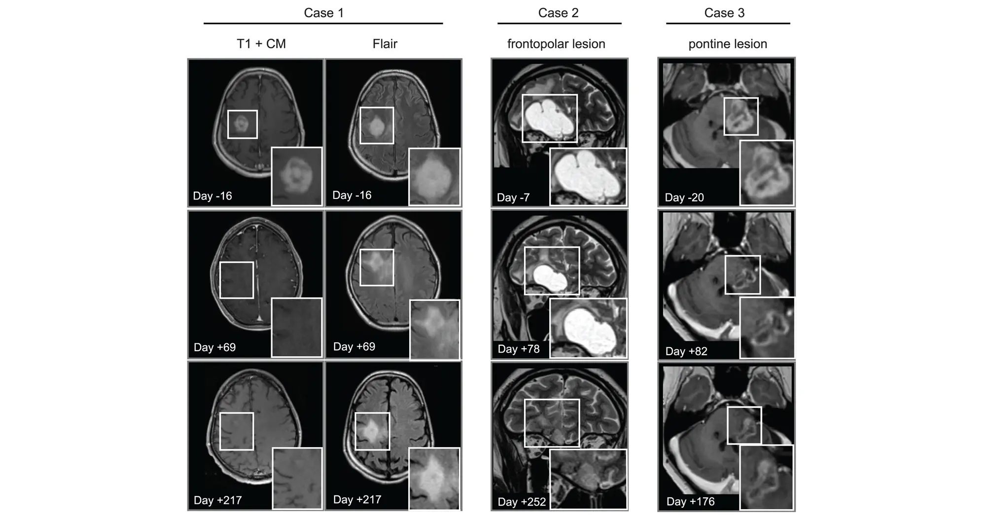
Scientists show that an established cancer drug travels to and shrinks some brain tumors, which may lead to new therapies for a disease with few treatments

Brain tumors are the leading cause of cancer-related death in childhood. The deadliest of these tumors are known as high-grade gliomas, with the grade referring to how quickly certain tumors grow and spread throughout the central nervous system.

Treatment options for high-grade gliomas are limited. Surgical removal is typically the first option depending on the tumor size and location. Radiation often follows to kill any remaining cancer cells to prevent another tumor from forming.

“Drug options to pair with surgery and/or radiation are few and far between,” said Lukas Chavez, PhD, associate professor in the Cancer Genome and Epigenetics Program at Sanford Burnham Prebys. “A big reason for this is the blood-brain barrier being as formidable a boundary as the mythological River Styx.”

The blood-brain barrier can, at times, mean the difference between life and death. It protects the brain and spinal cord from potential toxins and pathogens circulating in the bloodstream. However, in its vigilance, it also blocks beneficial drugs from reaching the brain. This presents a major challenge, since most medications are designed to travel through the bloodstream after being ingested or injected.

Scientists from an international team including Sanford Burnham Prebys, the University of Michigan, Dana Farber Cancer Institute, the Medical University of Vienna and many other institutions published findings March 13, 2025, in Cancer Cell demonstrating that the drug avapritinib could treat certain brain tumor cells. And, like the Styx’s ferryman Charon, the medicine is one of the rare few that can cross the blood-brain barrier known to prevent the passage of more than 98% of small molecule drugs.

The researchers selected avapritinib—which is approved by the Food and Drug Administration for treating gastrointestinal and other cancers—after finding it was the strongest commercially available drug for inhibiting the gene Platelet-derived growth factor receptor alpha (PDGFRA), which is found to be mutated in 15% of high-grade gliomas.

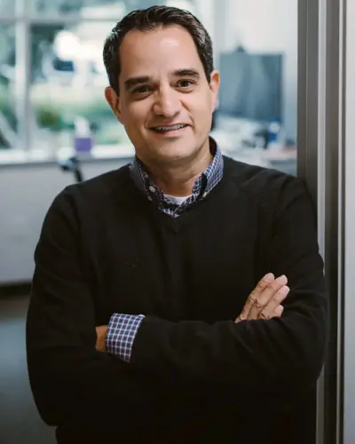
Lukas Chavez, PhD

Lukas Chavez, PhD, is an associate professor in the Cancer Genome and Epigenetics Program at Sanford Burnham Prebys.

In addition to showing that avapritinib inhibited PDGFRA in cancer cells and mouse brain tumors, the research team tested its effects on eight human pediatric and young adult high-grade glioma patients through a compassionate-use program. The treatment was found to be safe and investigators observed that the drug caused tumors to shrink in three patients.

“More research is needed to better understand how to best repurpose this drug for high-grade gliomas,” said Chavez. “We’ll learn a lot from the ongoing Rover study, a phase 1/2 multicenter trial of avapritinib based on these findings that will include more participants.”

The authors of the new study also highlighted the need to study combining multiple targeted therapies to overcome acquired resistance to any single treatment.


Mariella G. Filbin, MD, PhD, assistant professor of Pediatrics at Harvard Medical School and research co-director of the Pediatric Neuro-Oncology Program at the Dana-Farber/Boston Children’s Cancer and Blood Disorders Center, is the lead contact on the study.

Carl Koschmann, MD, ChadTough Defeat DIPG Research Professor and associate professor of Pediatric Neuro-Oncology at the University of Michigan Medical School, and Johannes Gojo, MD, PhD, head of Pediatric Precision Oncology CNS and ITCC-Lab/Clinical Trials Unit at the Medical University of Vienna, are corresponding authors along with Filbin.

Lisa Mayr, Sina Neyazi, Kallen Schwark and Maria Trissal share first authorship of the study.

Additional authors include:

  • Owen Chapman, Sunita Sridhar, Rishaan Kenkre, Aditi Dutta, Shanqing Wang, and Jessica Wang from Sanford Burnham Prebys
  • Jenna Labelle, Sebastian K. Eder, Joana G. Marques, Carlos A.O. de Biagi-Junior, Costanza Lo Cascio, Olivia Hack, Andrezza Nascimento, Cuong M. Nguyen, Sophia Castellani, Jacob S. Rozowsky, Andrew Groves, Eshini Panditharatna, Gustavo Alencastro Veiga Cruzeiro, Rebecca D. Haase, Kuscha Tabatabai, Alicia Baumgartner, Frank Dubois, Pratiti Bandopadhayay and Keith Ligon from the Dana-Farber/Boston Children’s Cancer and Blood Disorder Center and Harvard Medical School
  • Liesa Weiler-Wichtl, Sibylle Madlener, Katharina Bruckner, Daniel Senfter, Anna Lammerer, Natalia Stepien, Daniela Lotsch-Gojo, Walter Berger, Ulrike Leiss, Verena Rosenmayr, Christian Dorfer, Karin Dieckmann, Andreas Peyrl, Amedeo A. Azizi, Leonhard Mullauer, Christine Haberler and Julia Furtner from the Medical University of Vienna
  • Jack Wadden, Tiffany Adam, Seongbae Kong, Madeline Miclea, Tirth Patel, Chandan Kumar-Sinha, Arul Chinnaiyan and Rajen Mody from the University of Michigan Medical School
  • Alexander Beck from Ludwig Maximilians University Munich
  • Jeffrey Supko and Hiroaki Wakimoto from Massachusetts General Hospital
  • Armin S. Guntner from Johannes Kepler University
  • Hana Palova, Jakub Neradil, Ondrej Slaby, Petra Pokorna and Jaroslav Sterba from Masaryk University
  • Louise M. Clark, Amy Cameron and Quang-De Nguyen from the Dana-Farber Cancer Institute
  • Noah F. Greenwald and Rameen Beroukhim from the Broad Institute of MIT and Harvard
  • Christof Kramm from University Medical Center Gottingen
  • Annika Bronsema from University Medical Center Hamburg-Eppendorf
  • Simon Bailey from Great North Children’s Hospital and Newcastle University
  • Ana Guerreiro Stucklin from University Children’s Hospital Zurich
  • Sabine Mueller from the University of California San Francisco
  • Mary Skrypek from Children’s Minnesota
  • Nina Martinez from Jefferson University
  • Daniel C. Bowers from the University of Texas Southwestern Medical Center
  • David T.W. Jones, Natalie Jager from Hopp Children’s Cancer Center Heidelberg
  • Chris Jones from the Institute of Cancer Research
Institute News

The implastic nature of plastic culture

AuthorScott LaFee
Date

November 4, 2024

There is an art (and science) to creating cell culture models that reflect the complexities of disease. Such models have long been indispensable to parsing the underlying mechanisms of pathology and to preclinical drug discovery.

But art, writes Kevin Tharp, PhD, assistant professor in the Cancer Metabolism and Microenvironment Program, doesn’t always imitate life — at least not when it comes to finding effective cancer therapeutics.

“Just like a machine-learning algorithm trained on irrelevant datasets, efforts to discover anticancer therapeutics are limited by the models we use,” Tharp writes in the British Journal of Pharmacology. “Our drug discovery pipeline works incredibly well but is applied to models that poorly recapitulate in vivo physiology. This may be why drug discovery approaches efficiently identify drugs that work in the context tested and yet often fail to translate into clinical success.”

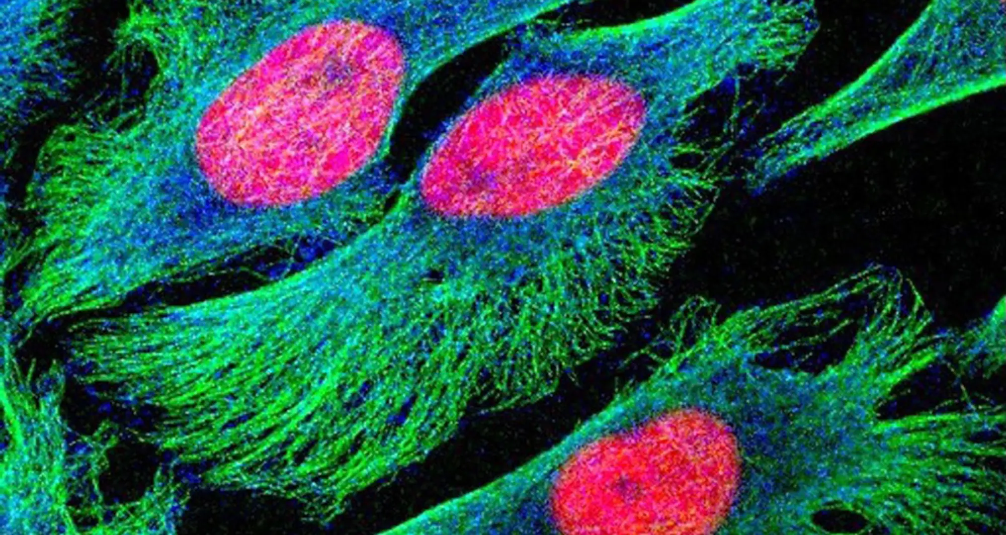
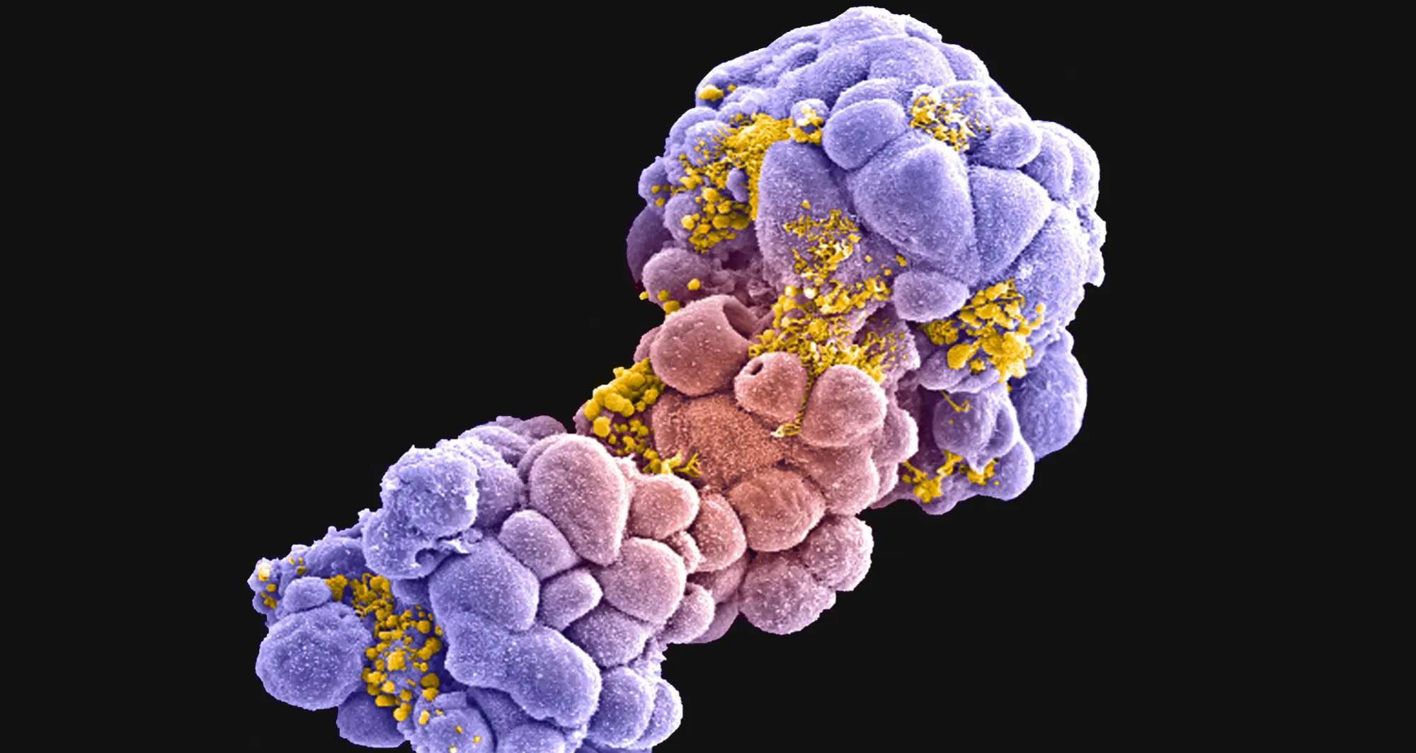
It’s a case of there’s no place like home. Cancer cell models are cultured on plastic in two-dimensions with limited or no diversity of neighbors. Cancer cells in vivo reside in three dimensions, with dynamic and complex interactions with neighboring cells and surroundings, i.e., the tumor microenvironment.

It’s like growing up on Disneyland’s Main Street versus a real-world urban city. Cultured cancer cells simply don’t look or behave exactly the same as cancer cells in an actual  tumor. Nor do the investigational molecules being tested as potential therapies.

Tharp suggests a multi-pronged approach: Initially culture target cells using conventional methods, then transfer the cells to new culture formats that enforce distinct, non-genomic cytoskeleton architectures and expression patterns that more closed mimic real life.

Institute News

Raising awareness of breast cancer research at Sanford Burnham Prebys

AuthorGreg Calhoun
Date

October 31, 2024

The October Science Connect Series event was themed around Breast Cancer Awareness Month and featured two cancer research experts.

The Sanford Burnham Prebys Wellness Ambassadors hosted a Science Connect event on Wednesday, October 30, 2024, featuring two faculty experts discussing their breast cancer research and its implications.

The Science Connect Series provides a forum for Sanford Burnham Prebys principal investigators to share their research with administrative personnel. Faculty members gain experience in communicating their science to a lay audience, and administrators gain a better understanding of research conducted at the institute so they can become better advocates and ambassadors of the shared mission to translate science into health.

Kelly Kersten, PhD, an assistant professor in the Cancer Metabolism and Microenvironment Program, opened the event by focusing on the importance of finding new treatments —such as immunotherapies — for the one-third of breast cancer patients that are diagnosed after the early stages of the disease when surgery is less effective.

The immune system is one of the main defenses of the human body to fend off harmful pathogens and invasive cells, such as cancer. Among all white blood cells, a particular cell type, called a T cell, can directly kill cancer cells and therefore plays an essential role in building anti-tumor immune responses.

Many types of cancer are confronted and infiltrated by T cells, only to be suppressed by the local tumor environment.

“While immunotherapies that boost the immune system have revolutionized the way we treat cancer, many patients do not respond to the treatments, and the mechanisms of resistance remain largely unclear,” said Kersten.

Kersten’s goal is to understand why T cells enter a state known as exhaustion and lose their tumor-killing capacity. This knowledge will help her team find potential future therapies that could prevent T-cell exhaustion and improve immunotherapies for cancer patients.

Kevin Tharp, PhD, also an assistant professor in the Cancer Metabolism and Microenvironment Program, shared that his lab’s focus is on how cancer cells adapt their metabolism to generate the energy needed to spread to other tissues through metastasis. He presented his team’s work with the Kersten lab on another aspect of potential resistance to immunotherapy in breast cancer.

Tharp and Kersten are studying the hypothesis that part of the reason why these therapies fail is due to tumor-associated fibrosis, the creation of a thick layer of fibrous collagen (like scar tissue) that acts as a barrier against the anti-tumor immune response. They published a paper on June 3, 2024, in Nature Cancer,  discussing how tumor-associated macrophages, a type of immune cell found abundantly in the tumor microenvironment, respond to the physical properties of fibrosis.

By synthesizing injury-associated collagens that facilitate wound closure, TAMs experience metabolic changes and generate metabolic byproducts that suppress the anti-tumor function of immune cells.

“The metabolic changes in the microenvironment present more of a challenge to anti-tumor responses than the physical barrier,” said Tharp. “Our study provides an alternative explanation for why anti-tumor immunity is impaired in fibrotic solid tumors.”

To follow up on these results, Tharp is collaborating with Sarah Blair, MD, a professor of surgery at the University of California San Diego, to fund and initiate a clinical trial testing the potential of dietary supplements to counteract the suppressive effects of TAM metabolic byproducts as an adjunct therapy to surgery.

Institute News

Using machines to personalize patient care

AuthorGreg Calhoun
Date

July 30, 2024

Artificial intelligence (AI) and other computational techniques are aiding scientists and physicians in their quest to create treatments for individuals rather than populations

The Human Genome Project captured the public’s imagination with its global quest to better understand the genetic blueprint stored on the DNA within our cells. The project succeeded in delivering the first-ever sequence of the human genome while foreshadowing a future for medicine once considered to be science fiction. The project presaged the possibility that health care could be personalized based on clues within a patient’s unique genetic code.

Chavez lab

The Chavez Lab

While many more people have undergone genetic testing through consumer genealogy and health services such as 23andMe and Ancestry than through health care systems, genomic sequencing has influenced clinical care in some specialties. Personalized medicine—also known as precision medicine or genomic medicine—has been especially helpful for people suffering from rare diseases that historically have been difficult to diagnose and treat.

Scientists at Sanford Burnham Prebys are employing new technologies and expertise to test ways to improve diagnoses and customize treatments for many diseases based on unique characteristics within tumors, blood samples and other biopsies.

AI and other computational techniques are enabling patient samples to be rapidly analyzed and compared to data from vast numbers of individuals who have been treated for the same condition. Physicians can use AI and other tools to identify subtypes of cancers and other conditions, as well as improve selection of eligible candidates for clinical trials.

“I think we’ve gotten a lot better at precision diagnostics,” says Lukas Chavez, PhD, an assistant professor in the Cancer Genome and Epigenetics Program at Sanford Burnham Prebys. “In my work at Rady Children’s Hospital in cancer, we can characterize a tumor based on mutations, including predicting how quickly different tumors will spread. What we too often lack, however, are better treatment approaches or medicines. That will be the next generation of precision medicine.”

Sanju Sinha, PhD, an assistant professor in the Cancer Molecular Therapeutics Program at Sanford Burnham Prebys, is developing projects to help bridge the gap between precision diagnostics and treatment. He is partnering with the National Cancer Institute on a first-of-its-kind computational tool to systematically predict patient response to cancer drugs at single-cell resolution.

A study published in the journal  Nature Cancer discussed how the tool, called PERCEPTION, was successfully validated by predicting the response to individual therapies and combination treatments in three independent published clinical trials for multiple myeloma, breast and lung cancer.

Lukas Chavez, PhD

Lukas Chavez, PhD, is an assistant professor in the Cancer Genome and Epigenetics Program at Sanford Burnham Prebys.

In each case, PERCEPTION correctly stratified patients into responder and non-responder categories. In lung cancer, it even captured the development of drug resistance as the disease progressed, a notable discovery with great potential.

Sanju Sinha, PhD

Sanju Sinha, PhD, is an assistant professor in the Cancer Molecular
Therapeutics Program at Sanford Burnham Prebys.

“The ability to monitor the emergence of resistance is the most exciting part for me,” says Sinha. “It has the potential to allow us to adapt to the evolution of cancer cells and even modify our treatment strategy.”

While PERCEPTION is not yet ready for clinics, Sinha hopes that widespread adoption of this technology will generate more data, which can be used to further develop and refine the technology for use by health care providers.

In another project, Sinha is focused on patients being treated for potential cancers that may never progress into dangerous conditions warranting treatment and its accompanying side effects.

“Many women who are diagnosed with precancerous changes in the breast seek early treatment,” says Sinha. “Most precancerous cells never lead to cancer, so it may be that as many as eight of 10 women with this diagnosis are being overtreated, which is a huge issue.”

To try and counter this phenomenon, Sinha is training AI models on images of biopsied samples in conjunction with multi-omics sequencing data. His team’s goal is to develop a tool capable of predicting which patients’ cancers would progress based on the imaged samples alone.

“In the field of precancer, insurance does not cover the cost of computing this omics data,” says Sinha. “Health care systems do routinely generate histopathological slides from patient biopsies, so we feel that a tool leveraging these images could be a scalable and accessible solution.”

If Sinha’s team is successful, an AI tool integrated into clinics would predict whether precancerous cells would progress within the next 10 years to guide treatment decisions and how patients are monitored.

“With precision medicine, our hope is not to just treat patients with better drugs, but also to make sure that patients are not unnecessarily treated and made to bear needless costs and side effects that disrupt their quality of life.”


Programming in a Petri Dish, an 8-part series

How artificial intelligence, machine learning and emerging computational technologies are changing biomedical research and the future of health care

  • Part 1 – Using machines to personalize patient care. Artificial intelligence and other computational techniques are aiding scientists and physicians in their quest to prescribe or create treatments for individuals rather than populations.
  • Part 2 – Objective omics. Although the hypothesis is a core concept in science, unbiased omics methods may reduce attachments to incorrect hypotheses that can reduce impartiality and slow progress.
  • Part 3 – Coding clinic. Rapidly evolving computational tools may unlock vast archives of untapped clinical information—and help solve complex challenges confronting health care providers.
  • Part 4 – Scripting their own futures. At Sanford Burnham Prebys Graduate School of Biomedical Sciences, students embrace computational methods to enhance their research careers.
  • Part 5 – Dodging AI and computational biology dangers. Sanford Burnham Prebys scientists say that understanding the potential pitfalls of using AI and other computational tools to guide biomedical research helps maximize benefits while minimizing concerns.
  • Part 6 – Mapping the human body to better treat disease. Scientists synthesize supersized sets of biological and clinical data to make discoveries and find promising treatments.
  • Part 7 – Simulating science or science fiction? By harnessing artificial intelligence and modern computing, scientists are simulating more complex biological, clinical and public health phenomena to accelerate discovery.
  • Part 8 – Acceleration by automation. Increases in the scale and pace of research and drug discovery are being made possible by robotic automation of time-consuming tasks that must be repeated with exhaustive exactness.
Institute News

The Cancer Letter covers collaboration between Sanford Burnham Prebys and the National Cancer Institute to precisely prescribe cancer drugs

AuthorGreg Calhoun
Date

May 14, 2024

The May 10 issue of The Cancer Letter details a recent publication explaining the investigation of a new AI tool that may be able to match cancer drugs more precisely to patients.

The Cancer Letter—a news organization and weekly publication based in Washington, D.C., that focuses on cancer research and clinical care—included an article in its May 10 issue about a partnership between scientists at Sanford Burnham Prebys and the National Cancer Institute (NCI).

Authored by Sanju Sinha, PhD, assistant professor in the Cancer Molecular Therapeutics Program at Sanford Burnham Prebys, and the NCI’s Eytan Ruppin, MD, PhD, the “Trials & Tribulations” feature describes a first-of-its-kind computational tool to systematically predict patient response to cancer drugs at single-cell resolution. The study regarding this new tool was published on April 18, 2024, in the journal  Nature Cancer.

The Cancer Letter was founded in 1973 and focuses its coverage on the development of cancer therapies, drug regulation, legislation, cancer research funding, health care finance and public health.

Institute News

Media coverage of AI study predicting responses to cancer therapy ranks top 5% among published research

AuthorScott LaFee, Susan Gammon and Greg Calhoun
Date

April 29, 2024

Last week, Sanford Burnham Prebys and the National Cancer Institute shared findings regarding a first-of-its-kind computational tool to systematically predict patient response to cancer drugs at single-cell resolution.

Many news outlets and trade publications took note of this study and the computational tool’s potential future use in hospitals and clinics. This coverage placed the paper in the top 5% of all manuscripts ranked by Altmetric—a service that tracks and analyzes online attention of published research to improve the understanding and value of research and how it affects people and communities.

The results from the highlighted study were published on April 18, 2024, in the journal Nature Cancer.

“Our goal is to create a clinical tool that can predict the treatment response of individual cancer patients in a systematic, data-driven manner. We hope these findings spur more data and more such studies, sooner rather than later,” says first author Sanju Sinha, PhD, assistant professor in the Cancer Molecular Therapeutics Program at Sanford Burnham Prebys.

Here are a few of the venues that helped spread the word about this research: 

  • AP News: “Researchers … suggest that such single-cell RNA sequencing data could one day be used to help doctors more precisely match cancer patients with drugs that will be effective for their cancer.”
  • Politico, fourth story in Future Pulse newsletter: “Our hope is that being able to characterize the tumors on a single-cell resolution will enable us to treat and target potentially the most resistant and aggressive [cells], which are currently missed.”
  • NIH.gov: “The researchers discovered that if just one clone were resistant to a particular drug, the patient would not respond to that drug, even if all the other clones responded.”
  • Inside Precision Medicine: “The model was validated by predicting the response to monotherapy and combination treatment in three independent, recently published clinical trials for multiple myeloma, breast, and lung cancer.”

“I’m very pleased with how many news outlets covered our work,” Sinha says. “It is important and will help us continue improving the tool with more data so it can one day benefit cancer patients.”

Institute News

New genome mapping tool may uncover secrets for treating blood cancers

AuthorGreg Calhoun
Date

February 1, 2024

The outlook for patients with acute myeloid leukemia (AML), a deadly set of blood cancers that is difficult to treat, has remained dire for decades, especially among patients who are not eligible for bone marrow transplantation.

More than 30% of treated patients will never achieve complete remission using current chemotherapies and, even when chemotherapy treatments work, most patients relapse within five years without a transplant.

While prior research has begun to unravel the genetic underpinnings of the disease, more inquiry is needed to understand the genetic variation within the roughly 15 AML subtypes and how that variation might affect treatment strategies.

“In addition to the one to eight average genetic mutations in AML patients found in traditional sequencing studies, experiments employing high-resolution optical genomic mapping have found approximately 40 to 80 rare genomic structural variants per patient,” says Kristiina Vuori, MD, PhD, Pauline and Stanley Foster Distinguished Chair and professor in the Sanford Burnham Prebys Cancer Center’s Cancer Molecular Therapeutics Program. “We wanted to take these structural variant findings in AML to the next level by connecting them with patients’ sensitivity or resistance to current cancer treatments.”

Kristiina Vuori, MD, PhD

In a paper published January 18, 2024, in Cancers, a multidisciplinary team of biologists, bioinformaticians and clinicians from Sanford Burnham Prebys, Bionano Genomics Inc. and Scripps MD Anderson were the first to associate genomic structural variants (SVs) in AML patients with drug sensitivities.

“SVs are changes to the genome in which sections of 50 or more base pairs in a strand of DNA have been errantly deleted, duplicated, inverted or translocated,” explains Darren (Ben) Finlay, PhD, first author on the manuscript and research associate professor in the Sanford Burnham Prebys Cancer Center.

“Such changes amount to different combinations of DNA gains, losses or rearrangements. When cells use these altered instructions in the DNA to make proteins or carry out other functions, it is like a chef trying to cook with a recipe that is missing steps, has them in the wrong order or includes more or less of the key ingredients.”

Darren (Ben) Finally, PhD

Scientists have become more able to find SVs as next-generation genomic analysis technologies and techniques have improved. Research has shown that SVs contribute to the development and progression of cancer, including blood cancers. Of particular concern among SVs are DNA changes that join two otherwise distant genes. This event, called gene fusion, is known to drive certain pediatric and blood cancers.

Finlay, Vuori and colleagues analyzed SVs in samples from 23 AML patients and found their genomes featured 16-45 extremely rare SVs within genes but not seen in healthy volunteers’ samples. The scientists detailed the patients’ SVs using a technique called optical genome mapping. This tool tags DNA in specific locations to create recognizable sequences, unwinds and straightens the genomic DNA for linear scanning, and converts the imaged sequences into digital representations of DNA molecules. Because it directly images DNA rather than relying on algorithmic analyses, optical genome mapping is better than next-generation sequencing at finding SVs throughout the entire genome, especially large SVs, the researchers said.

To begin building the connection between SVs and drug sensitivity, the scientists tested samples from each patient with 120 FDA-approved drugs, and experimental treatments currently in phase III clinical trials. This allowed the researchers to map out how strongly each patient’s sample reacted with each drug.

Next, the investigators used statistical analysis to compare the SVs within the optical genome mapping results with the findings from the drug sensitivity tests. The team found 61 statistically significant interactions between SVs and existing cancer therapies. In one interaction, the group demonstrated that a commonly used AML drug, Idarubicin, and two similar compounds (Daunorubicin and Epirubicin) were more effective in leukemia samples with a specific insertion in a gene that carries the instructions for a signaling enzyme that helps nerves communicate with muscles. These and other examples lend support to the scientists’ hypothesis that optical genome mapping could be used to develop personalized treatment plans that account for patients’ SVs.

“In this pilot study, our hope was to identify structural variants that could be used as new biomarkers for current AML drugs as well as to identify other drugs that could be repurposed to treat leukemia patients,” says Finlay.

“Ensuring patients receive the most effective drugs on the market through personalized treatment and identifying new potential therapies for AML are critically important,” adds Vuori, senior author on the study. “Especially for patients who do not achieve remission with current standard chemotherapies or who are ineligible for bone marrow transplants or clinical trials.”
 

Cancers 2024, 16(2), 418; https://doi.org/10.3390/cancers16020418

Institute News

The “Eph” system may pave the way for novel cancer therapies

AuthorSusan Gammon
Date

November 27, 2023

Over the past three decades, researchers have been investigating an important cell communication system called the “Eph system,” and the evidence implicating the system in cancer is staggering.

The Eph system is comprised of multiple Eph receptors and their ligands—ephrins—and are involved in contact-dependent communication between cells. They play essential roles in regulating various cellular processes.

Modern studies have shed light on the Eph system’s role in tumor expansion, invasiveness, metastasis, cancer stem cell maintenance and therapy resistance.

This month, Elena Pasquale, PhD, published a review in Nature Reviews Cancer that summarizes the current state of research on the Eph system and its links to cancer progression and drug resistance.

“The Eph system has many critical functions during the development of tissues and organs, but it also has the capacity to either promote or suppress cancer progression and malignancy” says Pasquale. “In cancer, the activities of the Eph system can differ depending on the circumstances—for example, which Eph receptors and ligands are present in a tumor cell, the types of tumor cells in which they function, and the characteristics of these cells.”

“It’s this remarkable versatility that makes the Eph system a compelling but also challenging target for potential therapies,” says Pasquale.

“The aims of this review were to comprehensively survey the large body of data regarding various aspects related to Eph signaling in tumors and to highlight potential strategies for therapeutic targeting,” says Pasquale. “Overall, while significant progress has been made in deciphering the Eph system in cancer, there is much more to learn.

“Gaining a deeper understanding of how the Eph system functions in cancer is challenging but will be essential for the development of targeted therapies and personalized treatment approaches for patients.”

Institute News

Scientists design potential drug for triple-negative breast cancer

AuthorMonica May
Date

February 16, 2021

Drug candidate blocks autophagy, a cellular recycling process that cancer cells hijack as a way to resist treatment

Scientists at Sanford Burnham Prebys Medical Discovery Institute have designed a next-generation drug, called SBP-7455, which holds promise as a treatment for triple-negative breast cancer—an aggressive cancer with limited treatment options. The drug blocks a cellular recycling process called autophagy, which cancer cells hijack as a way to resist treatment. The proof-of-concept study was published in the Journal of Medicinal Chemistry.

“Scientists are now learning that autophagy is one of the main ways that cancer cells are able to survive, even in the presence of growth-blocking treatments,” says Huiyu Ren, a graduate student in the laboratory of Nicholas Cosford, PhD, at Sanford Burnham Prebys, and first author of the study. “If all goes well, we hope this compound will stop cancer cells from turning on autophagy and allow people with triple-negative breast cancer to benefit from their treatment for as long as possible.”

Cells normally use autophagy as a way to recycle waste products. However, when cancer cells’ survival is threatened by a growth-blocking treatment, this process is often “revved up” so the cancer cell can continue to receive nutrients and keep growing. Certain cancers are more likely to rely on the autophagy process for survival, including breast, pancreatic, prostate and lung cancers.

“While this study focused on triple-negative breast cancer, an area of great unmet need, we are actively testing this drug’s potential against more cancer types,” says Cosford, professor and deputy director in the National Cancer Institute (NCI)-designated Cancer Center at Sanford Burnham Prebys and senior author of the study. “An autophagy-inhibiting drug that stops treatment resistance from taking hold would be a great addition to an oncologist’s toolbox.”

About 15% to 20% of all breast cancers are triple negative, which means they do not respond to hormonal therapy or targeted treatments. The cancer is currently treated with surgery, chemotherapy and radiation, and is deadlier than other breast cancer types. If the tumor returns, other treatments such as PARP inhibitors or immunotherapy are considered. People under the age of 50 are more likely to have triple-negative breast cancer, as well as women who are Black, Hispanic, and/or have an inherited BRCA mutation.

An optimized drug

In this study, the scientists optimized a first-generation drug they created in 2015. The result is a compound called SBP-7455 that blocks two autophagy proteins, ULK1 and ULK2. SBP-7455 exhibits promising bioavailability in mice and reduces autophagy levels in triple-negative breast cancer cells, resulting in cell death. Importantly, combining the drug with PARP inhibitors, which are currently used to treat people with recurrent triple-negative breast cancer, makes the drug even more effective.

“We are hopeful that we have found a new potential therapy for people living with triple-negative breast cancer,” says Reuben Shaw, PhD, a study author and professor in the Molecular and Cell Biology Laboratory and director of the NCI-designated Cancer Center at the Salk Institute. “We envision this drug being used in combination with targeted therapies, such as PARP inhibitors, to prevent cancer cells from becoming treatment resistant.”

Next, the scientists plan to test the drug in mouse models of triple-negative breast cancer to confirm that the compound can stop tumor growth in an animal model. In parallel, they will continue optimization efforts to ensure the drug has the greatest chance of clinical success.

“Triple-negative breast cancer is one of the hardest cancers to treat today,” says Ren. “I hope that our research marks the start of a path to successful treatment that helps more people survive this aggressive cancer.”


Additional study authors include Nicole A. Bakas, Mitchell Vamos, Allison S. Limpert, Carina D. Wimer, Lester J. Lambert, Lutz Tautz, Maria Celeridad and Douglas J. Sheffler of Sanford Burnham Prebys; Apirat Chaikuad and Stefan Knapp of the Buchmann Institute for Molecular Life Sciences and Goethe-University Frankfurt; and Sonja N. Brun of the Salk Institute.

This work was supported by the National Institutes of Health (P30CA030199, T32CA211036), Epstein Family Foundation, Larry L. Hillblom Foundation (2019-A-005-NET), Pancreatic Cancer Action Network (19-65-COSF), SGC—a registered charity that receives funds from AbbVie, Bayer Pharma AG, Boehringer Ingelheim, Canada Foundation for Innovation, Eshelman Institute for Innovation, Genome Canada through Ontario Genomics Institute [OGI-196], EU/EFPIA/OICR/McGill/KTH/Diamond, Innovative Medicines Initiative 2 Joint Undertaking (875510), Janssen, Merck KGaA, Merck & Co, Pfizer, São Paulo Research Foundation-FAPESP, Takeda, and Wellcome.

The study’s DOI is 0.1021/acs.jmedchem.0c00873.