NIH funding Archives - Sanford Burnham Prebys
Institute News

Cancer drug finds new purpose in the brain

AuthorGreg Calhoun
Date

April 14, 2025

Scientists show that an established cancer drug travels to and shrinks some brain tumors, which may lead to new therapies for a disease with few treatments

Brain tumors are the leading cause of cancer-related death in childhood. The deadliest of these tumors are known as high-grade gliomas, with the grade referring to how quickly certain tumors grow and spread throughout the central nervous system.

Treatment options for high-grade gliomas are limited. Surgical removal is typically the first option depending on the tumor size and location. Radiation often follows to kill any remaining cancer cells to prevent another tumor from forming.

“Drug options to pair with surgery and/or radiation are few and far between,” said Lukas Chavez, PhD, associate professor in the Cancer Genome and Epigenetics Program at Sanford Burnham Prebys. “A big reason for this is the blood-brain barrier being as formidable a boundary as the mythological River Styx.”

The blood-brain barrier can, at times, mean the difference between life and death. It protects the brain and spinal cord from potential toxins and pathogens circulating in the bloodstream. However, in its vigilance, it also blocks beneficial drugs from reaching the brain. This presents a major challenge, since most medications are designed to travel through the bloodstream after being ingested or injected.

Scientists from an international team including Sanford Burnham Prebys, the University of Michigan, Dana Farber Cancer Institute, the Medical University of Vienna and many other institutions published findings March 13, 2025, in Cancer Cell demonstrating that the drug avapritinib could treat certain brain tumor cells. And, like the Styx’s ferryman Charon, the medicine is one of the rare few that can cross the blood-brain barrier known to prevent the passage of more than 98% of small molecule drugs.

The researchers selected avapritinib—which is approved by the Food and Drug Administration for treating gastrointestinal and other cancers—after finding it was the strongest commercially available drug for inhibiting the gene Platelet-derived growth factor receptor alpha (PDGFRA), which is found to be mutated in 15% of high-grade gliomas.

Lukas Chavez, PhD

Lukas Chavez, PhD, is an associate professor in the Cancer Genome and Epigenetics Program at Sanford Burnham Prebys.

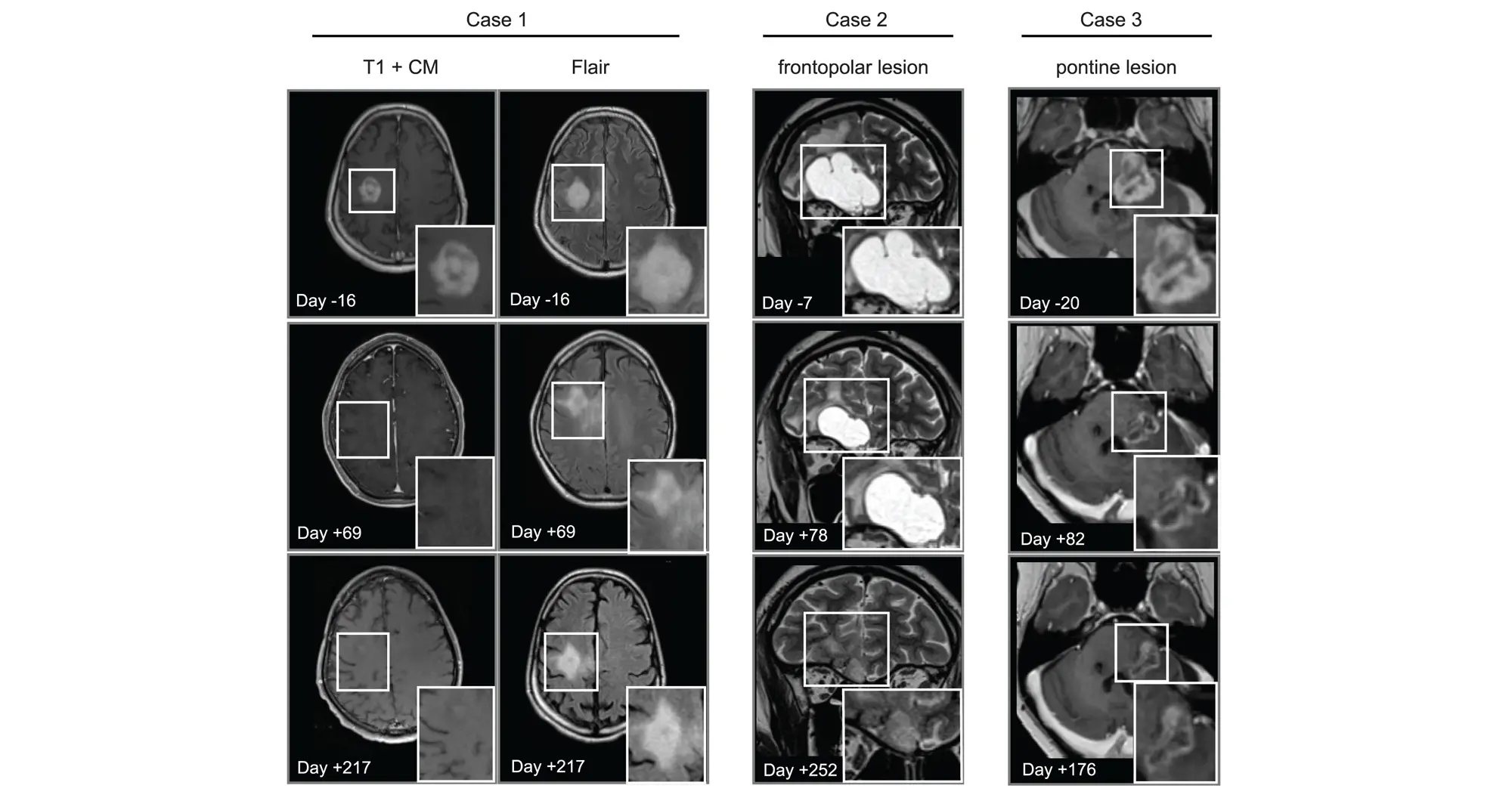
In addition to showing that avapritinib inhibited PDGFRA in cancer cells and mouse brain tumors, the research team tested its effects on eight human pediatric and young adult high-grade glioma patients through a compassionate-use program. The treatment was found to be safe and investigators observed that the drug caused tumors to shrink in three patients.

“More research is needed to better understand how to best repurpose this drug for high-grade gliomas,” said Chavez. “We’ll learn a lot from the ongoing Rover study, a phase 1/2 multicenter trial of avapritinib based on these findings that will include more participants.”

The authors of the new study also highlighted the need to study combining multiple targeted therapies to overcome acquired resistance to any single treatment.


Mariella G. Filbin, MD, PhD, assistant professor of Pediatrics at Harvard Medical School and research co-director of the Pediatric Neuro-Oncology Program at the Dana-Farber/Boston Children’s Cancer and Blood Disorders Center, is the lead contact on the study.

Carl Koschmann, MD, ChadTough Defeat DIPG Research Professor and associate professor of Pediatric Neuro-Oncology at the University of Michigan Medical School, and Johannes Gojo, MD, PhD, head of Pediatric Precision Oncology CNS and ITCC-Lab/Clinical Trials Unit at the Medical University of Vienna, are corresponding authors along with Filbin.

Lisa Mayr, Sina Neyazi, Kallen Schwark and Maria Trissal share first authorship of the study.

Additional authors include:

  • Owen Chapman, Sunita Sridhar, Rishaan Kenkre, Aditi Dutta, Shanqing Wang, and Jessica Wang from Sanford Burnham Prebys
  • Jenna Labelle, Sebastian K. Eder, Joana G. Marques, Carlos A.O. de Biagi-Junior, Costanza Lo Cascio, Olivia Hack, Andrezza Nascimento, Cuong M. Nguyen, Sophia Castellani, Jacob S. Rozowsky, Andrew Groves, Eshini Panditharatna, Gustavo Alencastro Veiga Cruzeiro, Rebecca D. Haase, Kuscha Tabatabai, Alicia Baumgartner, Frank Dubois, Pratiti Bandopadhayay and Keith Ligon from the Dana-Farber/Boston Children’s Cancer and Blood Disorder Center and Harvard Medical School
  • Liesa Weiler-Wichtl, Sibylle Madlener, Katharina Bruckner, Daniel Senfter, Anna Lammerer, Natalia Stepien, Daniela Lotsch-Gojo, Walter Berger, Ulrike Leiss, Verena Rosenmayr, Christian Dorfer, Karin Dieckmann, Andreas Peyrl, Amedeo A. Azizi, Leonhard Mullauer, Christine Haberler and Julia Furtner from the Medical University of Vienna
  • Jack Wadden, Tiffany Adam, Seongbae Kong, Madeline Miclea, Tirth Patel, Chandan Kumar-Sinha, Arul Chinnaiyan and Rajen Mody from the University of Michigan Medical School
  • Alexander Beck from Ludwig Maximilians University Munich
  • Jeffrey Supko and Hiroaki Wakimoto from Massachusetts General Hospital
  • Armin S. Guntner from Johannes Kepler University
  • Hana Palova, Jakub Neradil, Ondrej Slaby, Petra Pokorna and Jaroslav Sterba from Masaryk University
  • Louise M. Clark, Amy Cameron and Quang-De Nguyen from the Dana-Farber Cancer Institute
  • Noah F. Greenwald and Rameen Beroukhim from the Broad Institute of MIT and Harvard
  • Christof Kramm from University Medical Center Gottingen
  • Annika Bronsema from University Medical Center Hamburg-Eppendorf
  • Simon Bailey from Great North Children’s Hospital and Newcastle University
  • Ana Guerreiro Stucklin from University Children’s Hospital Zurich
  • Sabine Mueller from the University of California San Francisco
  • Mary Skrypek from Children’s Minnesota
  • Nina Martinez from Jefferson University
  • Daniel C. Bowers from the University of Texas Southwestern Medical Center
  • David T.W. Jones, Natalie Jager from Hopp Children’s Cancer Center Heidelberg
  • Chris Jones from the Institute of Cancer Research
Institute News

How basic science fuels economic gain

AuthorScott LaFee
Date

April 1, 2025

A recent story in The New York Times describes how new and changed federal policies pose long-term risk to science and the economic gains it creates.

Highlighted in the article is the story of Hudson Freeze, PhD, director of the Sanford Children’s Health Research Center, who as an undergraduate student at Indiana University was involved in the discovery of heat-resistant microbes.

Those microbes would prove critical to the development of polymerase chain reaction (PCR), a DNA replication tool that is now ubiquitous in genetic science and beyond. 

Institute News

Acceleration by automation

AuthorGreg Calhoun
Date

September 5, 2024

Increases in the scale and pace of research and drug discovery are being made possible by robotic automation of time-consuming tasks that must be repeated with exhaustive exactness.

Humans have long been fascinated by automata, objects that can or appear to move and act of their own volition. From the golems of Jewish folklore to Pinocchio and Frankenstein’s Creature—among the subjects of many other tales—storytellers have long explored the potential consequences of creating beings that range from obedient robots to sentient saboteurs.

While the power of our imagination preceded the available technology for such feats of automation, many scientists and engineers throughout history succeeded in creating automata that were as amusing as they were examples of technical mastery. Three doll automata made by inventor Pierre Jaquet-Droz traveled around the world to delight kings and emperors by writing, drawing and playing music, and they now fascinate visitors to the Musée d’Art et d’Histoire of Neuchâtel, Switzerland.

While these more whimsical machinations can be found in collections from the House on the Rock in Spring Green, Wis., to the Hermitage Museum in Saint Petersburg, Russia, applications in certain forms of labor have made it so more modern automation is located in factories and workshops. There is no comparing the level of automation at research institutions to that of many manufacturing facilities more than 110 years since the introduction of the assembly line, nor should there be given the differing aims. However, the mechanization of certain tasks in the scientific process has been critical to increasing the accessibility of the latest biomedical research techniques and making current drug discovery methods possible.

researcher at work in Prebys Center

As a premier drug discovery center, the Conrad Prebys Center for Chemical Genomics team is well-versed in using automation to enable the testing of hundreds of thousands of chemicals to find new potential medicines.

“Genomic sequencing has become a very important procedure for experiments in many labs,” says Ian Pass, PhD, director of High-Throughput Screening at the Conrad Prebys Center for Chemical Genomics (Prebys Center) at Sanford Burnham Prebys. “Looking back just 20-30 years, the first sequenced human genome required the building of a robust international infrastructure and more than 12 years of active research. Now, with how we’ve refined and automated the process, I could probably have my genome sampled and sequenced in an afternoon.”

While many tasks in academic research labs require hands-on manipulation of pipettes, petri dishes, chemical reagents and other tools of the trade, automation has been a major factor enabling omics and other methods that process and sequence hundreds or thousands of samples to capture incredible amounts of information in a single experiment. Many of these sophisticated experiments would be simply too labor-intensive and expensive to conduct by hand.

Where some of the automation of yore would play a tune, enact a puppet show or tell a vague fortune upon inserting a coin, scientists now prepare samples for instruments equipped with advanced robotics, precise fluid handling technologies, cameras and integrated data analysis capabilities. Automation in liquid handling has enabled one of the biggest steps forward as it allows tests to be miniaturized. This not only results in major cost savings, but also it allows experiments to have many replicas, generating very high-quality, reliable data. These characteristics in data are a critical underpinning for ensuring the integrity of the scientific community’s findings and maintaining the public’s trust.

Ian Pass headshot

Ian Pass, PhD, is the director of High-Throughput Screening at the Conrad Prebys Center for Chemical Genomics.

“At their simplest, many robotic platforms amount to one or more arms that have a grip that can be programmed to move objects around,” explains Pass. “If a task needs to be repeated just a few times, then it probably isn’t worth the effort to deploy a robot. But, once that step needs to be repeated thousands of times at precise intervals, and handled the exact same way each time, then miniaturization and automation are the answers.”

As a premier drug discovery center, the Prebys Center team is well-versed in using automation to enable the testing of hundreds of thousands of chemicals to find new potential medicines. The center installed its first robotics platform, affectionately called “big yellow,” in the late 2000s to enable what is known as ultra-high-throughput screening (uHTS). Between 2009 and 2014, this robot was the workhorse for completing over 100 uHTS of a large chemical library. It generated tens of millions of data points as part of an initiative funded by the National Institutes of Health (NIH) called the Molecular Libraries Program that involved more than 50 research institutions across the US. The output of the program was the identification of hundreds of chemical probes that have been used to accelerate drug discovery and launch the field of chemical biology.

“Without automation, we simply couldn’t have done this,” says Pass. “If we were doing it manually, one experiment at a time, we’d still be on the first screen.”

Over the past 10 years the Center has shifted focus from discovering chemical probes to discovering drugs. Fortunately, much of the process is the same, but the scale of the experiments is even bigger, with screens of over 750,000 chemicals. To screen such large libraries, highly miniaturized arrays are used in which 1536 tests are conducted in parallel. Experiments are miniaturized to such an extent that hand pipetting is not possible and acoustic dispensing (i.e. sound waves) are used to precisely move the tiny amounts of liquid in a touchless, tipless automated process. In this way, more than 250,000 tests can be accomplished in a single day, allowing chemicals that bind to the drug target to be efficiently identified. Once the Prebys Center team identifies compounds that bind, these prototype drugs are then improved by the medicinal chemistry team, ultimately generating drugs with properties suitable for advancing to phase I clinical trials in humans.

Within the last year, the Prebys Center has retired “big yellow” and replaced it with three acoustic dispensing enabled uHTS robotic systems using 1536 well high-density arrays that can run fully independently.

“We used to use big yellow for just uHTP library screening, but now, with the new line up of robots, we use them for everything in the lab we can,” notes Pass. “It has really changed how we use automation to support and accelerate our science. Having multiple systems allows us to run simultaneous experiments and avoid scheduling conflicts. It also allows us to stay operational if one of the systems requires maintenance.”

One of the many drug discovery projects at the Prebys Center focuses on the national epidemic of opioid addiction. In 2021, fentanyl and other synthetic opioids accounted for nearly 71,000 of 107,000 fatal drug overdoses in the U.S. By comparison, in 1999 drug-involved overdose deaths totaled less than 20,000 among all ages and genders.

Like other addictive substances, opioids are intimately related to the brain’s dopamine-based reward system. Dopamine is a neurotransmitter that serves critical roles in memory, movement, mood and attention. Michael Jackson, PhD, senior vice president of Drug Discovery and Development at the Prebys Center and co-principal investigator Lawrence Barak, MD, PhD, at Duke University, have been developing a completely new class of drugs that works by targeting a receptor on neurons called neurotensin 1 receptor or NTSR1, that regulates dopamine release.

The researchers received a $6.3 million award from NIH and the National Institute on Drug Abuse (NIDA) in 2023 to advance their addiction drug candidate, called SBI-810, to the clinic. SBI-810 is an improved version of SBI-533, which previously had been shown to modulate NTSR1 signaling and demonstrated robust efficacy in mouse models of addiction without adverse side effects.

Michael Jackson profile photo

Michael Jackson, PhD, is the senior vice president of Drug Discovery and Development at the Conrad Prebys Center for Chemical Genomics.

Prebys Center researchers at work

The funding from the NIH and NIDA will be used to complete preclinical studies and initiate a Phase 1 clinical trial to evaluate safety in humans.

“The novel mechanism of action and broad efficacy of SBI-810 in preclinical models hold the promise of a truly new, first-in-class treatment for patients affected by addictive behaviors,” says Jackson.


Programming in a Petri Dish, an 8-part series

How artificial intelligence, machine learning and emerging computational technologies are changing biomedical research and the future of health care

  • Part 1 – Using machines to personalize patient care. Artificial intelligence and other computational techniques are aiding scientists and physicians in their quest to prescribe or create treatments for individuals rather than populations.
  • Part 2 – Objective omics. Although the hypothesis is a core concept in science, unbiased omics methods may reduce attachments to incorrect hypotheses that can reduce impartiality and slow progress.
  • Part 3 – Coding clinic. Rapidly evolving computational tools may unlock vast archives of untapped clinical information—and help solve complex challenges confronting health care providers.
  • Part 4 – Scripting their own futures. At Sanford Burnham Prebys Graduate School of Biomedical Sciences, students embrace computational methods to enhance their research careers.
  • Part 5 – Dodging AI and computational biology dangers. Sanford Burnham Prebys scientists say that understanding the potential pitfalls of using AI and other computational tools to guide biomedical research helps maximize benefits while minimizing concerns.
  • Part 6 – Mapping the human body to better treat disease. Scientists synthesize supersized sets of biological and clinical data to make discoveries and find promising treatments.
  • Part 7 – Simulating science or science fiction? By harnessing artificial intelligence and modern computing, scientists are simulating more complex biological, clinical and public health phenomena to accelerate discovery.
  • Part 8 – Acceleration by automation. Increases in the scale and pace of research and drug discovery are being made possible by robotic automation of time-consuming tasks that must be repeated with exhaustive exactness.
Institute News

Caroline Kumsta awarded $2.9M to study how short-term stress improves health and life expectancy

AuthorSusan Gammon
Date

July 11, 2024

By learning how small amounts of stress activate autophagy, researchers may create new approaches to combat age-related disease

Assistant Professor Caroline Kumsta, Ph.D., has been awarded a five-year, $2.9 million grant from the National Institute on Aging (NIA), part of the National Institutes of Health (NIH). The funding will advance research to better understand how the body’s cellular recycling system (autophagy) needs to be activated to produce long-term health benefits.

“This award will enable us to take a deeper dive into the fascinating concept of hormesis, where mild, sublethal stress leads to improved health and a longer lifespan,” says Kumsta. “Our goal with this grant is to learn more about how this is regulated, which may lead to healthier aging and improved treatments for age-related conditions.”

Like many researchers, Kumsta uses C. elegans—a tiny roundworm—as a model organism to reveal important lessons about aging and autophagy. C. elegans is a powerful tool for biological research because it shares many of the same anatomic and cell functions as humans, and their short lifespan (average 17 days) enables researchers to study how genes are activated and measure the effects in just two to three weeks.

Kumsta’s lab has previously shown how brief exposure to heat shock (stress) early in life triggers autophagy, which is crucial for maintaining cellular health and function. They identified two key transcription factors, HLH-30/TFEB and HSF-1, proteins that help turn specific genes on or off, which play a significant role in regulating autophagy and are required for these long-term benefits.

“Next, we aim to pinpoint the exact timing and specific tissues where autophagy must be activated to achieve these long-term health benefits,” says Kumsta. “We will investigate how heat shock affects autophagy-related genes over time and uncover new regulators of HLH-30/TFEB.

“Our preliminary data suggest that certain autophagy genes maintain elevated transcript levels for several days post-heat shock, indicating a sustained beneficial effect. We will use cutting-edge techniques like single-cell RNA sequencing to identify these long-term transcriptional changes and determine their roles in promoting longevity and improved proteostasis,” adds Kumsta.

By understanding the precise spatiotemporal requirements for autophagy activation, Kumsta hopes to develop innovative strategies, such as heat therapy, to enhance cellular health during aging and treat age-related diseases.

The grant, awarded by the National Institute on Aging, is titled, “Hormetic regulation of Autophagy in Aging” R01 AG083373).

Institute News

How a protein component of nuclear pore complexes regulates development of blood cells and may contribute to myeloid disorders

AuthorCommunications
Date

June 5, 2024

Nuclear pore complexes (NPCs) are channels composed of multiple proteins that ferry molecules in and out of the nucleus, regulating many critical cellular functions, such as gene expression, chromatin organization and RNA processes that influence cell survival, proliferation, and differentiation.

In recent years, new studies, including work by Maximiliano D’Angelo, PhD, associate professor in the Cancer Metabolism and Microenvironment Program at Sanford Burnham Prebys, have noted that NPCs in cancer cells are different, but how these alterations contribute to malignancy and tumor development—or even how NPCs function in normal cells—is poorly understood.

In a new paper, published June 5, 2024 in Science Advances, D’Angelo with first author Valeria Guglielmi, PhD, and co-author Davina Lam, uncover Nup358, one of roughly 30 proteins that form the NPCs, as an early player in the development of myeloid cells, blood cells that if not formed or working properly leads to myeloid disorders such as leukemias.

The researchers found that when they eliminated Nup358 in a mouse model, the animals experienced a severe loss of mature myeloid cells, a group of critical immune cells responsible for fighting pathogens that are also responsible for several human diseases including cancer. Notably, Nup358 deficient mice showed an abnormal accumulation of early progenitors of myeloid cells referred as myeloid-primed multipotent progenitors (MPPs).

“MPPs are one of the earliest precursors of blood cells,” said D’Angelo. “They are produced in the bone marrow from hematopoietic stem cells, and they differentiate to generate the different types of blood cells.

Maximiliano D’Angelo and Valeria Guglielmi

“There are different populations of MPPs that are responsible for producing specific blood cells and we found that in the absence of Nup358, the MPPs that generate myeloid cells, which include red blood cells and key components of the immune system, get stuck in the differentiation process.”

Fundamentally, said Gugliemi, Nup358 has a critical function in the early stages of myelopoiesis (the production of myeloid cells). “This is a very important finding because it provides insights into how blood cells develop, and can help to establish how alterations in Nup358 contribute to blood malignancies.”

The findings fit into D’Angelo’s ongoing research to elucidate the critical responsibilities of NPCs in healthy cells and how alterations to them contribute to immune dysfunction and the development and progression of cancer.

“Our long-term goal is to develop novel therapies targeting transport machinery like NPCs,” said D’Angelo, who recently received a two-year, $300,000 Discovery Grant from the American Cancer Society to advance his work.


This research was supported in part by a Research Scholar Grant from the American Cancer Society (RSG-17-148-01), the Department of Defense (grant W81XWH-20-1-0212) and the National Institutes of Health (AI148668).

The study’s DOI is 10.1126/sciadv.adn8963.

Institute News

Time to talk about aging research

AuthorGreg Calhoun
Date

February 29, 2024

Hundreds of scientists gather in San Diego and virtually to share knowledge on the science of aging

For scientists in San Diego and across the United States, March 6-7, 2024, is an important time to talk about developments in aging research. To kick off two scientific meetings on the subject, the NIH-funded San Diego Nathan Shock Center, a collaboration among the Salk Institute for Biological Studies, Sanford Burnham Prebys and the University of California San Diego, will host its 2024 symposium focused on the center’s primary research area, “The Heterogeneity of Aging,” on Wednesday, March 6 at the Salk Institute for Biological Studies in the Conrad T. Prebys Auditorium in La Jolla.

Just as people and organisms age at different rates, scientists have demonstrated that tissues also age at their own speeds – even some cells within tissues age at a unique pace. This phenomenon, known as heterogeneity of aging, is of great interest to researchers as it may hold clues for how to develop interventions that enable people to lead healthier lives as they age. to discuss this topic.

Caroline Kumsta, PhD, assistant professor in the Development, Aging and Regeneration Program at Sanford Burnham Prebys and associate dean of student affairs of the Institute’s Graduate School of Biomedical Sciences, will speak at the 2024 symposium about heterogeneity of aging within the process cells use to recycle or dispose of damaged DNA and other waste products. Kumsta recently coauthored a manuscript in Nature Aging that found new functions for genes involved in this waste management process, known as autophagy. Gaining a better understanding of autophagy is important as scientists have demonstrated that autophagy genes are responsible for prolonged life span in a variety of long-lived organisms. Kumsta received a pilot award from the San Diego Nathan Shock Center in 2022 to support her research on the subject.

“We’re excited to once again offer the La Jolla Aging Meeting on the next day, as we have found that many people like to attend both, and that both meetings help each other,” says Alessandra Sacco, PhD, cohost of both events, director of and professor in the Development, Aging and Regeneration Program at Sanford Burnham Prebys, and dean of the Institute’s Graduate School of Biomedical Sciences.

The 7th annual La Jolla Aging Meeting will be held on Thursday, March 7, also in Salk’s Conrad T. Prebys Auditorium. The meeting was organized by Sacco and Peter Adams, PhD, director of and professor in the Cancer Genome and Epigenetics Program at Sanford Burnham Prebys, and by Jan Karlseder, PhD, Donald and Darlene Shiley Chair, senior vice president and chief science officer at the Salk Institute. The event features mostly short talks from San Diego-based postdoctoral fellows and students researching the biology of aging. The meeting’s goal is to enable participants to meet other researchers and start new collaborations.

“The La Jolla Aging Meeting has more of a focus on early career development, so the events complement each other very well,” adds Sacco.

Three members of Sanford Burnham Prebys will be presenting at the La Jolla Aging Meeting, including Xiao Tian, PhD, who recently joined the Institute as an assistant professor in the Degenerative Diseases Program. Tian focuses on epigenomic changes and deterioration that influence age-related diseases by studying the remarkable traits of naked mole rats: They rarely get cancer. They are resistant to some types of pain. They can survive up to 18 minutes without oxygen. And compared to their rodent peers, naked mole rats age very slowly. Tian’s goal is to unravel the molecular basis of aging and develop strategies to promote a healthier, more vital lifespan.

Last year, more than 400 academics, students and trainees at every career stage gathered in person and virtually from 20 states and several countries to participate in the San Diego Nathan Shock Center “The Heterogeneity of Aging” Symposium and the La Jolla Aging Meeting.

About the San Diego Nathan Shock Center
The San Diego Nathan Shock Center (SD-NSC), led by Gerald Shadel, PhD, Audrey Geisel Chair in Biomedical Science and professor in the Molecular and Cell Biology Laboratory at the Salk Institute, was established in the fall of 2020 with the overall goal of understanding the heterogeneity of aging in order to allow development of personalized interventions to increase the number of years of healthy life. To this end, the center provides three novel scientific Research Resource Cores to develop new human cell models of aging and enable the integrated analysis of molecular, cellular and tissue heterogeneity. The SD-NSC also supports and advocates basic biology of aging research in general through the development, training and mentoring activities of a Research Development Core and robust outreach efforts. All of these activities are accomplished via a consortium of three premier research institutions on the La Jolla Research Mesa: the Salk Institute for Biological Studies, Sanford Burnham Prebys and the University of California San Diego.

Alessandra Sacco serves as director of the SD-NSC Research Development Core and Peter Adams serves as codirector of the SD-NSC Heterogeneity of Aging Core.

Institute News

The heterogeneity of aging, a symposium

AuthorScott LaFee
Date

February 9, 2024

Aging research is always timely, and here’s a date: On March 6, the San Diego Nathan Shock Center, a consortium of Sanford Burnham Prebys, Salk and UC San Diego, will hold its second annual Symposium on the Heterogeneity of Aging at the Salk Institute.

The day-long, hybrid program will include scientific presentations from a diverse group of researchers focused on the biology and secrets of aging, including Caroline Kumsta, PhD, co-author of a recently published paper that revealed possible novel functions for various autophagy genes, which may control different forms of disposal including misfolded proteins — and ultimately affect aging.

For more information about the symposium and to register, click here.

Institute News

New genome mapping tool may uncover secrets for treating blood cancers

AuthorGreg Calhoun
Date

February 1, 2024

The outlook for patients with acute myeloid leukemia (AML), a deadly set of blood cancers that is difficult to treat, has remained dire for decades, especially among patients who are not eligible for bone marrow transplantation.

More than 30% of treated patients will never achieve complete remission using current chemotherapies and, even when chemotherapy treatments work, most patients relapse within five years without a transplant.

While prior research has begun to unravel the genetic underpinnings of the disease, more inquiry is needed to understand the genetic variation within the roughly 15 AML subtypes and how that variation might affect treatment strategies.

“In addition to the one to eight average genetic mutations in AML patients found in traditional sequencing studies, experiments employing high-resolution optical genomic mapping have found approximately 40 to 80 rare genomic structural variants per patient,” says Kristiina Vuori, MD, PhD, Pauline and Stanley Foster Distinguished Chair and professor in the Sanford Burnham Prebys Cancer Center’s Cancer Molecular Therapeutics Program. “We wanted to take these structural variant findings in AML to the next level by connecting them with patients’ sensitivity or resistance to current cancer treatments.”

Kristiina Vuori, MD, PhD

In a paper published January 18, 2024, in Cancers, a multidisciplinary team of biologists, bioinformaticians and clinicians from Sanford Burnham Prebys, Bionano Genomics Inc. and Scripps MD Anderson were the first to associate genomic structural variants (SVs) in AML patients with drug sensitivities.

“SVs are changes to the genome in which sections of 50 or more base pairs in a strand of DNA have been errantly deleted, duplicated, inverted or translocated,” explains Darren (Ben) Finlay, PhD, first author on the manuscript and research associate professor in the Sanford Burnham Prebys Cancer Center.

“Such changes amount to different combinations of DNA gains, losses or rearrangements. When cells use these altered instructions in the DNA to make proteins or carry out other functions, it is like a chef trying to cook with a recipe that is missing steps, has them in the wrong order or includes more or less of the key ingredients.”

Darren (Ben) Finally, PhD

Scientists have become more able to find SVs as next-generation genomic analysis technologies and techniques have improved. Research has shown that SVs contribute to the development and progression of cancer, including blood cancers. Of particular concern among SVs are DNA changes that join two otherwise distant genes. This event, called gene fusion, is known to drive certain pediatric and blood cancers.

Finlay, Vuori and colleagues analyzed SVs in samples from 23 AML patients and found their genomes featured 16-45 extremely rare SVs within genes but not seen in healthy volunteers’ samples. The scientists detailed the patients’ SVs using a technique called optical genome mapping. This tool tags DNA in specific locations to create recognizable sequences, unwinds and straightens the genomic DNA for linear scanning, and converts the imaged sequences into digital representations of DNA molecules. Because it directly images DNA rather than relying on algorithmic analyses, optical genome mapping is better than next-generation sequencing at finding SVs throughout the entire genome, especially large SVs, the researchers said.

To begin building the connection between SVs and drug sensitivity, the scientists tested samples from each patient with 120 FDA-approved drugs, and experimental treatments currently in phase III clinical trials. This allowed the researchers to map out how strongly each patient’s sample reacted with each drug.

Next, the investigators used statistical analysis to compare the SVs within the optical genome mapping results with the findings from the drug sensitivity tests. The team found 61 statistically significant interactions between SVs and existing cancer therapies. In one interaction, the group demonstrated that a commonly used AML drug, Idarubicin, and two similar compounds (Daunorubicin and Epirubicin) were more effective in leukemia samples with a specific insertion in a gene that carries the instructions for a signaling enzyme that helps nerves communicate with muscles. These and other examples lend support to the scientists’ hypothesis that optical genome mapping could be used to develop personalized treatment plans that account for patients’ SVs.

“In this pilot study, our hope was to identify structural variants that could be used as new biomarkers for current AML drugs as well as to identify other drugs that could be repurposed to treat leukemia patients,” says Finlay.

“Ensuring patients receive the most effective drugs on the market through personalized treatment and identifying new potential therapies for AML are critically important,” adds Vuori, senior author on the study. “Especially for patients who do not achieve remission with current standard chemotherapies or who are ineligible for bone marrow transplants or clinical trials.”
 

Cancers 2024, 16(2), 418; https://doi.org/10.3390/cancers16020418

Institute News

A potential new weapon against a deadly, drug-resistant bacterial pathogen

AuthorScott LaFee
Date

January 8, 2024

Carbapenems are a class of highly effective antibiotics that are often used to treat severe bacterial infections. They are usually reserved for known or suspected bacterial infections resistant to other drugs.

Carbapenem-resistant Acinetobacter baumannii (CRAB) is, as the name suggests, impervious to carbapenems; and it has become a major global pathogen, particularly in hospital settings and conflict zones. No new antibiotic chemical class with activity against A. baumannii has successfully emerged in more than 50 years.

In a paper published January 3, 2024, in Nature, a multi-institutional team including Andrei Osterman, PhD, at Sanford Burnham Prebys, with colleagues at Roche—the Swiss-based pharmaceutical/healthcare company—and others, describe a novel class of small-molecule tethered macrocyclic peptide (MCP) antibiotics with potent antibacterial activity against CRAB. Osterman’s lab provided critical data and discoveries related to the drug target and mapping of drug-resistant mutations.

Developing a new class of antibiotics effective against CRAB is critical. The bacterium is resistant to nearly all antibiotics and is difficult to remove from the environment. It poses a particular health threat to hospitalized patients and nursing home residents, with an estimated mortality rate in invasive cases of 40–60%.

The World Health Organization and the Centers for Disease Control (CDC) have both categorized multidrug-resistant A. baumannii as a top-priority pathogen and public health threat.

In the new study, Osterman and colleagues applied an experimental evolution approach to help identify the drug target (the LPS transporter complex) of a new class of antibiotics—a macrocyclic peptide called Zosurabalpin—and elucidate the dynamics and mechanisms of acquired drug resistance in four distinct strains of A. baumannii.

They used an integrative workflow that employs continuous bacterial culturing in an “evolution machine” (morbidostat) followed by time-resolved, whole-genome sequencing and bioinformatics analysis to map resistance-inducing mutations.

In addition to a mechanistic understanding (crucial from a regulatory perspective), the new information also helped reveal the drug-binding site. A related paper in the same issue experimentally verified the findings.

“This comprehensive mapping of the drug-resistance landscape yields valuable insights for a variety of practical applications,” says Osterman, “from therapy optimization via genomics-based assessment of drug resistance/susceptibility of bacterial pathogens to a rational development of novel drugs with minimized resistibility potential.”

A commentary in Nature said the research was “cause for cautious celebration” and urged further development.

Institute News

Sanford Burnham Prebys research plays a key role in developing microbiome-directed complementary food to help save malnourished children

AuthorScott LaFee
Date

January 4, 2024

Among the consequences of childhood malnutrition is the underdevelopment of their gut microbiomes, critical to human health, from innate immunity to appetite and energy metabolism.

Although malnourished children gain some weight and grow better when fed a nutrient-rich diet, they do not catch up to their well-fed counterparts—and their gut microbiomes do not recover.

In a 2021 clinical trial, researchers at Washington University School of Medicine showed how a newly designed therapeutic food—a unique mix of peanuts, bananas and other foods dubbed microbiome-directed complementary food, or MDCF—more effectively nourished healthy gut microbial communities than standard dietary supplements.

Now, with bioinformatics support from Andrei L. Osterman, PhD, professor in the Immunity and Pathogenesis and Cancer Metabolism and Microenvironment programs at Sanford Burnham Prebys  and his colleagues Dmitry Rodionov, PhD, and Alex Arzamasov, the multi-institutional scientific team has published new research that identifies and describes the bioactive elements of microbiome-directed food.

“These are naturally occurring carbohydrate structures that could, in theory, be recovered in large quantities from the by-product streams of food manufacturing and be used to produce prebiotics,” said senior study author Jeffrey I. Gordon, MD, the Dr. Robert J. Glaser Distinguished University Professor at Washington University.

“We also have identified the microbes that process these food components, and in theory, there is potential for the organisms themselves to be part of a therapeutic intervention in children completely lacking these beneficial gut microbes.”

Osterman’s lab contributed bioinformatics analyses of 1,000 new metagenomically assembled genomes, or MAGs, representing the gut microbiomes of healthy Bangladeshi infants. The analyses included genome-based inference of the presence or absence in these MAGs of functional metabolic pathways for 106 major nutrients and intermediary metabolites.

“These predictions enabled the assessment of the microbiome-wide representation or enrichment of dietary carbohydrate utilization capabilities across numerous biospecimens from a randomized, controlled trial of MDCF in Bangladeshi children with moderate acute malnutrition,” said Osterman.

“The analyses helped elucidate glycan components of MDCF metabolized by bacterial taxa that are positively associated with healthy weight growth. The knowledge will help guide the therapeutic use of current MDCF and enable development of new formulations.”

Childhood undernutrition is a global scourge. In 2020, an estimated 149 million children under the age of 5 had stunted growth (low height for age), and 45 million exhibited stunting (low weight for height). More than 30 million children worldwide have moderate, acute malnutrition.

Undernutrition and its consequences are the leading causes of disease and death for children in this age range. An estimated 3 million children die each year due to poor nutrition and hunger.