Stem Cells Archives - Sanford Burnham Prebys
Institute News

From child neurology to stem cells: An interview with Evan Snyder

AuthorMiles Martin
Date

November 18, 2021

What do Evan Snyder and Sigmund Freud have in common? Both radically changed how we see the human brain.

In a first for San Diego, Sanford Burnham Prebys Professor Evan Y. Snyder, MD, PhD has been featured in the second edition of Child Neurology: Its Origins, Founders, Growth and Evolution, a collection of biographies detailing the lives of innovators in child neurology throughout history. 

To celebrate this honor, we caught up with Snyder to discuss how his work in child neurology led him to make foundational discoveries in the fields of stem cell biology and regenerative medicine, as well as where these fields are headed in the future.

Plasticity, both at the “macro” sociological level and at the “micro” clinical level, always fascinated me. And that fascination with resilience and my curiosity about its source led to my early discoveries surrounding stem cells in the brain.

How does it feel to be included in this book?
Snyder: Ever since I was a student, I thumbed through the first edition and was inspired by all these old images of people from the early days of the field. To see my chapter in there along with the likes of Sigmund Freud is mind-blowing. It means a lot to be included. 

How did your work in child neurology lead you to work on stem cells?
Snyder: I was always interested in resilience. As a kid, I worked at a camp for children with various challenges: poverty, behavioral issues, disrupted home lives. These children were being raised in terribly deprived environments with challenging family situations, but they adjusted and bore up without complaint. When we took them out of that environment, even for a short time, they flourished. I never forgot that lesson. 

Later, doing my pediatric and neurology residencies and neonatology fellowship at Boston Children’s Hospital, I would care for babies in the newborn intensive care unit with seemingly devastating brain injuries. As a neurologist, I would often follow up with those kids months or even years later.

Despite the horrible injuries they had sustained as newborns, by the time they were children, some had recovered to the point where it was hard to notice a deficit—they were achieving their developmental milestones, were seizure-free off medications, and playing and interacting like normal kids.

It astounded me that some children could have that resilience while I knew that adults with those same injuries would likely be incapacitated. Plasticity, both at the “macro” sociological level and at the “micro” clinical level, always fascinated me. And that fascination with resilience and my curiosity about its source led to my early discoveries surrounding stem cells in the brain.

There was no notion that there could be such regenerative cells in a solid organ, particularly one that was thought to be as static as the brain.

How did your early work shape the stem cell field?
Snyder: Before I, and a few others, began examining the molecular biology of cell types that composed the brain, there really was no research area called the “stem cell field”—at least not for solid organs like the brain. What was known clinically about stem cells in the early ’80s was confined to the making of blood cells for bone-marrow transplants.

The goal of hematologists back then was to identify the youngest cell in the bone marrow that could give rise to other types of blood cells. That cell, called the hematopoietic stem cell, was difficult to isolate and identify, but it was always assumed to exist because a healthy person’s blood is always turning over.

There was no notion that there could be such regenerative cells in a solid organ, particularly one that was thought to be as static as the brain.

My postdoctoral project at Harvard unexpectedly forced me to challenge that notion. I set out to study how the brain is put together by isolating all of its different cell types into separate containers, then building a miniature brain in a dish, cell by cell.

But from the very beginning, I had difficulty making containers of cells with a single specific identity, even if I started with what seemed to be a single cell and its identical sibling cells. The young cells I isolated always seemed to be changing their identities, even though they started out looking the same.

Colleagues observing my initial results assumed it was just an odd tissue culture artifact, but then I thought back to those kids with brain injuries for whom I’d cared, and I started thinking that maybe what I was observing wasn’t an artifact at all; maybe these cell types were one of the repositories of plasticity in the brain I had puzzled over—a “stem cell” of sorts.

To prove that notion, I quietly transplanted those stem cells into a mouse brain and waited two years to examine the brain. The cells I’d transplanted were not only still there but they had integrated into the fabric of the brain and taken on different identities depending on which part of the brain they took up residence.

This observation—which I made alone, in the middle of the night under a microscope—gave me chills that I still recall today.

I shuddered with excitement at the implication of what I was seeing. The central nervous system was always thought to be a part of the body that could not regenerate at all, yet here I had isolated cells from one mouse brain and used them to populate another brain with multiple cell types in multiple locations. I could make pots of those donor cells to be transplanted whenever and wherever I liked, like a bone-marrow transplant in the brain.

The potential seemed enormous, and my curiosity to see where that potential might lead in terms of understanding and healing the human brain became the focus of my lab—first at Harvard and then at Sanford Burnham Prebys.

Finding how we could address unmet medical needs became somewhat like looking for a lock for which we already had a key.

How did regenerative medicine enter the picture?
Snyder: Other people started finding stem cells, not only in the brain but everywhere in the body. In the mid-1990s, the focus then became figuring out how to exploit them. Those efforts helped give rise to the new field of regenerative medicine, where the emphasis is on repairing and regrowing tissues rather than just treating the symptoms of a disease. 

We found that we could put these neural stem cells almost anywhere in the nervous system, particularly if that region was diseased or injured—and hit the “reset button” for that region. I was more interested in studying the overall biology of this new cell type rather than focusing on any one illness. So, while learning about this cell, we found many different ways that its biology could be exploited therapeutically.

Finding how we could address unmet medical needs became somewhat like looking for a lock for which we already had a key. Looking for those locks—the therapeutic obstacles that stem cell biology might help circumvent—is where my clinical experience proved handy. 

One example was brain tumors. I’m not an oncologist, but I’d learned from my studies that stem cells will naturally go to regions where pathology exists in the brain, even over long distances from their point of implantation. So, by putting a therapeutic gene into a neural stem cell, I could use that cell like a “heat-seeking missile” to deliver that gene and its product to where it might be needed.

Brain tumors are often so difficult to cure because we can’t safely access all the brain areas where the tumor has infiltrated. The neural stem cell seemed a perfect way to deliver a tumor-killing gene to those disseminated tumor cells simply by harnessing their natural powers. That strategy has now moved into clinical trials, and the use of stem cells to deliver therapeutic genes is being used for other conditions as well.

What came next in the history of stem cells?
Snyder: If we fast-forward a few decades from the beginnings of regenerative medicine, we now know a lot more about the different “flavors” of stem cells, how they work, and how they can be therapeutic.

One of the new flavors of stem cells is induced pluripotent stem cells, or iPSCs. One can take a mature skin or blood cell and push it back to a state where it loses all of its organ identity, and then force it to become a completely different mature cell type.

In other words, plasticity can go in both directions: an immature cell type can take on multiple identities going forward in development, and a mature cell type can be pushed back in developmental time to the point where it can now make different choices. 

Importantly, that induced stem cell retains many of the characteristics of the person from which it originally came, including diseases and genetic defects that individual may have had. This realization gave rise to the idea of modeling diseases in a dish. 

This approach of using iPSCs works best for diseases where we know the specific cell type, pathway, gene, or protein that causes the disease, but we wondered if iPSCs could help for diseases where we have no clue which gene, protein, cell type, or pathway is causing a particular disease. 

Psychiatric disorders are the poster children for such complex and mysterious diseases. To date, there’s no psychiatric disease that we can attribute to a specific defect in a particular gene or protein. Nevertheless, I felt confident that we could use iPSCs to discover the underlying cause of challenging diseases where we have no prior knowledge of what has gone wrong.

I decided to zero in on bipolar disorder because, as a physician, I knew that this condition did have an effective treatment—lithium—which had been accidentally discovered many years ago without knowing why it seemed to help some patients.

I reasoned that If we knew lithium’s target, we could work backward, recognizing that whatever lithium was changing was the underlying cause of bipolar disorder. That strategy worked. We used the iPSCs to map the lithium-response pathway, and we learned that errors in the regulation of that pathway were the likely cause of bipolar disorder. With that in mind, we could discover drugs better than lithium.

In this branch of regenerative medicine, it’s not the stem cell that goes into the patient, but rather the drug discovered by the stem cell that goes into the patient, but this would not be possible if we could not use stem cells to give us those patient-specific disease models.

Organs and their diseases never involve just one type of cell. If we want to improve the way we model diseases, we need to re-create and target that complexity.

Where is the field going from here?
Snyder: Our models for diseases are only going to get better. We’re moving away from looking at small numbers of homogenous cells in a flat dish toward creating more complex three-dimensional mini-organs from a patient’s stem cells, directing those cells to become the many different interacting cell types that make an organ.

Organs and their diseases never involve just one type of cell. If we want to improve the way we model diseases, we need to re-create and target that complexity.

As far as using stem cells directly for therapy, we’re becoming much more sophisticated in deriving the cell type we need and directing it to the specific regions in need of repair. Furthermore, as our three-dimensional representations of organs in a dish become more sophisticated, these mini-organs may themselves become the material we transplant.

I’m also certain that there are applications for stem cells we haven’t conceived of yet.

Every generation of scientists needs to be reminded that one is constantly going to be surprised; one’s initial hypotheses, based on old understandings, are likely going to be wrong. One must be prepared to revise those hypotheses when the data—even if counter to expectations—leads one in an unconventional or inconvenient direction.

Although we’ve made much progress in the stem cell field over the past 30 years, there’s so much we don’t know. And, even when we think we know something, we probably don’t. None of my work would have been possible if I’d kept my blinders on and not been able to see the connections between different areas in which I was working—including allowing my work in the clinic and as a social worker to inform my scientific vision. That mindset is critical to the future of science.

Institute News

Our top 10 discoveries of 2020

AuthorMonica May
Date

December 14, 2020

This year required dedication, patience and perseverance as we all adjusted to a new normal—and we’re proud that our scientists more than rose to the occasion.

Despite the challenges presented by staggered-shift work and remote communications, our researchers continued to produce scientific insights that lay the foundation for achieving cures.

Read on to learn more about our top 10 discoveries of the year—which includes progress in the fight against COVID-19, insights into treating deadly cancers, research that may help children born with a rare condition, and more.
 

  1. Nature study identifies 21 existing drugs that could treat COVID-19

    Sumit Chanda, PhD, and his team screened one of the world’s largest drug collections to find compounds that can stop the replication of SARS-CoV-2. This heroic effort was documented by the New York Times, the New York Times Magazine, TIME, NPR and additional outlets—and his team continues to work around the clock to advance these potential treatment options for COVID-19 patients.


  2. Fruit flies reveal new insights into space travel’s effect on the heart

    Wife-and-husband team Karen Ocorr, PhD, and Rolf Bodmer, PhD, shared insights that hold implications for NASA’s plan to build a moon colony by 2024 and send astronauts to Mars.


  3. Personalized drug screens could guide treatment for children with brain cancer

    Robert Wechsler-Reya, PhD, and Jessica Rusert, PhD, demonstrated the power of personalized drug screens for medulloblastoma, the most common malignant brain cancer in children.


  4. Preventing pancreatic cancer metastasis by keeping cells “sheltered in place”

    Cosimo Commisso, PhD, identified druggable targets that hold promise as treatments that stop pancreatic cancer’s deadly spread.


  5. Prebiotics help mice fight melanoma by activating anti-tumor immunity

    Ze’ev Ronai, PhD, showed that two prebiotics, mucin and inulin, slowed the growth of melanoma in mice by boosting the immune system’s ability to fight cancer.


  6. New test for rare disease identifies children who may benefit from a simple supplement

    Hudson Freeze, PhD, helped create a test that determines which children with CAD deficiency—a rare metabolic disease—are likely to benefit from receiving a nutritional supplement that has dramatically improved the lives of other children with the condition.


  7. Drug guides stem cells to desired location, improving their ability to heal

    Evan Snyder, MD, PhD, created the first drug that can lure stem cells to damaged tissue and improve treatment efficacy—a major advance for regenerative medicine.


  8. Scientists identify a new drug target for dry age-related macular degeneration (AMD)

    Francesca Marassi, PhD, showed that the blood protein vitronectin is a promising drug target for dry age-related macular degeneration (AMD), a leading cause of vision loss in Americans 60 years of age and older.


  9. Scientists uncover a novel approach to treating Duchenne muscular dystrophy

    Pier Lorenzo Puri, MD, PhD, collaborated with scientists at Fondazione Santa Lucia IRCCS and Università Cattolica del Sacro Cuore in Rome to show that pharmacological (drug) correction of the content of extracellular vesicles released within dystrophic muscles can restore their ability to regenerate muscle and prevent muscle scarring.


  10. New drug candidate reawakens sleeping HIV in the hopes of a functional cure

    Sumit Chanda, PhD, Nicholas Cosford, PhD, and Lars Pache, PhD, created a next-generation drug called Ciapavir (SBI-0953294) that is effective at reactivating dormant human immunodeficiency virus (HIV)—an approach called “shock and kill.”

Institute News

COVID-19: Using “mini lungs” to understand why some people fare worse than others

AuthorMonica May
Date

April 24, 2020

Evan Snyder tells us how he is using lung organoids to find effective treatments for COVID-19. 

As the novel coronavirus races around the globe, mystifying patterns are emerging. The virus seems to hit some people hard—including men, people over the age of 65 and individuals with pre-existing medical conditions. For others, particularly children, the symptoms may be mild or even nonexistent.

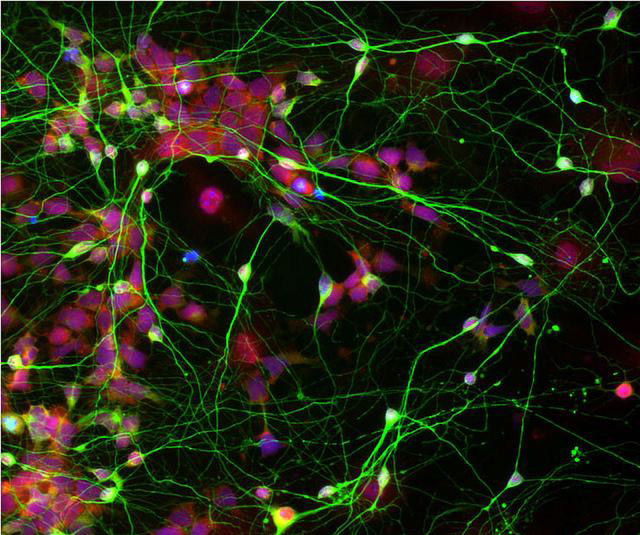
To understand why these disparities occur, stem cell expert Evan Snyder, MD, PhD, director of Sanford Burnham Prebys’ Center for Stem Cells and Regenerative Medicine, is turning to lung organoids—3D structures that replicate human lungs. 

We caught up with Snyder to learn more about these “mini lungs” in a dish—and how the outbreak has impacted his lab.

Why did you create these organoids?
A neonatologist who works in my lab, Sandra Leibel, MD, originally developed these organoids to see if we could help babies who have trouble breathing. Premature babies, and some full-term babies born with genetic conditions, can’t make surfactant—the material that allows the lungs to be flexible and breathe. The severe respiratory distress that is thought to kill some people with severe COVID-19 is actually the adult version of what these babies experience. 

I’m happy to report that, using this model, Sandra was able to find a therapy that might help these infants with the previously untreatable genetic condition. I’m hoping we can learn from this success to help people with severe COVID-19 and “adult respiratory distress syndrome.” 

How are these lung organoids made? 
We start with human induced pluripotent cells (hiPSC) created from skin cells and turn these into lung cells using the same chemical signals our body uses. Then the exciting part comes: We transform these cells from flat, two-dimensional layers into three-dimensional spheres that grow the way a lung would—aka “mini lungs in a dish.” Sandra, along with technician Alicia Winquist and Sanford Burnham Prebys graduate student Rachael McVicar, figured out the “secret sauce” the cells need to complete this process. 

What makes these lung organoids so special?
Lungs are very complicated organs. They have many cell types and structures that all interact with each other: air sac cells that make surfactant, airway cells, immune cells, blood vessel cells; and cells with cilia, tail-like organelles that whip around and clear out debris. Our organoids contain all these different parts, so they can be used to model human disease more closely than single layers of cells. They’re also easier to scrutinize, manipulate and test treatments in animal models. These organoids let us ask and answer all sorts of interesting questions.

What do you hope to learn about COVID-19 using this model? 
We want to learn the basics about the virus that causes COVID-19, called SARS-CoV-2. For example, how does the virus infect human lung cells? What does the virus do after it enters the lung cell? How does the virus move from one lung cell to another? The answers to these questions will allow us to find effective treatments for COVID-19. 

In addition, we can use this model to determine why some people fare worse than others. We can compare organoids created from men and women; younger or older people; people exposed to various environmental toxins from smoking or vaping; people with diabetes, heart or kidney disease; and even people of different racial backgrounds or individuals who have genetic variations that affect their ability to fight infection. 

If we figure out why the virus affects some people differently, we can potentially create tailored treatments. For example, we can ask whether any disparities we observe can be compensated for simply by increasing a dose of a drug or by adding another drug. With our model, we may be able to find these answers relatively quickly so that as many people as possible can benefit from any breakthroughs.

Will you partner with additional Sanford Burnham Prebys scientists on this research? 
Yes, we are in a fortunate position to collaborate with Sumit Chanda, PhD, who just completed a heroic (and I do mean heroic) effort to screen 12,000 known drugs for COVID-19. He pinpointed 30 promising drugs, and now we will help him study exactly how these drugs are working in a system that is clinically relevant. For example, in what stage of the virus’s life are they disrupting? Which part of the cell’s function is being rescued? Using lung organoids, we can rank the effectiveness of each drug—and help Sumit winnow the list to the most promising drugs for close-to-immediate use.

The best approach to treat COVID-19 will most likely be with a drug cocktail, similar to how we treat other RNA viruses. For this reason, it’s important to know how each drug is working, because we want to attack the virus at multiple points in its life cycle and block toxic downstream effects. Our model will help us map this out and advance the most promising drug combinations. 

How has the outbreak affected your lab work? 
I have time-sensitive, patient-relevant work ongoing, so I am still coming into the lab. In addition to our COVID-19 work, we are preparing to launch a clinical trial using stem cells to help newborns who are at risk for cerebral palsy

However, things are very different now. When I do go in, there are only one or two people instead of dozens, and we work in shifts. We limit our time to a few hours and only come in a few days a week. What I really miss is the meeting of the minds. Some of the most inspirational science comes from sitting around in a group and sharing ideas. Or having someone walk over and look through your microscope at the primary data. We do our best with Zoom. But the in-person human interactions are the part of science I really miss. 
 

Institute News

Breakthrough in understanding how stem cells become specialized

AuthorJessica Moore
Date

August 4, 2016

Scientists at Sanford Burnham Prebys Medical Discovery Institute (SBP) have made a major advance in understanding how the cells of an organism, which all contain the same genetic information, come to be so diverse. A new study published in Molecular Cell shows that a protein called OCT4 narrows down the range of cell types that stem cells can become. The findings could impact efforts to produce specific types of cells for future therapies to treat a broad range of diseases, as well as aid the understanding of which cells are affected by drugs that influence cell specialization.

“We found that the stem cell-specific protein OCT4 primes certain genes that, when activated, cause the cell to differentiate, or become more specialized,” said Laszlo Nagy, MD, PhD, professor and director of the Genomic Control of Metabolism Program at SBP’s Lake Nona campus and senior author of the study. “This priming customizes stem cells’ responses to signals that induce differentiation and makes the underlying genetic process more efficient.”

Differentiation matters

As an organism—such as a human—develops from its simplest, earliest form into maturity, its cells transition from a highly flexible state—stem cells—to more specialized types that make up its tissues. Many labs are trying to recapitulate this process to generate specific types of cells that could be transplanted into patients to treat disease. For example, pancreatic beta cells could treat diabetes, and neurons that produce dopamine could treat Parkinson’s.

What OCT4 does

OCT4 is a transcription factor—a protein that regulates gene activity—that maintains stem cells’ ability to give rise to any tissue in the body. OCT4 works by sitting on DNA and recruiting factors that either help initiate or repress the reading of specific genes.

The new study shows that, at certain genes, OCT4 also collaborates with transcription factors that are activated by external signals, such as the retinoic acid (vitamin A) receptor (RAR) and beta-catenin, to turn on their respective genes. Vitamin A converts stem cells to neuronal precursors, and activation of beta-catenin by Wnt can either support pluripotency or promote non-neural differentiation, depending on what other signals are present. Recruitment of these factors ‘primes’ a subset of the genes that the signal-responsive factors can activate.

The big picture

“Our findings suggest a general principle for how the same differentiation signal induces distinct transitions in various types of cells,” added Nagy. “Whereas in stem cells, OCT4 recruits the RAR to neuronal genes, in bone marrow cells, another transcription factor would recruit RAR to genes for the granulocyte program. Which factors determine the effects of differentiation signals in bone marrow cells—and other cell types—remains to be determined.”

Next steps

“In a sense, we’ve found the code for stem cells that links the input—signals like vitamin A and Wnt—to the output—cell type,” said Nagy. “Now we plan to explore whether other transcription factors behave similarly to OCT4—that is, to find the code in more mature cell types.

“If other factors also have this dual function—both maintaining the current state and priming certain genes to respond to external signals—that would answer a key question in developmental biology and advance the field of stem cell research.”

The paper is available online here.

Institute News

What can salamanders teach us about regeneration?

AuthorJessica Moore
Date

July 11, 2016

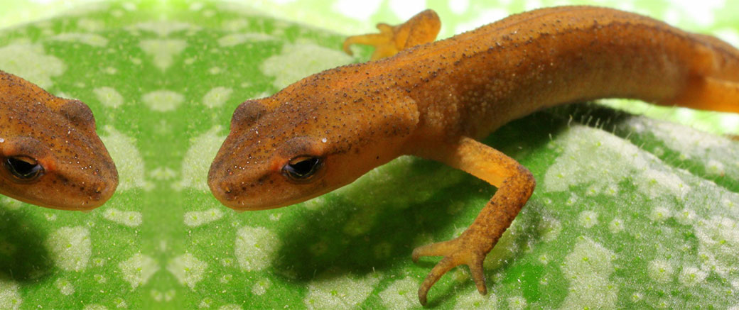
Salamanders can regenerate whole limbs, even as adults. While this amphibian superpower might seem irrelevant to human health, investigating the underlying biology may impart vital lessons about how the healing process can be redirected from scarring to replacing lost tissue. For this reason, Alessandra Dall’Agnese, a graduate student in the laboratory of Pier Lorenzo Puri, MD, PhD, professor in the Development, Aging, and Regeneration Program, recently wrote a review, published in BioEssays, comparing healing in salamanders to that in mammals.

“If we can figure out the means by which injured salamander limbs turn on developmental programs, we may be able to use that knowledge to create treatments that help the human body heal itself,” said Puri.

The review describes the key difference between how salamanders and humans heal following a major injury to a limb. Salamanders heal the wound, then form a regenerative center of proliferative, stem-like cells. In contrast, a regenerative center only forms in humans if the injury affects the tip of a finger or toe distal to the nail bed—otherwise, wound healing is followed by scarring.

“The important thing we’ve learned from salamanders is that there’s not an inherent limit to how much of a limb can be regrown,” explained Dall’Agnese. “The fact that mammals can only regrow the tips of digits instead suggests that there may be some property of the nail bed that fosters regeneration.

“From research in salamanders and mice, we know some of the factors that have to be turned on or off to enable regeneration. But we need a more detailed picture before we can start to develop therapies.”

“The possibility that we could discover salamanders’ secret to regeneration is a good example of why we should study a wide range of organisms,” Puri commented. “If we only studied animals closely related to us, we wouldn’t learn how to help our bodies do things they can’t normally do.”

The review is available online here.

Institute News

SBP supports opening of stem cell exhibit at the Reuben H. Fleet Science Center

Authorjmoore
Date

January 29, 2016

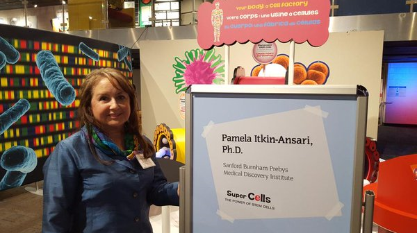
Pamela Itkin-Ansari, PhD, adjunct assistant professor in the Development, Aging, and Regeneration Program at SBP, participated in the grand opening event for the Super Cells exhibit at the Fleet on Jan. 28. She served as an expert on the current understanding of stem cells, answering questions and explaining what stem cell researchers do. Continue reading “SBP supports opening of stem cell exhibit at the Reuben H. Fleet Science Center”

Institute News

New Department of Defense grant funds efforts to treat ALS with stem cells

Authorjmoore
Date

January 28, 2016

Evan Snyder, MD, PhD, director of the Center for Stem Cells and Regenerative Medicine and a professor in SBP’s Human Genetics Program, was awarded a grant to develop a stem cell treatment for amyotrophic lateral sclerosis (ALS). ALS, also known as Lou Gehrig’s disease, involves degeneration and death of motor neurons (which control voluntary muscles), causing difficulty speaking, swallowing, and eventually breathing. No available treatments can slow the progression of ALS, which affects approximately 20,000 people in the U.S.

As new therapies are urgently needed, the Department of Defense announced a funding program for new therapeutic ideas in ALS in 2015. Snyder received one of eight grants awarded in this competitive program.

This grant will support research on human neural stem cells (hNSCs) as an approach to support the survival and function of existing motor neurons. The Snyder lab is modifying hNSCs so that they can be administered via the bloodstream and home to the spinal cord. This strategy allows the stem cells to become distributed throughout the spinal cord, overcoming a previous limitation. Snyder’s team has already shown that transplanted NSCs improved motor performance, respiratory function, and symptom-free survival in a mouse model of ALS.

hNSCs are already in phase I clinical trials for ALS, which have shown that this therapy is safe. These trials were possible in part because of the Snyder lab’s pioneering work in preclinical models. The current research promises to lead to an improved version of this therapy.

Institute News

Stem cell model reveals molecular cues critical to neurovascular unit formation

Authorsgammon
Date

June 1, 2015

Crucial bodily functions we depend on but don’t consciously think about — things like heart rate, blood flow, breathing, and digestion — are regulated by the neurovascular unit. The neurovascular unit is made up of blood vessels and smooth muscles under the control of autonomic neurons. Yet how the nervous and vascular systems come together during development to coordinate these functions is not well understood. Using human embryonic stem cells, researchers at Sanford-Burnham, University of California, San Diego School of Medicine, and Moores Cancer Center created a model that allows them to track cellular behavior during the earliest stages of human development in real-time. The model reveals, for the first time, how autonomic neurons and blood vessels come together to form the neurovascular unit. The study was published May 21 by Stem Cell Reports. Continue reading “Stem cell model reveals molecular cues critical to neurovascular unit formation”

Institute News

11th annual Christopher Reeve “Hot Topics” in stem cell biology

Authorsgammon
Date

November 11, 2014

On November 17, 2014, an evening of data “blitzes” on stem cell science will be presented at the 11th Annual Christopher Reeve Satellite Symposium at the Society for Neuroscience (SFN). The Symposium is a three-hour event with presentations by thought leaders delivered in a rapid, enjoyable, no-nonsense fashion with the latest controversies and insights in stem cell biology—with emphasis on fundamental biology. Continue reading “11th annual Christopher Reeve “Hot Topics” in stem cell biology”