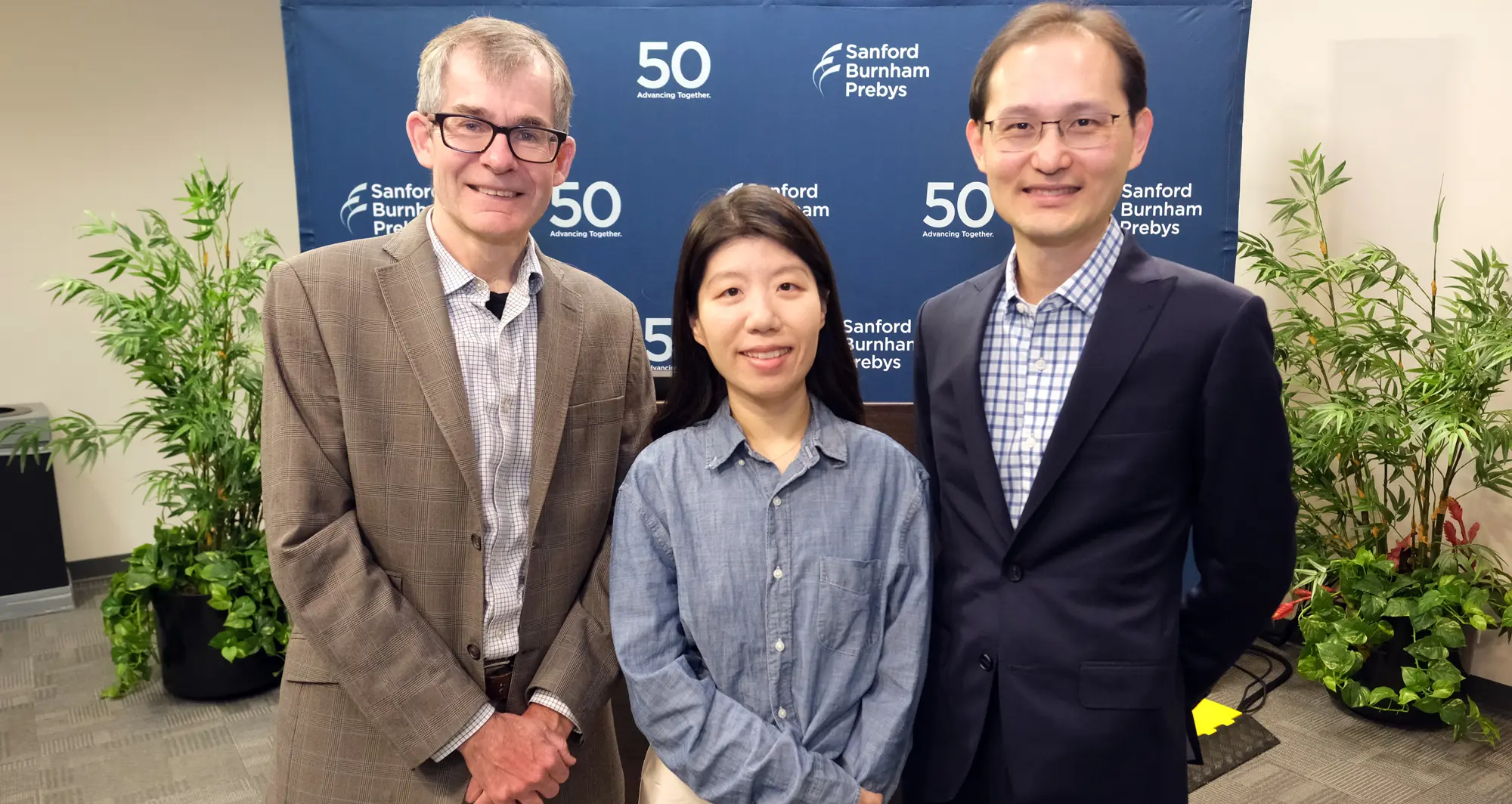
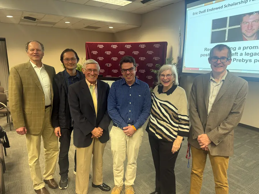
Peter Adams, PhD, with scholarship recipient Shanshan Yin, PhD, and Kevin Yip, PhD.
Yin, a postdoctoral associate at Sanford Burnham Prebys, received the honor in recognition of her achievements in research on cancer and aging
Shanshan Yin, PhD, was named the 2025 recipient of The Eric Dudl Endowed Scholarship at Sanford Burnham Prebys Medical Discovery Institute.
The scholarship fund was established at the institute to remember Eric Dudl, a postdoctoral researcher whose life was tragically cut short by cancer at the age of 33. Since 2007, 18 postdoctoral scientists have received support for their research from the endowed scholarship fund.
Yin is a postdoctoral associate in the lab of Peter Adams, PhD, director of the Cancer Genome and Epigenetics Program at Sanford Burnham Prebys. She wants to understand why the incidence of cancer increases with age. Yin studies changes in gene expression and immune system activity in breast cancer tumors as mice age.
Her research has shown that part of the reason that breast cancer is more common with age is because of an impaired immune system. Immune dysfunction due to aging allows the tumors to grow more frequently and more rapidly. Additional research on these findings may guide future preventive treatments.
Yin has garnered recognition throughout her scientific career, including the 2022 Lenka Finci and Erna Viterbi Fishman Fund Award from Sanford Burnham Prebys.
“I’m grateful for the Dudl family’s kindness and generosity,” said Yin. “I am moved by Eric’s determination, bravery and love for life and for science, and I would like to carry his example with me going forward and do my best to honor his legacy.”
“It’s been a real pleasure working with Shanshan over the years,” said Adams. “She truly does embody Eric Dudl’s commitment to and passion for science which is expressed so well by this inspirational award.”
The Eric Dudl Endowed Scholarship at Sanford Burnham Prebys was established at the institute to remember Eric, a postdoctoral researcher whose life was tragicallycut shortby cancerat the age of 33.
For more information on setting up a scholarship or to learn more about our philanthropy program, please contact giving@sbpdiscovery.org.
Institute News
Women in Science Lecture series showcases public health and nutrition policy leader
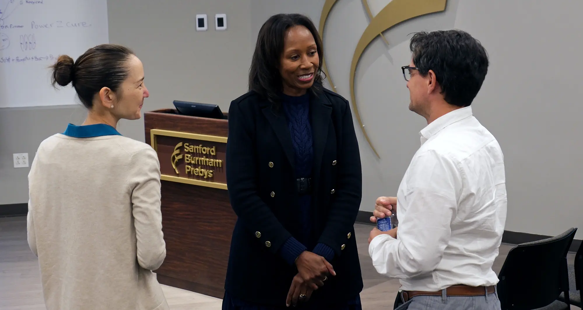
From left to right: physician-scientist Angela Liou, MD; public health and nutrition policy expert Cheryl A.M. Anderson, PhD, MPH, MS; and researcher Lukas Chavez, PhD, MS.
The series highlights the groundbreaking work and unique perspectives of women leaders in the biomedical sciences
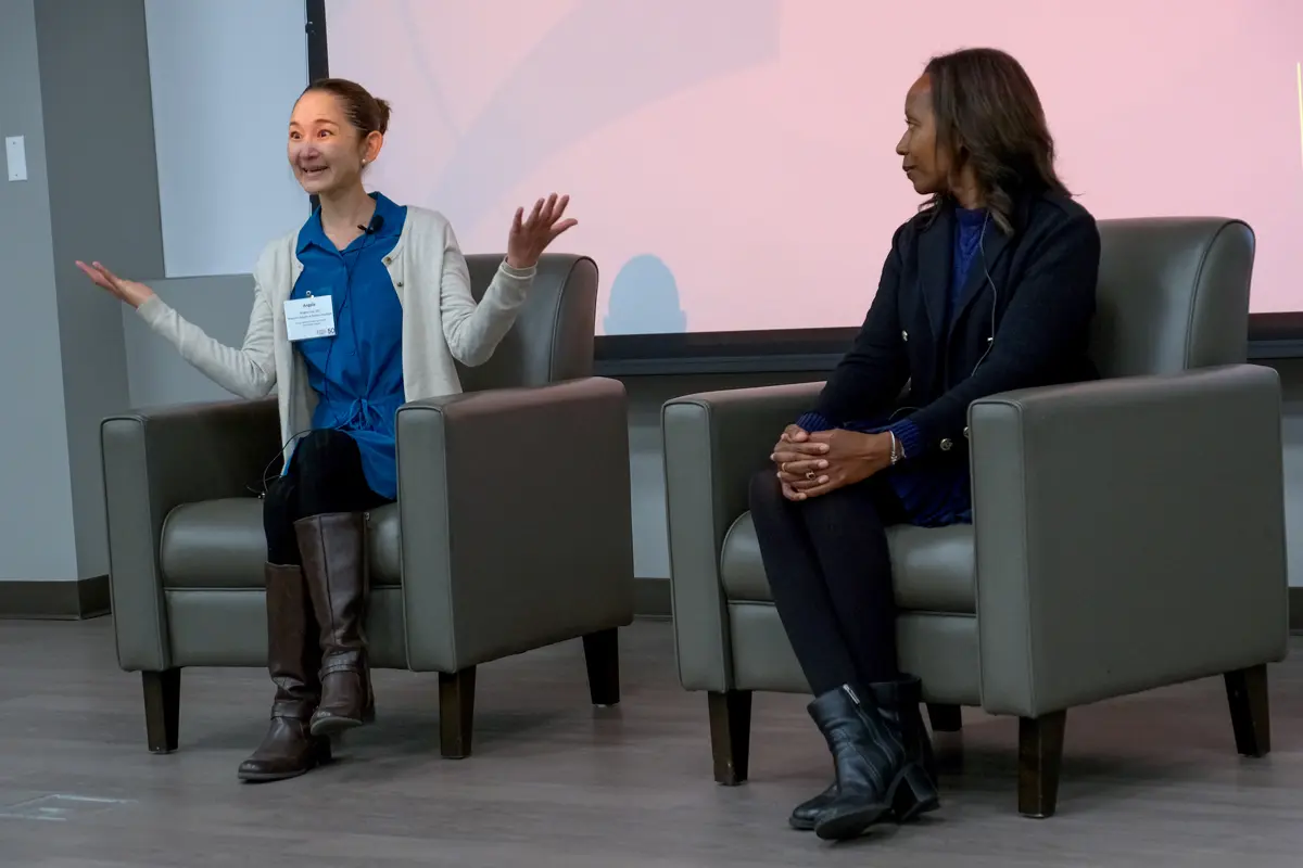
On February 11, 2026, Sanford Burnham Prebys Medical Discovery Institute hosted the second event in the Women in Science Lecture Series. The occasion opened with a presentation by Cheryl A.M. Anderson, PhD, MPH, MS, professor and dean of the Herbert Wertheim School of Public Health and Human Longevity Science at the University of California San Diego and director of the UCSD Center of Excellence in Health Promotion and Equity.
Anderson introduced attendees to some of the pivotal findings of her mentors studying the effects of nutrition on public health, including the landmark dietary approaches to stop hypertension (DASH) clinical trial. Because of the challenges in achieving significant heart disease prevention benefits outside of the controlled environments used in studies such as the DASH trial, Anderson was determined to explore other approaches.
“I put together this concept that instead of asking the individual to figure it all out from our dietary recommendations, maybe we could figure out how to have a healthy, sustainable food system,” said Anderson.
“I see a sustainable food system as one that maintains our ability to get lots and lots of nutrition and where you meet the current population’s needs without compromising what future generations might also need.”
In addition to discussing her scientific journey, Anderson provided insight into her experience serving with other experts to provide input into two different iterations of the federal government’s Dietary Guidelines for Americans. These guidelines from the Department of Health and Human Services and Department of Agriculture set the standards for food in federally funded programs such as public school and day care lunches as well as the Women, Infants and Children (WIC) special supplemental nutrition program. Anderson shared her experience working collaboratively to provide science-based counsel in an ecosystem that also contains political considerations such as the interests of industries involved in agriculture and food production.
Anderson (at right) opened the event discussing her career journey focused on how to develop a healthy, sustainable food system. The event also featured a fireside chat and audience question-and-answer session with Anderson and Liou.
Lukas Chavez, PhD, MS, associate professor in the Cancer Genome and Epigenetics Program at Sanford Burnham Prebys and scientific director of the Pediatric Neuro-Oncology Molecular Tumor Board at Rady Children’s Institute for Genomic Medicine, then moderated a fireside chat and audience question-and-answer session with Anderson and Angela Liou, MD, physician-scientist and pediatric oncologist with a dual appointment at Rady Children’s Health and the Cancer Genome and Epigenetics Program at Sanford Burnham Prebys. Topics included: how new scientific insights are translated to reduce population-level health risks or guide care for children facing serious illnesses; how new technologies change the way you conduct research and deliver patient care; what can be done to ensure that scientific discoveries can be equitably accessed and lead to better outcomes for all; and what do future clinicians and scientists need in terms of skills, mindset and institutional support to succeed as public health researchers and physician-scientists.
The Women in Science Lecture Series, featuring quarterly events that are free and open to the public, is part of broader efforts at Sanford Burnham Prebys to foster an environment that nurtures the success of individuals from all backgrounds. The series is hosted by the Office of Workforce Engagement & Belonging and highlights the groundbreaking work and unique perspectives of women leaders in the biomedical sciences, while fostering mentorship and collaboration across the Torrey Pines Mesa.
Lukas Chavez, PhD, Associate Professor
Cancer Genome and Epigenetics Program.
Credit: Sanford Burnham Prebys
Lukas Chavez, PhD, associate professor in the Cancer Genome and Epigenetics Program at Sanford Burnham Prebys, has been named a standing member of the Cancer Genetics Study Section at the National Institutes of Health.
Study sections are groups of invited experts from across the country who are tasked with assessing the scientific merits of grant applications. Their reviews are a major influence in the NIH’s decisions about which proposed research projects to fund.
Jayanta Bhattacharya, MD, PhD, director of the NIH, invited Chavez to join the cancer genetics study section because of his “demonstrated competence and achievement in his scientific discipline as evidenced by the quality of his research accomplishments, publications in scientific journals and other significant scientific activities, achievements, and honors.”
Chavez studies pediatric brain cancer and specifically, the role of extrachromosomal DNA as a driver of aggressive tumors. His study section term begins immediately and runs through June 30, 2029.
Institute News
Ani Deshpande promoted to professor in the Cancer Genome and Epigenetics Program at Sanford Burnham Prebys
The newly promoted scientist will continue studying how blood cancers sabotage stem cells’ special features to grow and spread
As of July 1, 2025, Ani Deshpande, PhD, was promoted to professor in the Cancer Genome and Epigenetics Program at Sanford Burnham Prebys.
The Deshpande lab studies developmental processes in stem cells that get hijacked by cancer, focusing specifically on acute myeloid leukemia (AML), one of the most common types of blood cancer. Several attributes of normal stem cells, including the ability to self-renew, are known to be co-opted or reactivated by cancer cells.
In addition, Deshpande collaborates within and beyond the institute on several large categories of AML research, including studying the genetics of AML, studying how the disease works in animal models and working to develop drugs that can target specific mutations associated with the disease, which are numerous.
“AML has many different subtypes, so it’s been difficult for researchers to make major advances to treat all cases of AML,” said Deshpande. “Most patients with AML are given the same treatments that have been used since the 1970s, which is why we want to look at AML from as many angles as possible.”
Deshpande joined the institute in 2015 and was promoted to associate professor in 2022. Prior to arriving at Sanford Burnham Prebys, he held positions at Memorial Sloan Kettering Cancer Center and Harvard Medical School. Recently, Deshpande and colleague Pamela Itkin-Ansari, PhD, launched The Discovery Dialogues Podcast, which explores groundbreaking discoveries in science and medicine.
“I’m deeply grateful for the incredible support of my trainees, mentors and colleagues,” said Deshpande. “And for all who made this scientific journey so meaningful and worthwhile.”
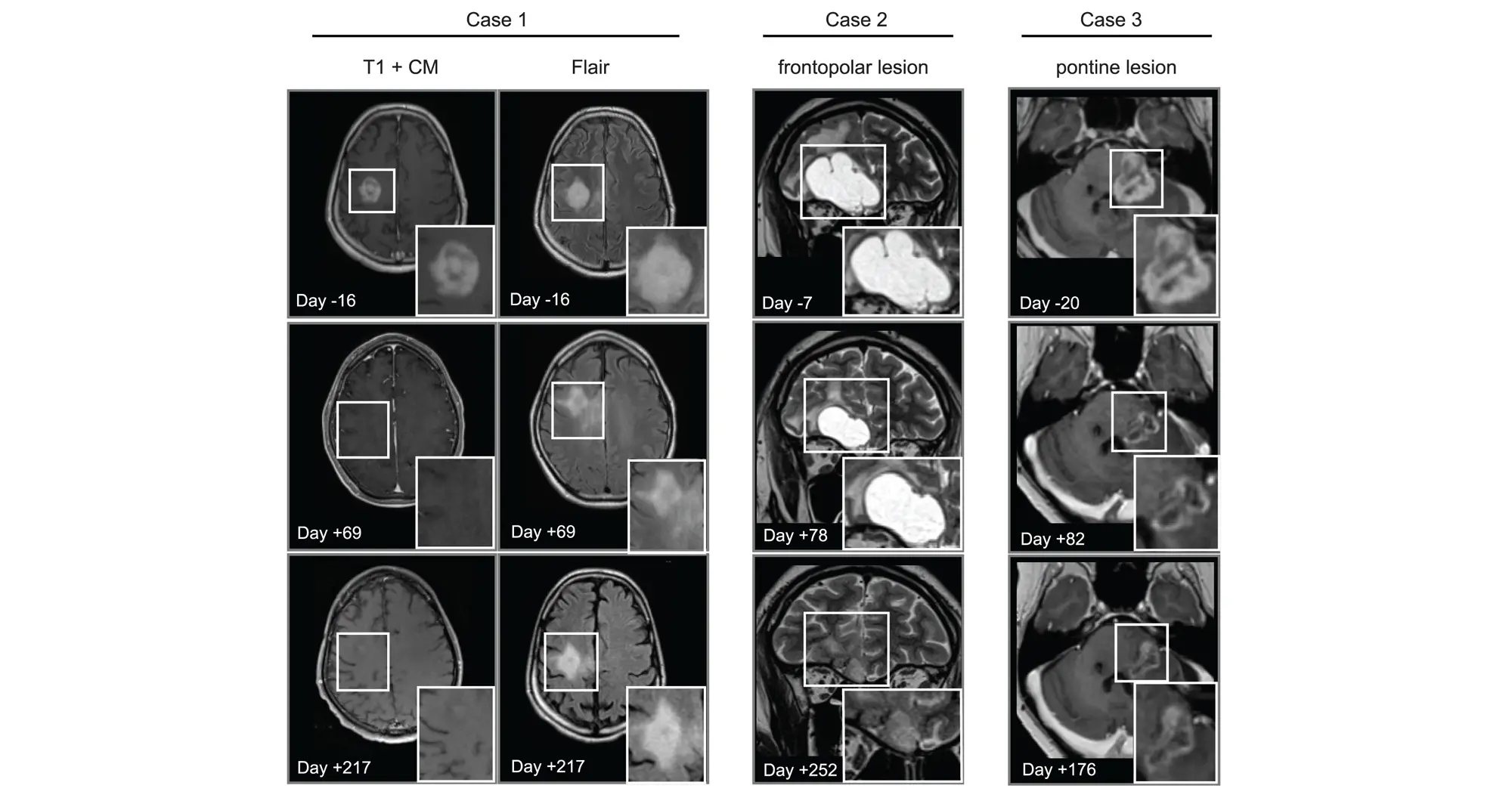
These representative MRIs of responding patients show tumors shrinking throughout the course of avapritinib therapy.
Scientists show that an established cancer drug travels to and shrinks some brain tumors, which may lead to new therapies for a disease with few treatments
Brain tumors are the leading cause of cancer-related death in childhood. The deadliest of these tumors are known as high-grade gliomas, with the grade referring to how quickly certain tumors grow and spread throughout the central nervous system.
Treatment options for high-grade gliomas are limited. Surgical removal is typically the first option depending on the tumor size and location. Radiation often follows to kill any remaining cancer cells to prevent another tumor from forming.
“Drug options to pair with surgery and/or radiation are few and far between,” saidLukas Chavez, PhD, associate professor in theCancer Genome and Epigenetics Programat Sanford Burnham Prebys. “A big reason for this is the blood-brain barrier being as formidable a boundary as the mythological River Styx.”
The blood-brain barrier can, at times, mean the difference between life and death. It protects the brain and spinal cord from potential toxins and pathogens circulating in the bloodstream. However, in its vigilance, it also blocks beneficial drugs from reaching the brain. This presents a major challenge, since most medications are designed to travel through the bloodstream after being ingested or injected.
Scientists from an international team including Sanford Burnham Prebys, the University of Michigan, Dana Farber Cancer Institute, the Medical University of Vienna and many other institutions published findings March 13, 2025, in Cancer Cell demonstrating that the drug avapritinib could treat certain brain tumor cells. And, like the Styx’s ferryman Charon, the medicine is one of the rare few that can cross the blood-brain barrier known to prevent the passage of more than 98% of small molecule drugs.
The researchers selected avapritinib—which is approved by the Food and Drug Administration for treating gastrointestinal and other cancers—after finding it was the strongest commercially available drug for inhibiting the gene Platelet-derived growth factor receptor alpha (PDGFRA), which is found to be mutated in 15% of high-grade gliomas.
Lukas Chavez, PhD, is an associate professor in the Cancer Genome and Epigenetics Program at Sanford Burnham Prebys.
In addition to showing that avapritinib inhibited PDGFRA in cancer cells and mouse brain tumors, the research team tested its effects on eight human pediatric and young adult high-grade glioma patients through a compassionate-use program. The treatment was found to be safe and investigators observed that the drug caused tumors to shrink in three patients.
“More research is needed to better understand how to best repurpose this drug for high-grade gliomas,” said Chavez. “We’ll learn a lot from the ongoing Rover study, a phase 1/2 multicenter trial of avapritinib based on these findings that will include more participants.”
The authors of the new study also highlighted the need to study combining multiple targeted therapies to overcome acquired resistance to any single treatment.
Mariella G. Filbin, MD, PhD, assistant professor of Pediatrics at Harvard Medical School and research co-director of the Pediatric Neuro-Oncology Program at the Dana-Farber/Boston Children’s Cancer and Blood Disorders Center, is the lead contact on the study.
Carl Koschmann,MD, ChadTough Defeat DIPG Research Professor and associate professor of Pediatric Neuro-Oncology at the University of Michigan Medical School, and Johannes Gojo, MD, PhD, head of Pediatric Precision Oncology CNS and ITCC-Lab/Clinical Trials Unit at the Medical University of Vienna, are corresponding authors along with Filbin.
Lisa Mayr, Sina Neyazi, Kallen Schwark and Maria Trissal share first authorship of the study.
Additional authors include:
Owen Chapman, Sunita Sridhar, Rishaan Kenkre, Aditi Dutta, Shanqing Wang, and Jessica Wang from Sanford Burnham Prebys
Jenna Labelle, Sebastian K. Eder, Joana G. Marques, Carlos A.O. de Biagi-Junior, Costanza Lo Cascio, Olivia Hack, Andrezza Nascimento, Cuong M. Nguyen, Sophia Castellani, Jacob S. Rozowsky, Andrew Groves, Eshini Panditharatna, Gustavo Alencastro Veiga Cruzeiro, Rebecca D. Haase, Kuscha Tabatabai, Alicia Baumgartner, Frank Dubois, Pratiti Bandopadhayay and Keith Ligon from the Dana-Farber/Boston Children’s Cancer and Blood Disorder Center and Harvard Medical School
Liesa Weiler-Wichtl, Sibylle Madlener, Katharina Bruckner, Daniel Senfter, Anna Lammerer, Natalia Stepien, Daniela Lotsch-Gojo, Walter Berger, Ulrike Leiss, Verena Rosenmayr, Christian Dorfer, Karin Dieckmann, Andreas Peyrl, Amedeo A. Azizi, Leonhard Mullauer, Christine Haberler and Julia Furtner from the Medical University of Vienna
Jack Wadden, Tiffany Adam, Seongbae Kong, Madeline Miclea, Tirth Patel, Chandan Kumar-Sinha, Arul Chinnaiyan and Rajen Mody from the University of Michigan Medical School
Alexander Beck from Ludwig Maximilians University Munich
Jeffrey Supko and Hiroaki Wakimoto from Massachusetts General Hospital
Armin S. Guntner from Johannes Kepler University
Hana Palova, Jakub Neradil, Ondrej Slaby, Petra Pokorna and Jaroslav Sterba from Masaryk University
Louise M. Clark, Amy Cameron and Quang-De Nguyen from the Dana-Farber Cancer Institute
Noah F. Greenwald and Rameen Beroukhim from the Broad Institute of MIT and Harvard
Christof Kramm from University Medical Center Gottingen
Annika Bronsema from University Medical Center Hamburg-Eppendorf
Simon Bailey from Great North Children’s Hospital and Newcastle University
Ana Guerreiro Stucklin from University Children’s Hospital Zurich
Sabine Mueller from the University of California San Francisco
Mary Skrypek from Children’s Minnesota
Nina Martinez from Jefferson University
Daniel C. Bowers from the University of Texas Southwestern Medical Center
David T.W. Jones, Natalie Jager from Hopp Children’s Cancer Center Heidelberg
Chris Jones from the Institute of Cancer Research
Institute News
Theo Tzaridis named 2024 recipient of Eric Dudl Endowed Scholarship
The scholarship fund was established at the institute to remember Eric Dudl, a postdoctoral researcher whose life was tragically cut short by cancer at the age of 33. Since 2007, 17 postdoctoral scientists have received support for their research from the endowed scholarship fund.
Tzaridis is a postdoctoral fellow in the lab of Peter Adams, PhD, director of the Cancer Genome and Epigenetics Program at Sanford Burnham Prebys. He studies ways to enhance immunotherapy for diffuse intrinsic pontine glioma (DIPG), the deadliest brain tumor in children.
Tzaridis found that targeting a checkpoint molecule called CD155 leads to an enhanced immune response and tumor control. He presented the work at the annual American Association for Cancer Research conference. There he established a collaboration with a company that produces the only available antibody against CD155, enabling Tzaridis to continue his research by testing the antibody’s potential efficacy for treating DIPG in order to pave the way for a clinical trial to improve survival for patients.
David Brenner, MD, Kevin Yip, PhD, and Peter Adams, PhD, with Robert James and Barbara Dudl and scholarship recipient Theo Tzaridis, MD.
The Eric Dudl Endowed Scholarship at Sanford Burnham Prebyswas established at the institute to remember Eric, a postdoctoral researcher whose life was tragically cut short by cancer at the age of 33.
Tzaridis has garnered recognition and extramural funding throughout his career as a physician-scientist, including the 2023 Lenka Finci and Erna Viterbi Fishman Fund Award from Sanford Burnham Prebys and the best oral presentation from the American Association of Immunologists during the 2024 La Jolla Immunology Conference. His career goal is to advance research findings into clinical trials that benefit patients, including trials regarding the effective use of immunotherapy as a treatment for brain cancer.
“I’m truly grateful for the support of The Eric Dudl Endowed Scholarship,” said Tzaridis. “Eric’s inspiring legacy as an immensely dedicated postdoctoral cancer researcher lives on through the important work the scholarship helps fund.”
“Theo is an outstanding physician and a superb scientist,” said Adams. “I have no doubt that he will advance the science of brain cancer while also contributing to meaningful improvements for patients and their families.”
For more information on setting up a scholarship or to learn more about our philanthropy program, please contact giving@sbpdiscovery.org.
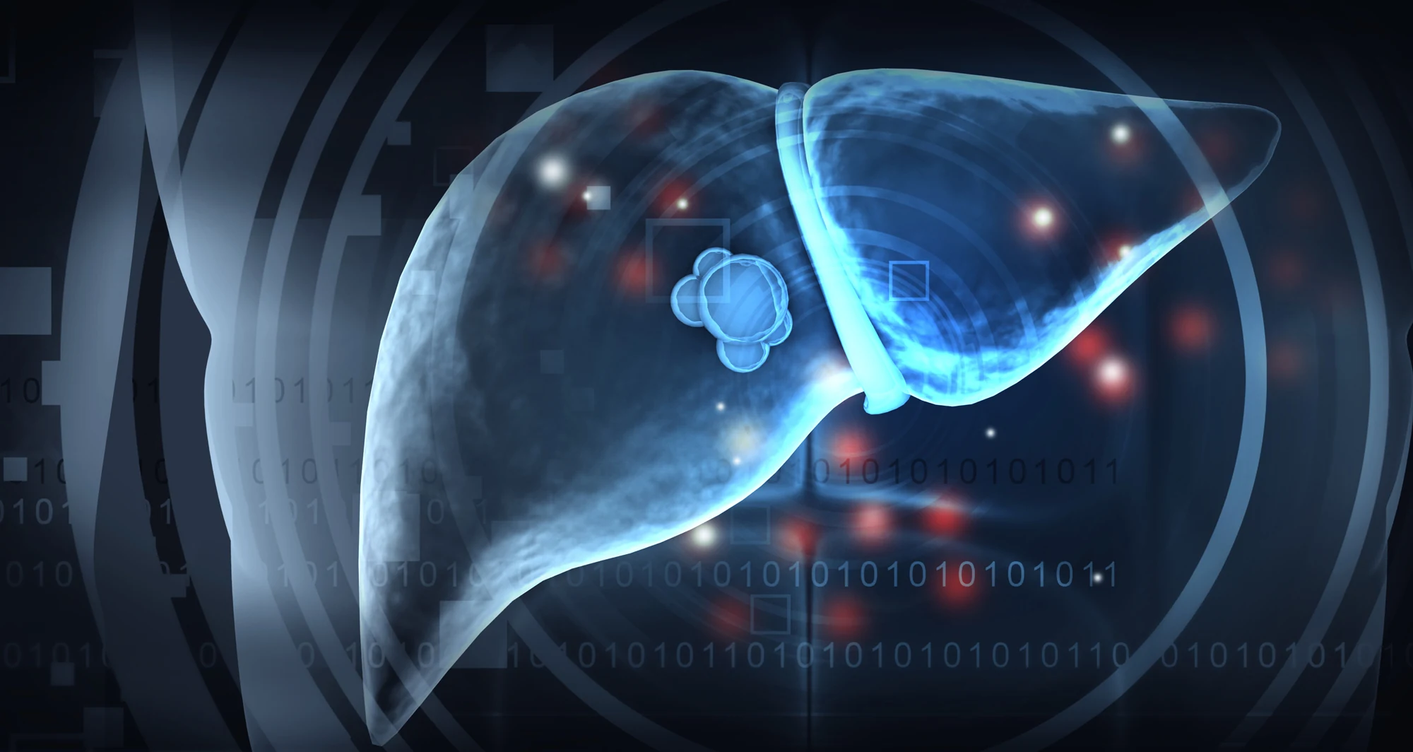
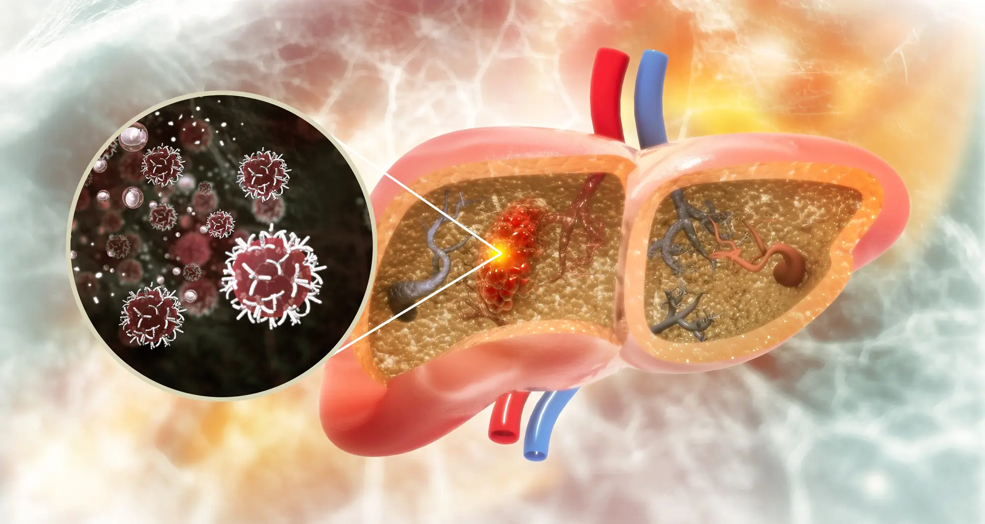
Scientists show how the advanced form of fatty liver disease has monstrous effects on liver cancer risk
Liver cancer has proven to be a tough beast to tame. Experts expected rates of the cancer to decrease following the development of the hepatitis B vaccine in the 1980s, which reduced one of the major risk factors for the disease.
Research in Taiwan showed that its universal infant hepatitis B vaccination program led to young adults experiencing a 35.9% reduction in cases of hepatocellular carcinoma (HCC), the most common liver cancer.
Despite innovation leading to the world’s first cancer-preventing vaccine, incidence of HCC has been on the rise due to a spike in fatty liver disease over recent decades. Lifestyle factors such as high-calorie diets, excessive alcohol consumption and minimal exercise — along with genetic predispositions — can lead to problematic changes in the liver, heart and kidneys.
Specifically in the liver, growing deposits of fat in the tissue can lead over time to an advanced form of fatty liver disease marked by chronic inflammation and the accumulation of thickened scar tissue, a condition known as metabolic-associated steatohepatitis (MASH). MASH significantly increases a patient’s risk of developing HCC.
Debanjan Dhar, PhD, is an associate professor in the Cancer Genome and Epigenetics Program.
In a paper published January 1, 2025, in Nature, scientists at Sanford Burnham Prebys, the University of California San Diego, Curtin University, the University of Pennsylvania and The Liver Cancer Collaborative, demonstrated that MASH damages the DNA of liver cells. The study also linked these changes to the development of liver cancer.
Peter Adams, PhD, is the director of the Cancer Genome and Epigenetics Program.
“DNA damage from MASH causes liver cells to stop dividing and enter a zombie-like state called senescence,” said Debanjan Dhar, PhD, associate professor in the Cancer Genome and Epigenetics Program at Sanford Burnham Prebys and coauthor on the study. “This study’s results demonstrate that some of these cells later exit senescence and are likely to become cancerous due to their accumulation of damage and mutations.”
“In the future, we can apply what we’ve learned to study potential opportunities to prevent or repair DNA damage from MASH to reduce patients’ risk of developing liver cancer,” said Peter Adams, PhD, director of the Cancer Genome and Epigenetics Program at Sanford Burnham Prebys and coauthor on the study.
Michael Karin, PhD, Distinguished Professor in the Department of Pharmacology at the University of California San Diego School of Medicine, is the senior and corresponding author on the study.
Li Gu, PhD, a former postdoctoral fellow in the Karin lab, shares first authorship of the study with visiting scientist Yahui Zhu.
Additional authors include:
Marcos Teneche and Souradipta Ganguly from Sanford Burnham Prebys
Shuvro Nandi, Maiya Lee, Kosuke Watari, Breanna Bareng, Masafumi Ohira, Yuxiao Liu, Sadatsugu Sakane, Mojgan Hosseini, Tatiana Kisseleva, Ludmil Alexandrov, Consuelo Sauceda and David Gonzalez from the University of California San Diego
Rodrigo Carlessi and Janina Tirnitz-Parker from Curtin Universit
The Liver Cancer Collaborative
M. Celeste Simon from the University of Pennsylvania
Institute News
Bile may be key to immunotherapy effectiveness in liver cancer
A 3D illustration of hepatocellular carcinoma, the most common liver cancer.
Understanding the crucial ingredient in bile may unlock the potential of treatments that help patients’ immune systems eliminate cancer
Hepatocellular carcinoma (HCC) is the most common liver cancer and a growing threat to public health across the globe due to the rising rate of fatty liver disease.
Liver cancer is difficult to treat as it often causes few if any symptoms early on, so it tends to be diagnosed at later, more aggressive stages. While immunotherapies that supercharge patients’ immune systems have proven effective in some cancers, this approach has had limited success in patients suffering from HCC or other forms of the disease.
Scientists are investigating the unique qualities of different tissues that may explain why the effectiveness of immunotherapy varies depending on the location of a tumor. The liver is known to have a flexible immune system capable of defending itself when necessary while not overreacting to a constant flood of foreign materials from digesting food, including metabolic byproducts from bacteria residing in the gut microbiome.
Transplant surgeons see the unique properties of the liver’s immune system firsthand when transplanted livers are typically integrated by recipients with only a low dose of immunosuppressive drugs. This ability to maintain immune tolerance, however, may reduce the ability of the liver’s immune system to find and destroy cancer cells, even when that capability is enhanced by immunotherapy.
In a paper published January 9, 2025, in Science, scientists at Sanford Burnham Prebys, the Salk Institute, the University of California San Diego, Columbia University Irving Medical Center, Memorial Sloan Kettering Cancer Center and the Geisel School of Medicine at Dartmouth, found that a critical ingredient in bile hinders the liver’s immune response against cancer.
Bile is a fluid made by the liver that assists in breaking down fats during digestion. This function is made possible by steroidal acids known as bile acids. The scientists found an increased amount of bile acids in tumor samples from patients with HCC. The team also found that genes involved in creating bile acids were being transcribed to make proteins and enzymes at an abnormally high rate in human samples and in mice genetically modified to develop liver cancer.
The authors went on to remove genes related to bile acid construction to demonstrate that mice without these blueprints developed fewer, smaller tumors. In addition, the liver’s T cells — the primary anti-tumor immune cells — were able to dig deeper into tumors and persist for longer without the immunosuppressive effects of certain bile acids.
“These findings underscore a new appreciation for the influence of bile acids on the liver’s immune system,” said Debanjan Dhar, PhD, associate professor in the Cancer Genome and Epigenetics Program at Sanford Burnham Prebys and coauthor on the study. More research is needed to test the potential use of drugs to directly inhibit certain bile acids or bile acid receptors as a therapeutic strategy to reduce liver cancer growth.
Debanjan Dhar, PhD, is an associate professor in the Cancer Genome and Epigenetics Program at Sanford Burnham Prebys.
Peter Adams, PhD, is the director of the Cancer Genome and Epigenetics Program at Sanford Burnham Prebys.
It may also be possible to achieve this effect through dietary changes that alter the microbiome and result in modified bile acid production. Based on their findings, the research team suggests that this could be done by using ursodeoxycholic acid, a bile acid that currently is used to treat an autoimmune condition called primary biliary cholangitis. The acid is found at high levels in bear bile, which has served for thousands of years as a treatment in traditional Chinese medicine.
“Given the safety profile of ursodeoxycholic acid and the limited effectiveness of immunotherapy on liver cancer, this study shows significant potential for testing this bile acid as a combination treatment for patients with HCC,” said Peter Adams, PhD, director of the Cancer Genome and Epigenetics Program at Sanford Burnham Prebys and coauthor on the study.
Susan Kaech, PhD, NOMIS Chair, professor and director of the NOMIS Center for Immunobiology and Microbial Pathogenesis at the Salk Institute is the senior and corresponding author on the study.
Siva Karthik Varanasi, PhD, assistant professor at the UMass Chan Medical School and a former postdoctoral fellow in the Kaech lab at the Salk Institute, is first author on the manuscript.
Additional authors include:
Souradipta Ganguly, Marcos G. Teneche and Aaron Havas, from Sanford Burnham Prebys
Dan Chen, Melissa A. Johnson, Kathryn Lande, Michael A. LaPorta, Filipe Araujo Hoffmann, Thomas H. Mann, Eduardo Casillas, Kailash C. Mangalhara, Varsha Mathew, Ming Sun, Yagmur Farsakoglu, Timothy Chen, Bianca Parisi, Shaunak Deota, H. Kay Chung, Satchidananda Panda, April E. Williams and Gerald S. Shadel, from the Salk Institute
Yingluo Liu, Cayla M. Miller, Jin Lee and Gen-Sheng Feng, from the University of California San Diego
Isaac J. Jensen and Donna L. Farber, from Columbia University Irving Medical Center
Andrea Schietinger from Memorial Sloan Kettering Cancer Center
Mark S. Sundrud from the Geisel School of Medicine at Dartmouth
Wolfram Goessling, MD, PhD, the Robert H. Ebert Associate Professor of Medicine and associate professor of Health Sciences and Technology at Harvard Medical School, authored a Perspective article on the new study in Science called, “Ena-bile-ing liver cancer growth.”
Institute News
A Conversation About Aging and Cancer at Sanford Burnham Prebys
Event recording now available for panel discussion with scientists held on October 9, 2024
David A. Brenner, MD, president and CEO of Sanford Burnham Prebys, welcomed attendees to the launch of a new community engagement program called “A Conversation About” in the institute’s Victor E. LaFave III Memorial Auditorium on October 9, 2024.
The initial panel discussion in the A Conversation About series focused on the connection between aging and cancer and included information about a current breast cancer research collaboration. A recording of the event is available online.
Reena Horowitz, the founder of Group of 12 and Friends at Sanford Burnham Prebys, provided introductory remarks. Brooke Emerling, PhD, director of the Cancer Metabolism and Microenvironment Program, moderated the discussion among three featured panelists:
Peter Adams, PhD, director of the Cancer Genome and Epigenetics Program, Sanford Burnham Prebys
Xiao Tian, PhD, assistant professor in the Degenerative Diseases Program, Sanford Burnham Prebys
Kay Yeung, MD, PhD, associate clinical professor in the Division of Hematology-Oncology, University of California San Diego Health
By bringing together community collaborators and clinicians with Sanford Burnham Prebys researchers, A Conversation About offers a unique perspective on how clinical research and practice can be used to inform fundamental and translational science.
Watch Event Recording
Institute News
Mammalian Genome Engineering Group holds 2024 symposium in San Diego
Participants at the 4th Mammalian Genome Engineering Symposium held at Sanford Burnham Prebys in La Jolla from September 12-15, 2024.
The four-day event included talks from experts from across North America and opportunities to discuss improving experimental methods and approaches to analyzing the resulting data.
Researchers convened at Sanford Burnham Prebys in La Jolla from September 12-15 to hear presentations from their peers and confer about the latest developments in modifying the genomes of mammalian animal models to advance biomedical research.
Anindya Bagchi, PhD, associate professor in the Institute’s Cancer Genome and Epigenetics Program, planned the 4th Mammalian Genome Engineering Symposium, which included 26 presentations from experts across the United States and Canada. Attendees asked many questions throughout, and numerous speakers commented on how valuable the conversation at the meeting was for refining planned experiments and considering new ideas and approaches.
“It was a truly enjoyable and thought-provoking meeting,” said Angela Liou, MD, an instructor in the Cancer Genome and Epigenetics Program at Sanford Burnham Prebys and pediatric oncologist and hematologist at Rady Children’s Hospital-San Diego. “It also was incredibly helpful in informing the next steps of my research project.”
“I’m so grateful for the invitation to attend this symposium,” said Praveen Raju, MD, PhD, the Nathan Gordon Chair in Neuro-Oncology and medical director of the Pediatric Neuro-Oncology Program at Rady Children’s Hospital-San Diego and director of the Pediatric Neuro-Oncology Program at the University of California San Diego School of Medicine.
Anindya Bagchi, PhD, is an associate professor in the Cancer Genome and Epigenetics Program.
“The presenters and attendees were welcoming and collaborative, and I certainly learned a lot.”
The symposium brings together the Mammalian Genome Engineering Group, which was formed by a small group of genome engineering enthusiasts including Bagchi, Nada Jabado, MD, PhD, professor of Pediatrics and Human Genetics at McGill University and a hematologist and oncologist at Montreal Children’s Hospital; David Largaespada, PhD, a professor of Pediatrics, Genetics, Cell Biology and Development at the University of Minnesota Medical School and the associate director for Basic Research in the Masonic Cancer Center; and Michael Taylor, MD, PhD, The Cyvia and Melvyn Wolff Chair of Pediatric Neuro-Oncology at Texas Children’s Cancer and Hematology Center and professor of Pediatrics (Hematology-Oncology) at Baylor College of Medicine.
The group is interested in developing functional models of genomic and epigenetic mutations associated with human diseases—especially cancers—that are difficult to recreate in animal models. The group’s first symposium was coordinated by Taylor in Napa, Calif., in 2014, followed by the 2nd symposium that was organized by Jabado in Montreal in 2015. After a hiatus, the group was revived in 2023 with the 3rd symposium hosted again by Taylor in Houston.
“We believe this symposium will, in the coming years, become a leading forum for discussing cutting-edge genomic and epigenomic approaches to tackle challenging genetic and epigenetic mutations,” said Bagchi. “These approaches are likely to become standard practice in the near future.”
The Sanford Burnham Prebys scientists that presented at the 4th Mammalian Genome Engineering Symposium were:
Liou, “Investigating the deposition of H3.3K27M oncohistone and its effect on retrotransposon reactivation in H3K27M pediatric diffuse midline glioma”
Ani Deshpande, PhD, associate professor in the Cancer Genome and Epigenetics Program and associate director of Diversity, Equity and Inclusion in the NCI-Designated Cancer Center, “Functional genomic approaches to identify selective dependencies in synovial sarcoma”
Peter D. Adams, PhD, the director of the Cancer Genome and Epigenetics Program, “The role of aging in cancer”
Lukas Chavez, PhD, associate professor in the Cancer Genome and Epigenetics Program, “Circular extrachromosomal DNA promotes tumor heterogeneity and enhancer rewiring”
Jerold Chun, MD, PhD, professor in the Degenerative Diseases Program, “Genetic mosaicism and somatic gene recombination in the brain”
Adarsh Rajesh, graduate student, Sanford Burnham Prebys, “CCND1-CDK6 complex inhibits DNA damage repair and promotes inflammation in senescence and the aged liver”
Additional speakers included:
Taylor, “Why does medulloblastoma love to be tetraploid and other nonsense”
Jabado, “Co-opting 3D structures to fuel tumorigenesis”
Tannishtha Reya, PhD, Herbert and Florence Irving Professor of Basic Science Research in Physiology and Cellular Biophysics, Columbia University, “New genetically engineered models to understand cancer heterogeneity and therapy resistance in pancreatic cancer”
Simona Dalin, PhD, postdoctoral fellow, Broad Institute of the Massachusetts Institute of Technology and Harvard University, “Contributions of perfect and imperfect homology to rearrangement formation in human and cancer genomes”
Alison M. Taylor, PhD, assistant professor of Pathology and Cell Biology, Columbia University, “Functional and computational approaches to uncover the consequences of chromosome arm aneuploidy in cancer”
Sean Eagan, PhD, senior scientist in the Cell Biology program at The Hospital for Sick Children, professor of Molecular Genetics, University of Toronto, “An update on Genetic analysis of 16q-syntenic block deletion in the mouse mammary gland – a tumor suppressor arm”
Claudia Kleinman, PhD, associate professor of Human Genetics, McGill University, investigator at the Lady Davis Institute for Medical Research, Jewish General Hospital, “Lineage programs and the 3D genome in pediatric brain tumors”
Branden Moriarity, PhD, associate professor of Pediatrics (Hematology and Oncology), University of Minnesota Medical School, “Next generation engineered immune effector cells for immunotherapy”
Beau Webber, PhD, associate professor of Pediatrics (Hematology and Oncology), University of Minnesota Medical School, “Building cancer in a dish: Sarcoma modeling using human pluripotent stem cells”
Sameer Agnihotri, PhD, associate professor of Neurological Surgery and director of the Brain Tumor Biology and Therapy Lab, University of Pittsburgh Medical Center Children’s Hospital of Pittsburgh, “Identifying genetic vulnerabilities by modeling Chromosome 9p loss”
Largaespada, “Loss of the polycomb repressor complex 2 (PRC2) alters the super-enhancer landscape, genome/epigenome stability, and therapeutic sensitivities of malignant peripheral nerve sheath tumors”
Teresa Davoli, PhD, assistant professor of Biochemistry and Molecular Pharmacology, New York University Langone Health, “Engineering chromosome specific aneuploidy by targeting human centromeres”
Rameen Beroukhim, MD, PhD, associate professor of Medicine, Dana-Farber Cancer Institute and Harvard Medical School, associate member of the Broad Institute of MIT and Harvard, “Detecting rearrangement signatures—naturally”
Quang Trinh, PhD, scientist, Ontario Institute for Cancer Research, “Perspectives and Challenges in PFA Integrative Analysis”
Taylor Gatesman, graduate student, University of Pittsburgh, “Genome Engineering: DREaming of and vCREating new models”
Joseph Skeate, PhD, postdoctoral fellow, University of Minnesota Medical School, “Targeted CAR integration and multiplex base editing in a single-step manufacturing process for enhanced cancer immunotherapies”