Bioblasts
Richard Altmann (1852-1900) was a German prosecutor-turned-anatomy professor who focused on advancing microscopy techniques in his day. Using a new staining technique, he observed filaments in nearly every cell type he viewed. These filaments developed from granules, which Altmann deduced were elementary living units.
He called them “bioblasts.”
His conclusions were not well-received by peers.
These days, it’s believed Altmann was describing mitochondria, a term coined by Carl Benda in 1898 replaced Altmann’s “bioblasts.” And no one disputes the significance of Altmann’s original observations.
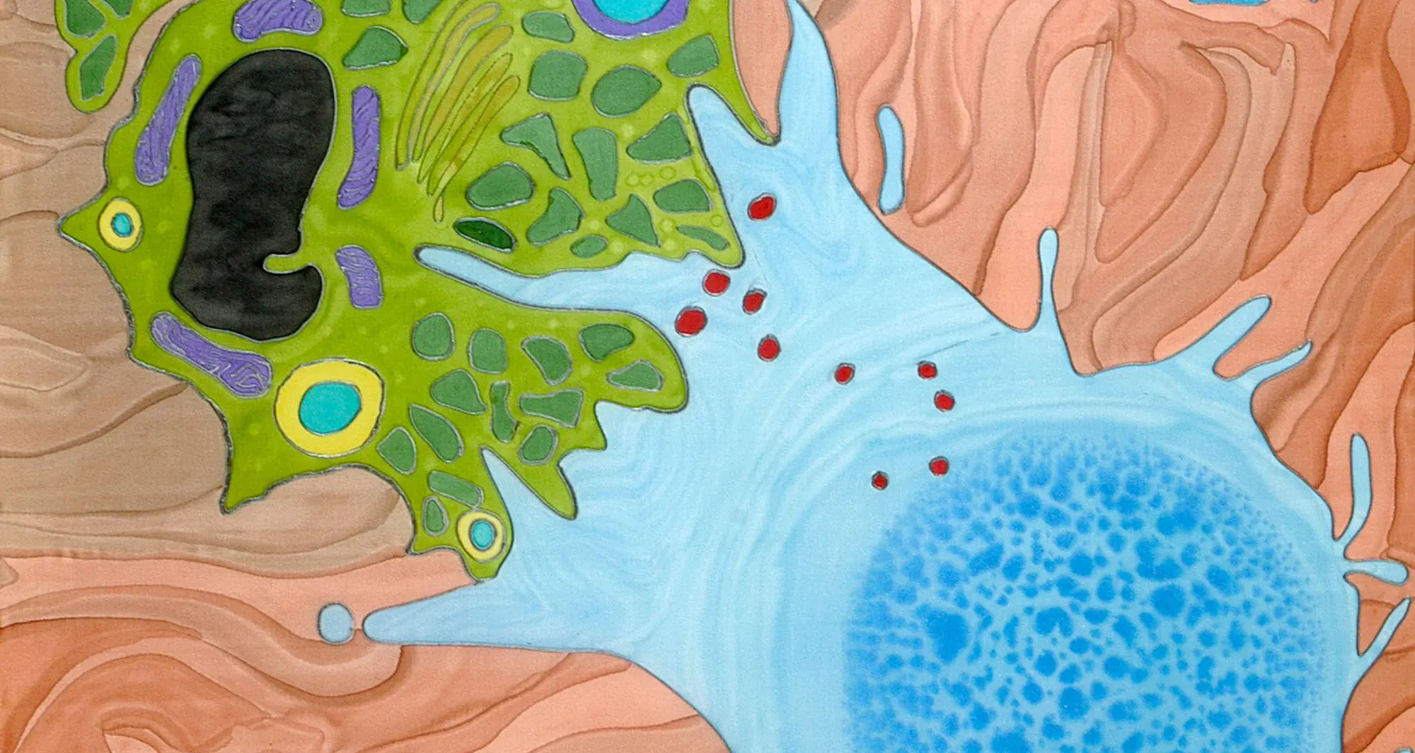
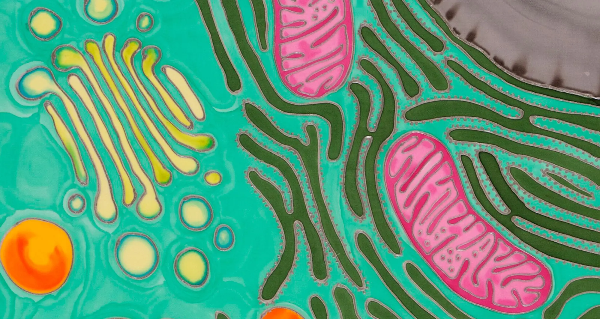
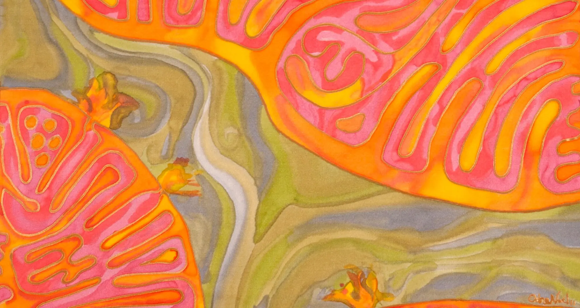
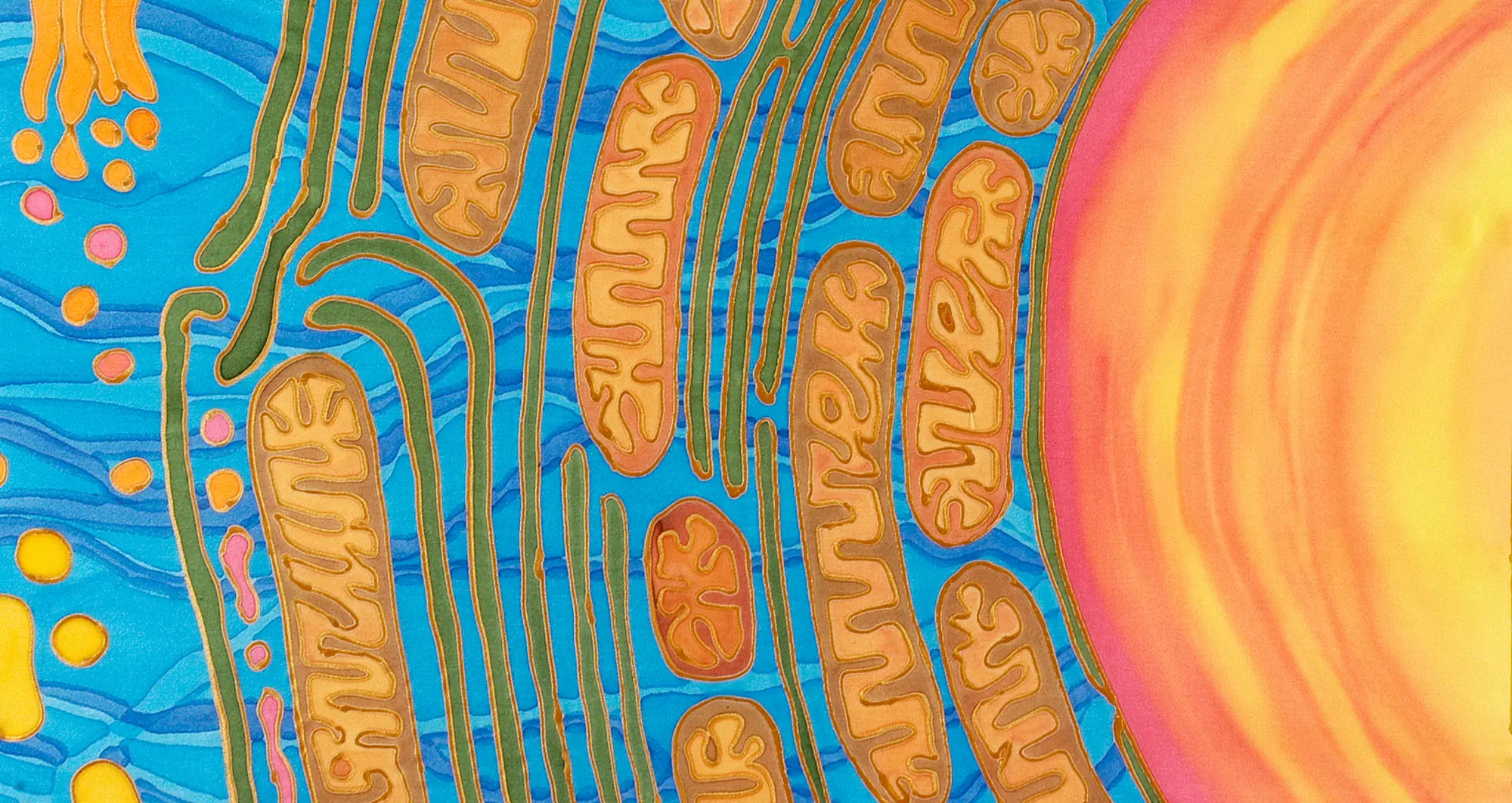
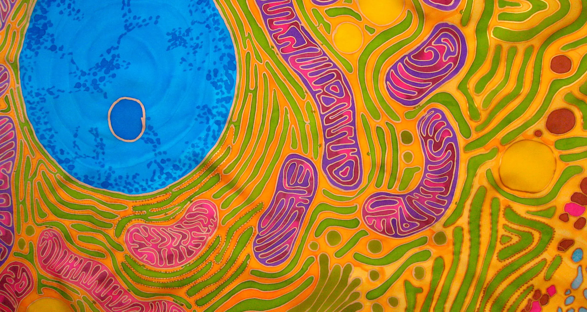
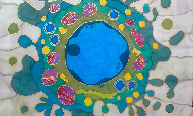
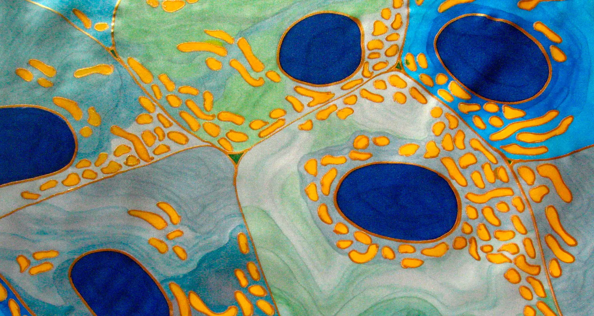
Mitochondria are cellular organelles that covert fuel into energy, the so-called “powerhouses of the cell.” They have many responsibilities and notably, possess their own genes apart from the cell’s nucleus. This fact supports the hypothesis that mitochondria were originally free-living prokaryotic cells lacking a nucleus that permanently fused with eukaryotic cells in the distant past.
Given their fundamental and expansive importance, mitochondria are the focus of much research. At Sanford Burnham Prebys, Peter Adams, PhD, with collaborators elsewhere, are exploring the role they play in cellular aging and immune responses, and particularly how they may affect the connection between aging and liver cancer.
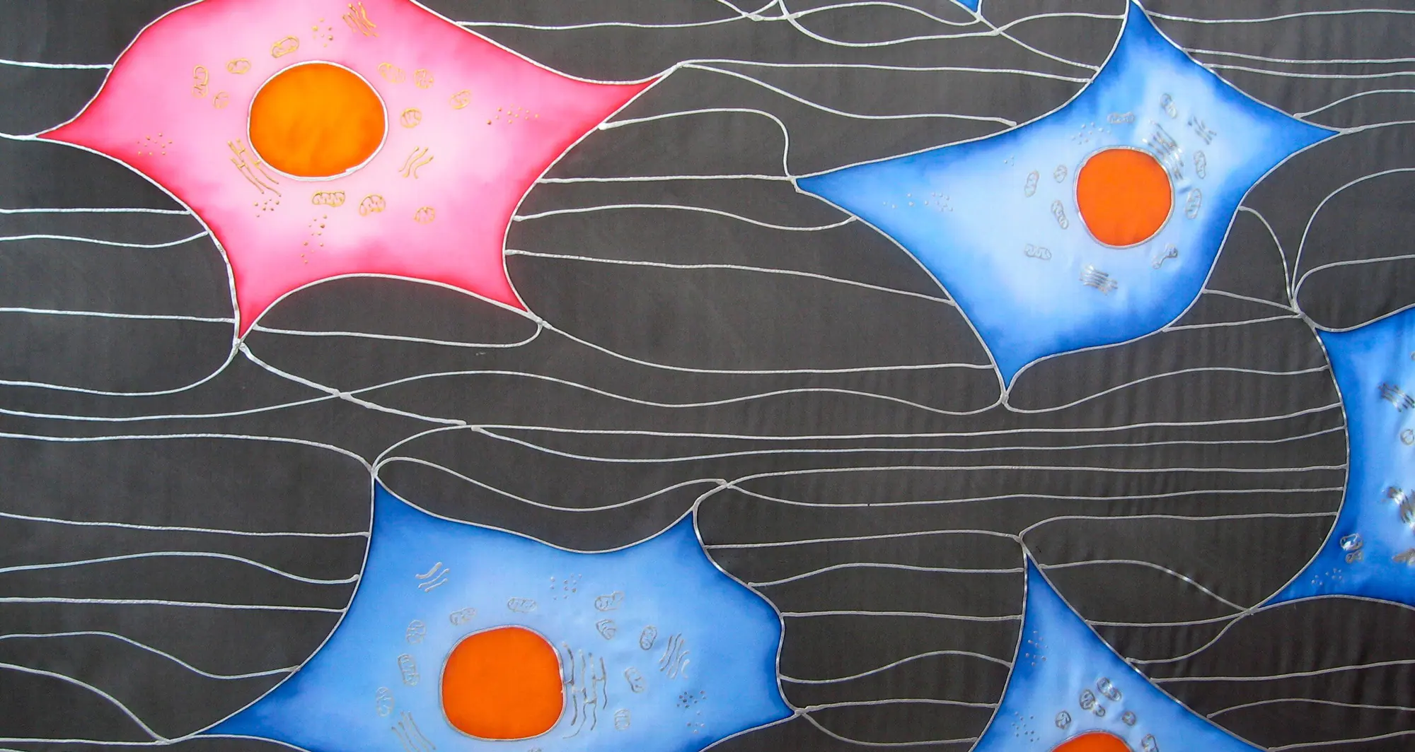
About the art: Odra Noel is a medical doctor and PhD in basic science, with additional degrees in aesthetics and music. Her silk paintings focus primarily on human biology, often informed by microscopy. Wellcome Collection.