cancer Archives - Page 2 of 11 - Sanford Burnham Prebys
Institute News

AI-driven cancer prediction tool makes NIH director’s highlights for 2024

AuthorScott LaFee
Date

January 3, 2025

On April 18, 2024, first author Sanju Sinha, PhD, an assistant professor in the Cancer Molecular Therapeutics Program at Sanford Burnham Prebys, and colleagues published details about a new artificial intelligence-powered tool called PERCEPTION (PERsonalized Single-Cell Expression-Based Planning for Treatments In ONcology).

PERCEPTION was proof-of-concept that AI could be used to predict a cancer’s treatment responses from bulk RNA. Sinha and colleagues built AI models for 44 drugs approved by the FDA and found that their tool “predicted the success of targeted treatments against cell lines with a high degree of accuracy.”

The paper was among six specifically highlighted by Monica Bertagnolli, MD, in her blog as director of the National Institutes of Health.

Institute News

Protein superfamily crucial to the immune system experiences Broadway-style revival

AuthorGreg Calhoun
Date

November 19, 2024

More than 25 years after targeting a member of this superfamily of proteins led to groundbreaking treatments for several autoimmune diseases including rheumatoid arthritis and Crohn’s disease, San Diego scientists note a resurgence of interest in research to find related new drug candidates.

In 1998, the same year “Footloose” debuted on Broadway, REMICADE® (infliximab) was approved by the FDA for the treatment of Crohn’s disease. This was the first monoclonal antibody ever used to treat a chronic condition, and it upended the treatment of Crohn’s disease.

Research published in February 2024 demonstrated better outcomes for patients receiving infliximab or similar drugs right after diagnosis rather than in a “step up” fashion after trying other more conservative treatments such as steroids.

Infliximab and ENBREL® (etanercept) — also approved in 1998 to treat rheumatoid arthritis — were the first FDA-approved tumor necrosis factor-α (TNF) inhibitors. TNF is part of a large family of signaling proteins known to play a key role in developing and coordinating the immune system.

The early success of infliximab and etanercept generated excitement among researchers and within the pharmaceutical industry at the possibility of targeting other members of this protein family. They were interested in finding new protein-based (biologics) drugs to alter inflammation that underlies the destructive processes in autoimmune diseases.

As “Footloose” made it back to Broadway in 2024 for the first time since its initial run, therapies targeting the TNF family are in the midst of their own revival. Carl Ware, PhD, a professor in the Immunity and Pathogenesis Program at Sanford Burnham Prebys, and collaborators at the La Jolla Institute for Immunology and biotechnology company Inhibrx, report in Nature Reviews Drug Discovery that there is a resurgence of interest and investment in these potential treatments.

“Many of these signaling proteins or their associated receptors are now under clinical investigation,” said Ware. “This includes testing the ability to target them to treat autoimmune and inflammatory diseases, as well as cancer.”

Today, there are seven FDA-approved biologics that target TNF family members to treat autoimmune and inflammatory diseases. There also are three biologics and two chimeric antigen receptor (CAR)-T cell-based therapies targeting TNF members for the treatment of cancer. This number is poised to grow as Ware and his colleagues report on the progress of research and many clinical trials to test new drugs in this field and repurpose currently approved drugs for additional diseases.

“The anticipation levels are high as we await the results of the clinical trials of these first-, second- and — in some cases — third-generation biologics,” said Ware.

Ware and his coauthors also weighed in on the challenges that exist as scientists and drug companies develop therapies targeting the TNF family of proteins, as well as opportunities presented by improvements in technology, computational analysis and clinical trial design.

Portrait of Carl Ware

Carl Ware, PhD, is a professor in the Immunity and Pathogenesis Program at Sanford Burnham Prebys.

“There are still many hurdles to get over before we truly realize the potential of these drugs,” noted Ware. “This includes the creation of more complex biologics that can engage several different proteins simultaneously, and the identification of patient subpopulations whose disease is more likely to depend on the respective proteins being targeted.

“It will be important for researchers to use computational analysis of genetics, biomarkers and phenotypic traits, as well as animal models that mimic these variables. This approach will likely lead to a better understanding of disease mechanisms for different subtypes of autoimmune conditions, inflammatory diseases, and cancer, enabling us to design better clinical trials where teams can identify the appropriate patients for each drug.”

Institute News

The implastic nature of plastic culture

AuthorScott LaFee
Date

November 4, 2024

There is an art (and science) to creating cell culture models that reflect the complexities of disease. Such models have long been indispensable to parsing the underlying mechanisms of pathology and to preclinical drug discovery.

But art, writes Kevin Tharp, PhD, assistant professor in the Cancer Metabolism and Microenvironment Program, doesn’t always imitate life — at least not when it comes to finding effective cancer therapeutics.

“Just like a machine-learning algorithm trained on irrelevant datasets, efforts to discover anticancer therapeutics are limited by the models we use,” Tharp writes in the British Journal of Pharmacology. “Our drug discovery pipeline works incredibly well but is applied to models that poorly recapitulate in vivo physiology. This may be why drug discovery approaches efficiently identify drugs that work in the context tested and yet often fail to translate into clinical success.”

It’s a case of there’s no place like home. Cancer cell models are cultured on plastic in two-dimensions with limited or no diversity of neighbors. Cancer cells in vivo reside in three dimensions, with dynamic and complex interactions with neighboring cells and surroundings, i.e., the tumor microenvironment.

It’s like growing up on Disneyland’s Main Street versus a real-world urban city. Cultured cancer cells simply don’t look or behave exactly the same as cancer cells in an actual  tumor. Nor do the investigational molecules being tested as potential therapies.

Tharp suggests a multi-pronged approach: Initially culture target cells using conventional methods, then transfer the cells to new culture formats that enforce distinct, non-genomic cytoskeleton architectures and expression patterns that more closed mimic real life.

Institute News

Raising awareness of breast cancer research at Sanford Burnham Prebys

AuthorGreg Calhoun
Date

October 31, 2024

The October Science Connect Series event was themed around Breast Cancer Awareness Month and featured two cancer research experts.

The Sanford Burnham Prebys Wellness Ambassadors hosted a Science Connect event on Wednesday, October 30, 2024, featuring two faculty experts discussing their breast cancer research and its implications.

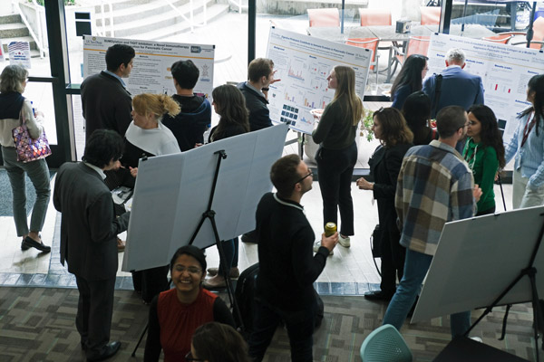
The Science Connect Series provides a forum for Sanford Burnham Prebys principal investigators to share their research with administrative personnel. Faculty members gain experience in communicating their science to a lay audience, and administrators gain a better understanding of research conducted at the institute so they can become better advocates and ambassadors of the shared mission to translate science into health.

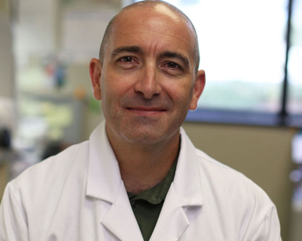
Kelly Kersten, PhD, an assistant professor in the Cancer Metabolism and Microenvironment Program, opened the event by focusing on the importance of finding new treatments —such as immunotherapies — for the one-third of breast cancer patients that are diagnosed after the early stages of the disease when surgery is less effective.

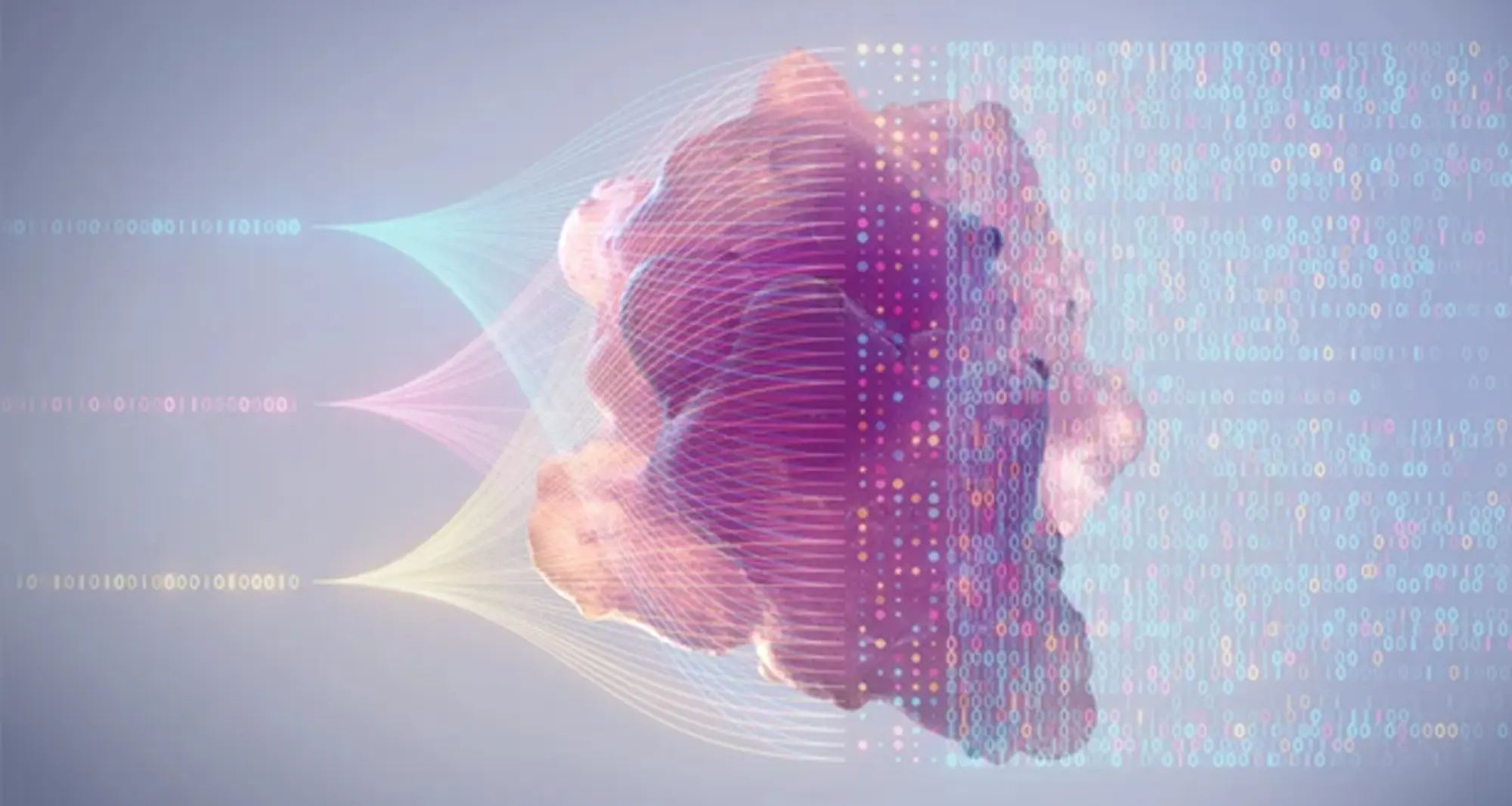
The immune system is one of the main defenses of the human body to fend off harmful pathogens and invasive cells, such as cancer. Among all white blood cells, a particular cell type, called a T cell, can directly kill cancer cells and therefore plays an essential role in building anti-tumor immune responses.

Many types of cancer are confronted and infiltrated by T cells, only to be suppressed by the local tumor environment.

“While immunotherapies that boost the immune system have revolutionized the way we treat cancer, many patients do not respond to the treatments, and the mechanisms of resistance remain largely unclear,” said Kersten.

Kersten’s goal is to understand why T cells enter a state known as exhaustion and lose their tumor-killing capacity. This knowledge will help her team find potential future therapies that could prevent T-cell exhaustion and improve immunotherapies for cancer patients.

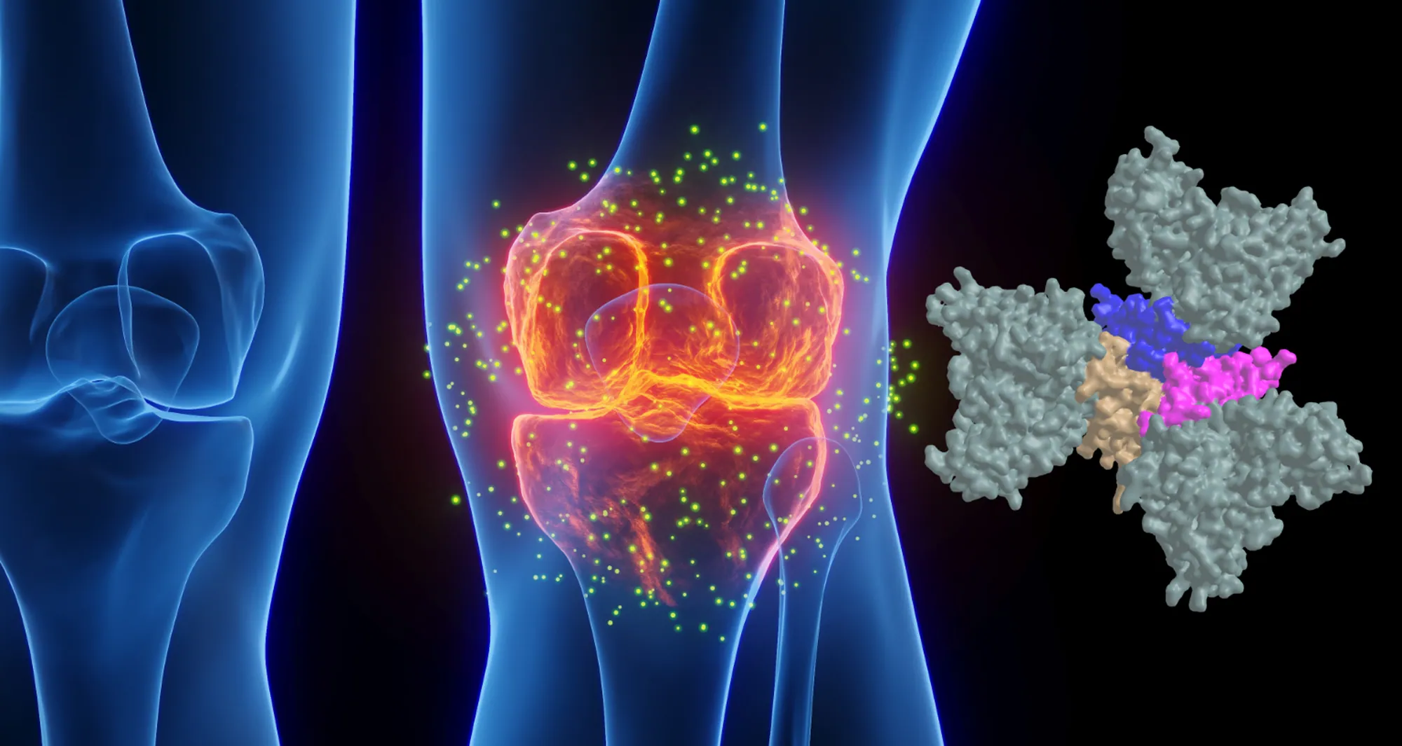
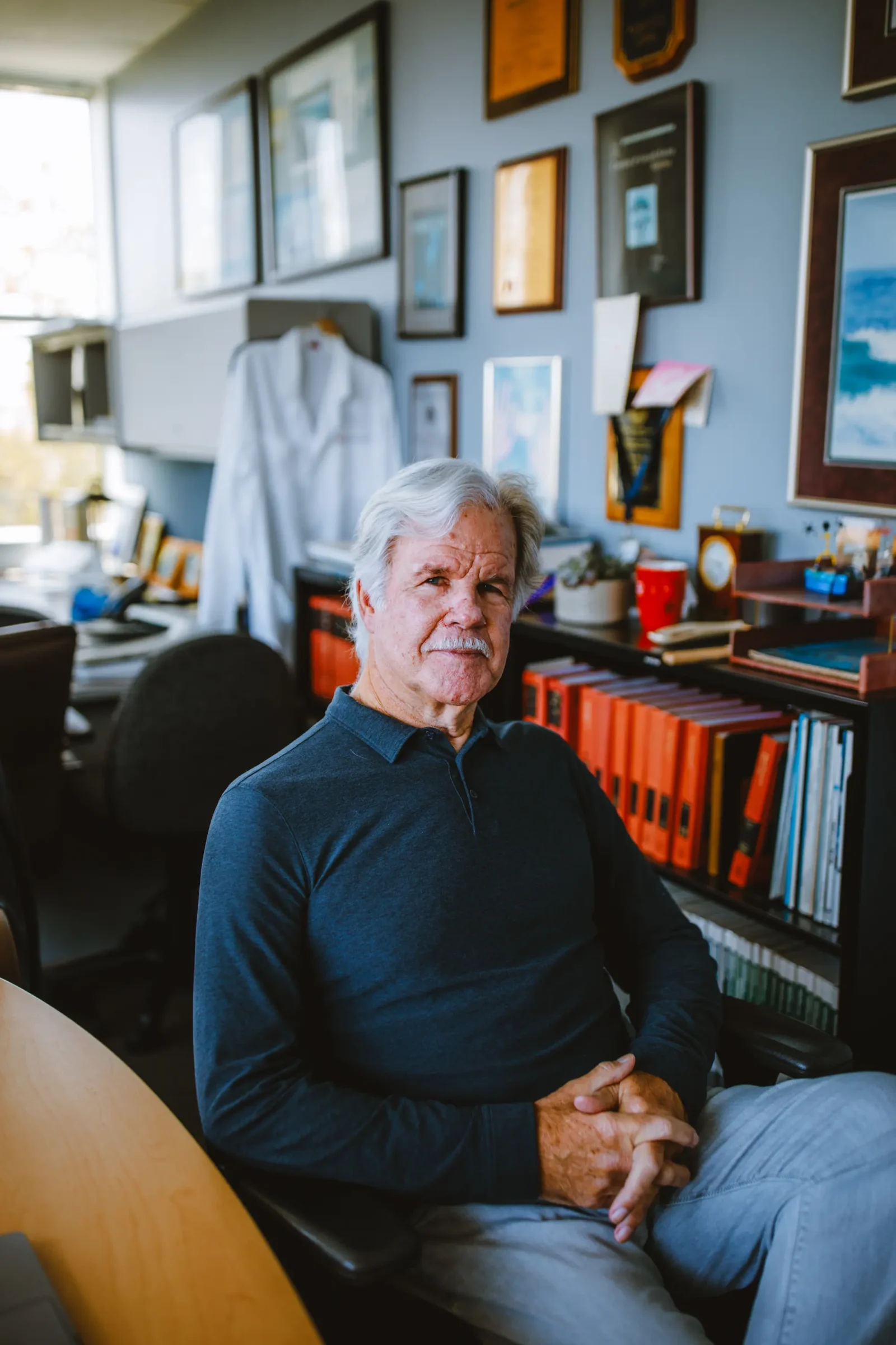
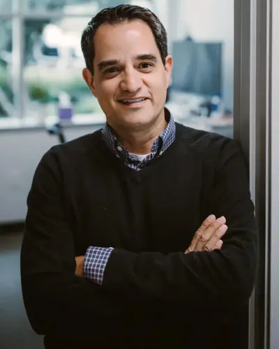
Kevin Tharp, PhD, also an assistant professor in the Cancer Metabolism and Microenvironment Program, shared that his lab’s focus is on how cancer cells adapt their metabolism to generate the energy needed to spread to other tissues through metastasis. He presented his team’s work with the Kersten lab on another aspect of potential resistance to immunotherapy in breast cancer.

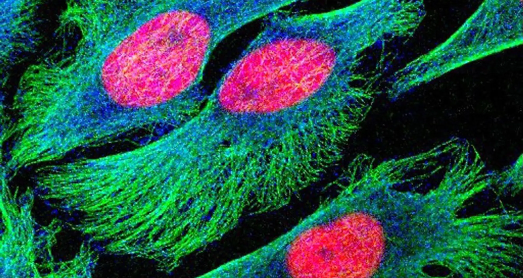
Tharp and Kersten are studying the hypothesis that part of the reason why these therapies fail is due to tumor-associated fibrosis, the creation of a thick layer of fibrous collagen (like scar tissue) that acts as a barrier against the anti-tumor immune response. They published a paper on June 3, 2024, in Nature Cancer,  discussing how tumor-associated macrophages, a type of immune cell found abundantly in the tumor microenvironment, respond to the physical properties of fibrosis.

By synthesizing injury-associated collagens that facilitate wound closure, TAMs experience metabolic changes and generate metabolic byproducts that suppress the anti-tumor function of immune cells.

“The metabolic changes in the microenvironment present more of a challenge to anti-tumor responses than the physical barrier,” said Tharp. “Our study provides an alternative explanation for why anti-tumor immunity is impaired in fibrotic solid tumors.”

To follow up on these results, Tharp is collaborating with Sarah Blair, MD, a professor of surgery at the University of California San Diego, to fund and initiate a clinical trial testing the potential of dietary supplements to counteract the suppressive effects of TAM metabolic byproducts as an adjunct therapy to surgery.

Institute News

A Conversation About Aging and Cancer at Sanford Burnham Prebys 

AuthorGreg Calhoun
Date

October 24, 2024

Event recording now available for panel discussion with scientists held on October 9, 2024

David A. Brenner, MD, president and CEO of Sanford Burnham Prebys, welcomed attendees to the launch of a new community engagement program called “A Conversation About” in the institute’s Victor E. LaFave III Memorial Auditorium on October 9, 2024.

The initial panel discussion in the A Conversation About series focused on the connection between aging and cancer and included information about a current breast cancer research collaboration. A recording of the event is available online.

Reena Horowitz, the founder of Group of 12 and Friends at Sanford Burnham Prebys, provided introductory remarks. Brooke Emerling, PhD, director of the Cancer Metabolism and Microenvironment Program, moderated the discussion among three featured panelists:

  • Peter Adams, PhD, director of the Cancer Genome and Epigenetics Program, Sanford Burnham Prebys
  • Xiao Tian, PhD, assistant professor in the Degenerative Diseases Program, Sanford Burnham Prebys
  • Kay Yeung, MD, PhD, associate clinical professor in the Division of Hematology-Oncology, University of California San Diego Health

By bringing together community collaborators and clinicians with Sanford Burnham Prebys researchers, A Conversation About offers a unique perspective on how clinical research and practice can be used to inform fundamental and translational science.

Watch Event Recording

Institute News

Mammalian Genome Engineering Group holds 2024 symposium in San Diego

AuthorGreg Calhoun
Date

September 25, 2024

The four-day event included talks from experts from across North America and opportunities to discuss improving experimental methods and approaches to analyzing the resulting data.

Researchers convened at Sanford Burnham Prebys in La Jolla from September 12-15 to hear presentations from their peers and confer about the latest developments in modifying the genomes of mammalian animal models to advance biomedical research.

Anindya Bagchi, PhD, associate professor in the Institute’s Cancer Genome and Epigenetics Program, planned the 4th Mammalian Genome Engineering Symposium, which included 26 presentations from experts across the United States and Canada. Attendees asked many questions throughout, and numerous speakers commented on how valuable the conversation at the meeting was for refining planned experiments and considering new ideas and approaches.

“It was a truly enjoyable and thought-provoking meeting,” said Angela Liou, MD, an instructor in the Cancer Genome and Epigenetics Program at Sanford Burnham Prebys and pediatric oncologist and hematologist at Rady Children’s Hospital-San Diego. “It also was incredibly helpful in informing the next steps of my research project.”

“I’m so grateful for the invitation to attend this symposium,” said Praveen Raju, MD, PhD, the Nathan Gordon Chair in Neuro-Oncology and medical director of the Pediatric Neuro-Oncology Program at Rady Children’s Hospital-San Diego and director of the Pediatric Neuro-Oncology Program at the University of California San Diego School of Medicine.

Anindya Bagchi, PhD, headshot

Anindya Bagchi, PhD, is an associate professor in the Cancer Genome and Epigenetics Program.

“The presenters and attendees were welcoming and collaborative, and I certainly learned a lot.”

The symposium brings together the Mammalian Genome Engineering Group, which was formed by a small group of genome engineering enthusiasts including Bagchi, Nada Jabado, MD, PhD, professor of Pediatrics and Human Genetics at McGill University and a hematologist and oncologist at Montreal Children’s Hospital; David Largaespada, PhD, a professor of Pediatrics, Genetics, Cell Biology and Development at the University of Minnesota Medical School and the associate director for Basic Research in the Masonic Cancer Center; and Michael Taylor, MD, PhD, The Cyvia and Melvyn Wolff Chair of Pediatric Neuro-Oncology at Texas Children’s Cancer and Hematology Center and professor of Pediatrics (Hematology-Oncology) at Baylor College of Medicine.

The group is interested in developing functional models of genomic and epigenetic mutations associated with human diseases—especially cancers—that are difficult to recreate in animal models. The group’s first symposium was coordinated by Taylor in Napa, Calif., in 2014, followed by the 2nd symposium that was organized by Jabado in Montreal in 2015. After a hiatus, the group was revived in 2023 with the 3rd symposium hosted again by Taylor in Houston.

“We believe this symposium will, in the coming years, become a leading forum for discussing cutting-edge genomic and epigenomic approaches to tackle challenging genetic and epigenetic mutations,” said Bagchi. “These approaches are likely to become standard practice in the near future.”

The Sanford Burnham Prebys scientists that presented at the 4th Mammalian Genome Engineering Symposium were:

  • Bagchi, “Why are MYC-driven cancers so lethal?” 
  • Liou, “Investigating the deposition of H3.3K27M oncohistone and its effect on retrotransposon reactivation in H3K27M pediatric diffuse midline glioma” 
  • Ani Deshpande, PhD, associate professor in the Cancer Genome and Epigenetics Program and associate director of Diversity, Equity and Inclusion in the NCI-Designated Cancer Center, “Functional genomic approaches to identify selective dependencies in synovial sarcoma” 
  • Peter D. Adams, PhD, the director of the Cancer Genome and Epigenetics Program, “The role of aging in cancer” 
  • Lukas Chavez, PhD, associate professor in the Cancer Genome and Epigenetics Program, “Circular extrachromosomal DNA promotes tumor heterogeneity and enhancer rewiring” 
  • Jerold Chun, MD, PhD, professor in the Degenerative Diseases Program, “Genetic mosaicism and somatic gene recombination in the brain” 
  • Adarsh Rajesh, graduate student, Sanford Burnham Prebys, “CCND1-CDK6 complex inhibits DNA damage repair and promotes inflammation in senescence and the aged liver”

Additional speakers included:

  • Taylor, “Why does medulloblastoma love to be tetraploid and other nonsense”
  • Jabado, “Co-opting 3D structures to fuel tumorigenesis”
  • Tannishtha Reya, PhD, Herbert and Florence Irving Professor of Basic Science Research in Physiology and Cellular Biophysics, Columbia University, “New genetically engineered models to understand cancer heterogeneity and therapy resistance in pancreatic cancer”
  • Simona Dalin, PhD, postdoctoral fellow, Broad Institute of the Massachusetts Institute of Technology and Harvard University, “Contributions of perfect and imperfect homology to rearrangement formation in human and cancer genomes”
  • Alison M. Taylor, PhD, assistant professor of Pathology and Cell Biology, Columbia University, “Functional and computational approaches to uncover the consequences of chromosome arm aneuploidy in cancer”
  • Sean Eagan, PhD, senior scientist in the Cell Biology program at The Hospital for Sick Children, professor of Molecular Genetics, University of Toronto, “An update on Genetic analysis of 16q-syntenic block deletion in the mouse mammary gland – a tumor suppressor arm”
  • Claudia Kleinman, PhD, associate professor of Human Genetics, McGill University, investigator at the Lady Davis Institute for Medical Research, Jewish General Hospital, “Lineage programs and the 3D genome in pediatric brain tumors”
  • Branden Moriarity, PhD, associate professor of Pediatrics (Hematology and Oncology), University of Minnesota Medical School, “Next generation engineered immune effector cells for immunotherapy”
  • Beau Webber, PhD, associate professor of Pediatrics (Hematology and Oncology), University of Minnesota Medical School, “Building cancer in a dish: Sarcoma modeling using human pluripotent stem cells”
  • Sameer Agnihotri, PhD, associate professor of Neurological Surgery and director of the Brain Tumor Biology and Therapy Lab, University of Pittsburgh Medical Center Children’s Hospital of Pittsburgh, “Identifying genetic vulnerabilities by modeling Chromosome 9p loss”
  • Largaespada, “Loss of the polycomb repressor complex 2 (PRC2) alters the super-enhancer landscape, genome/epigenome stability, and therapeutic sensitivities of malignant peripheral nerve sheath tumors”
  • Teresa Davoli, PhD, assistant professor of Biochemistry and Molecular Pharmacology, New York University Langone Health, “Engineering chromosome specific aneuploidy by targeting human centromeres”
  • Rameen Beroukhim, MD, PhD, associate professor of Medicine, Dana-Farber Cancer Institute and Harvard Medical School, associate member of the Broad Institute of MIT and Harvard, “Detecting rearrangement signatures—naturally”
  • Quang Trinh, PhD, scientist, Ontario Institute for Cancer Research, “Perspectives and Challenges in PFA Integrative Analysis”
  • Taylor Gatesman, graduate student, University of Pittsburgh, “Genome Engineering: DREaming of and vCREating new models”
  • Joseph Skeate, PhD, postdoctoral fellow, University of Minnesota Medical School, “Targeted CAR integration and multiplex base editing in a single-step manufacturing process for enhanced cancer immunotherapies”
Institute News

Using machines to personalize patient care

AuthorGreg Calhoun
Date

July 30, 2024

Artificial intelligence (AI) and other computational techniques are aiding scientists and physicians in their quest to create treatments for individuals rather than populations

The Human Genome Project captured the public’s imagination with its global quest to better understand the genetic blueprint stored on the DNA within our cells. The project succeeded in delivering the first-ever sequence of the human genome while foreshadowing a future for medicine once considered to be science fiction. The project presaged the possibility that health care could be personalized based on clues within a patient’s unique genetic code.

Chavez lab

The Chavez Lab

While many more people have undergone genetic testing through consumer genealogy and health services such as 23andMe and Ancestry than through health care systems, genomic sequencing has influenced clinical care in some specialties. Personalized medicine—also known as precision medicine or genomic medicine—has been especially helpful for people suffering from rare diseases that historically have been difficult to diagnose and treat.

Scientists at Sanford Burnham Prebys are employing new technologies and expertise to test ways to improve diagnoses and customize treatments for many diseases based on unique characteristics within tumors, blood samples and other biopsies.

AI and other computational techniques are enabling patient samples to be rapidly analyzed and compared to data from vast numbers of individuals who have been treated for the same condition. Physicians can use AI and other tools to identify subtypes of cancers and other conditions, as well as improve selection of eligible candidates for clinical trials.

“I think we’ve gotten a lot better at precision diagnostics,” says Lukas Chavez, PhD, an assistant professor in the Cancer Genome and Epigenetics Program at Sanford Burnham Prebys. “In my work at Rady Children’s Hospital in cancer, we can characterize a tumor based on mutations, including predicting how quickly different tumors will spread. What we too often lack, however, are better treatment approaches or medicines. That will be the next generation of precision medicine.”

Sanju Sinha, PhD, an assistant professor in the Cancer Molecular Therapeutics Program at Sanford Burnham Prebys, is developing projects to help bridge the gap between precision diagnostics and treatment. He is partnering with the National Cancer Institute on a first-of-its-kind computational tool to systematically predict patient response to cancer drugs at single-cell resolution.

A study published in the journal  Nature Cancer discussed how the tool, called PERCEPTION, was successfully validated by predicting the response to individual therapies and combination treatments in three independent published clinical trials for multiple myeloma, breast and lung cancer.

Lukas Chavez, PhD

Lukas Chavez, PhD, is an assistant professor in the Cancer Genome and Epigenetics Program at Sanford Burnham Prebys.

In each case, PERCEPTION correctly stratified patients into responder and non-responder categories. In lung cancer, it even captured the development of drug resistance as the disease progressed, a notable discovery with great potential.

Sanju Sinha, PhD

Sanju Sinha, PhD, is an assistant professor in the Cancer Molecular
Therapeutics Program at Sanford Burnham Prebys.

“The ability to monitor the emergence of resistance is the most exciting part for me,” says Sinha. “It has the potential to allow us to adapt to the evolution of cancer cells and even modify our treatment strategy.”

While PERCEPTION is not yet ready for clinics, Sinha hopes that widespread adoption of this technology will generate more data, which can be used to further develop and refine the technology for use by health care providers.

In another project, Sinha is focused on patients being treated for potential cancers that may never progress into dangerous conditions warranting treatment and its accompanying side effects.

“Many women who are diagnosed with precancerous changes in the breast seek early treatment,” says Sinha. “Most precancerous cells never lead to cancer, so it may be that as many as eight of 10 women with this diagnosis are being overtreated, which is a huge issue.”

To try and counter this phenomenon, Sinha is training AI models on images of biopsied samples in conjunction with multi-omics sequencing data. His team’s goal is to develop a tool capable of predicting which patients’ cancers would progress based on the imaged samples alone.

“In the field of precancer, insurance does not cover the cost of computing this omics data,” says Sinha. “Health care systems do routinely generate histopathological slides from patient biopsies, so we feel that a tool leveraging these images could be a scalable and accessible solution.”

If Sinha’s team is successful, an AI tool integrated into clinics would predict whether precancerous cells would progress within the next 10 years to guide treatment decisions and how patients are monitored.

“With precision medicine, our hope is not to just treat patients with better drugs, but also to make sure that patients are not unnecessarily treated and made to bear needless costs and side effects that disrupt their quality of life.”


Programming in a Petri Dish, an 8-part series

How artificial intelligence, machine learning and emerging computational technologies are changing biomedical research and the future of health care

  • Part 1 – Using machines to personalize patient care. Artificial intelligence and other computational techniques are aiding scientists and physicians in their quest to prescribe or create treatments for individuals rather than populations.
  • Part 2 – Objective omics. Although the hypothesis is a core concept in science, unbiased omics methods may reduce attachments to incorrect hypotheses that can reduce impartiality and slow progress.
  • Part 3 – Coding clinic. Rapidly evolving computational tools may unlock vast archives of untapped clinical information—and help solve complex challenges confronting health care providers.
  • Part 4 – Scripting their own futures. At Sanford Burnham Prebys Graduate School of Biomedical Sciences, students embrace computational methods to enhance their research careers.
  • Part 5 – Dodging AI and computational biology dangers. Sanford Burnham Prebys scientists say that understanding the potential pitfalls of using AI and other computational tools to guide biomedical research helps maximize benefits while minimizing concerns.
  • Part 6 – Mapping the human body to better treat disease. Scientists synthesize supersized sets of biological and clinical data to make discoveries and find promising treatments.
  • Part 7 – Simulating science or science fiction? By harnessing artificial intelligence and modern computing, scientists are simulating more complex biological, clinical and public health phenomena to accelerate discovery.
  • Part 8 – Acceleration by automation. Increases in the scale and pace of research and drug discovery are being made possible by robotic automation of time-consuming tasks that must be repeated with exhaustive exactness.
Institute News

Pancreatic cancer symposium celebrates 10th anniversary in San Diego

AuthorGreg Calhoun
Date

May 22, 2024

The 2024 PancWest Symposium brought more than 120 scientists to the Sanford Burnham Prebys campus in San Diego to discuss the latest advances in pancreatic cancer research.

More than 120 pancreatic cancer researchers from the West Coast traveled to San Diego from as far as Vancouver to attend the 2024 PancWest Symposium on May 17. The PancWest Symposium was founded in 2014 to regularly bring the scientific community studying pancreatic cancer together to discuss advances in the field and foster new collaborations.

The PancWest Symposium is held every two years in a different city to showcase expert scientists who are making important contributions to the field of pancreatic cancer research, including tumorigenesis, tumor progression and the discovery of novel therapeutic paradigms, such as immunomodulation and metabolic targeting.

The 2024 event was held on the Sanford Burnham Prebys campus in the Fishman Auditorium and was hosted by Cosimo Commisso, Ph.D., director of, and associate professor in, the Institute’s Cancer Metabolism and Microenvironment Program; and Pamela Itkin-Ansari, Ph.D., adjunct professor in the Institute’s Development, Aging and Regeneration Program.

“While pancreatic cancer accounts for only three percent of cancer cases, it has the highest mortality rate among major cancers and is the third leading cause of cancer-related death in the U.S.,” says Commisso.

PancWest Symposium poster presentations in Chairmen's Hall

The symposium’s events included a keynote address, 12 featured speakers, a poster session and a series of “power talks” providing attendees a chance to hear two-minute oral presentations from selected poster presenters.

“Unless we find ways to better diagnose and treat this disease, it is projected to become the second most deadly cancer in less than 20 years,” adds Itkin-Ansari. “That is why events such as PancWest are so important to enhance innovation and foster collaboration.”

Rosalie C. Sears, Ph.D., professor of Molecular and Medical Genetics, co-director of the Brenden-Colson Center for Pancreatic Care and Krista L. Lake Chair in Cancer Research at Oregon Health & Science University in Portland, gave the symposium’s keynote address.

Additional events at the symposium included 12 featured speakers, a poster session and a series of “power talks” providing attendees a chance to hear two-minute oral presentations from selected poster presenters.

“Being a part of PancWest has been a transformative experience,” shares Itkin-Ansari. “The exchange of groundbreaking research and innovative ideas among leading experts advanced our scientific understanding.”

“It also paved the way for new therapeutic strategies, ultimately offering hope and improved outcomes for patients battling pancreatic cancer,” adds Commisso.

More information about the symposium and featured speakers is available on the event’s webpage.

Institute News

Seminar Series: extrachromosomal DNA and the metabolic circuits of cancer immune suppression

AuthorScott LaFee
Date

March 25, 2024

The ongoing Sanford Burnham Prebys seminar series will feature a pair of speakers on March 27, from noon to 1p.m., in the Fishman Auditorium. They will be presenting on two topics: extrachromosomal DNA and the tumor microenvironment.

First, Owen Chapman, PhD, a postdoctoral research scientist in the lab of Lukas Chavez, PhD, will discuss clinical and genomic features of circular extrachromosomal DNA (ecDNA) in medulloblastomas, a type of brain tumor.

EcDNA is DNA found off chromosomes, either inside or outside the nucleus of a cell. In a study published last year, Chavez (senior author), Chapman (first author) and colleagues reported that patients with medulloblastomas containing ecDNA are twice as likely to relapse after treatment and three times as likely to die within five years of diagnosis.

The second presentation will be by Kevin Tharp, PhD assistant professor in the Cancer Metabolism and Microenvironment Program. Tharp, who joined Sanford Burnham Prebys in December 2023, studies how tumors manipulate their mitochondria to improve survivability and how those cellular mechanics can be leveraged to create more effective therapies.

Institute News

New genome mapping tool may uncover secrets for treating blood cancers

AuthorGreg Calhoun
Date

February 1, 2024

The outlook for patients with acute myeloid leukemia (AML), a deadly set of blood cancers that is difficult to treat, has remained dire for decades, especially among patients who are not eligible for bone marrow transplantation.

More than 30% of treated patients will never achieve complete remission using current chemotherapies and, even when chemotherapy treatments work, most patients relapse within five years without a transplant.

While prior research has begun to unravel the genetic underpinnings of the disease, more inquiry is needed to understand the genetic variation within the roughly 15 AML subtypes and how that variation might affect treatment strategies.

“In addition to the one to eight average genetic mutations in AML patients found in traditional sequencing studies, experiments employing high-resolution optical genomic mapping have found approximately 40 to 80 rare genomic structural variants per patient,” says Kristiina Vuori, MD, PhD, Pauline and Stanley Foster Distinguished Chair and professor in the Sanford Burnham Prebys Cancer Center’s Cancer Molecular Therapeutics Program. “We wanted to take these structural variant findings in AML to the next level by connecting them with patients’ sensitivity or resistance to current cancer treatments.”

Kristiina Vuori, MD, PhD

In a paper published January 18, 2024, in Cancers, a multidisciplinary team of biologists, bioinformaticians and clinicians from Sanford Burnham Prebys, Bionano Genomics Inc. and Scripps MD Anderson were the first to associate genomic structural variants (SVs) in AML patients with drug sensitivities.

“SVs are changes to the genome in which sections of 50 or more base pairs in a strand of DNA have been errantly deleted, duplicated, inverted or translocated,” explains Darren (Ben) Finlay, PhD, first author on the manuscript and research associate professor in the Sanford Burnham Prebys Cancer Center.

“Such changes amount to different combinations of DNA gains, losses or rearrangements. When cells use these altered instructions in the DNA to make proteins or carry out other functions, it is like a chef trying to cook with a recipe that is missing steps, has them in the wrong order or includes more or less of the key ingredients.”

Darren (Ben) Finally, PhD

Scientists have become more able to find SVs as next-generation genomic analysis technologies and techniques have improved. Research has shown that SVs contribute to the development and progression of cancer, including blood cancers. Of particular concern among SVs are DNA changes that join two otherwise distant genes. This event, called gene fusion, is known to drive certain pediatric and blood cancers.

Finlay, Vuori and colleagues analyzed SVs in samples from 23 AML patients and found their genomes featured 16-45 extremely rare SVs within genes but not seen in healthy volunteers’ samples. The scientists detailed the patients’ SVs using a technique called optical genome mapping. This tool tags DNA in specific locations to create recognizable sequences, unwinds and straightens the genomic DNA for linear scanning, and converts the imaged sequences into digital representations of DNA molecules. Because it directly images DNA rather than relying on algorithmic analyses, optical genome mapping is better than next-generation sequencing at finding SVs throughout the entire genome, especially large SVs, the researchers said.

To begin building the connection between SVs and drug sensitivity, the scientists tested samples from each patient with 120 FDA-approved drugs, and experimental treatments currently in phase III clinical trials. This allowed the researchers to map out how strongly each patient’s sample reacted with each drug.

Next, the investigators used statistical analysis to compare the SVs within the optical genome mapping results with the findings from the drug sensitivity tests. The team found 61 statistically significant interactions between SVs and existing cancer therapies. In one interaction, the group demonstrated that a commonly used AML drug, Idarubicin, and two similar compounds (Daunorubicin and Epirubicin) were more effective in leukemia samples with a specific insertion in a gene that carries the instructions for a signaling enzyme that helps nerves communicate with muscles. These and other examples lend support to the scientists’ hypothesis that optical genome mapping could be used to develop personalized treatment plans that account for patients’ SVs.

“In this pilot study, our hope was to identify structural variants that could be used as new biomarkers for current AML drugs as well as to identify other drugs that could be repurposed to treat leukemia patients,” says Finlay.

“Ensuring patients receive the most effective drugs on the market through personalized treatment and identifying new potential therapies for AML are critically important,” adds Vuori, senior author on the study. “Especially for patients who do not achieve remission with current standard chemotherapies or who are ineligible for bone marrow transplants or clinical trials.”
 

Cancers 2024, 16(2), 418; https://doi.org/10.3390/cancers16020418