Cancer Center Archives - Page 8 of 11 - Sanford Burnham Prebys
Institute News

Sanford Burnham Prebys joins the fight to end cancer at Padres Pedal the Cause fundraiser

AuthorMonica May
Date

November 18, 2019

Nearly everyone knows someone who has been affected by cancer: One in three Americans will be diagnosed in their lifetime. In San Diego, it’s the number one cause of death.

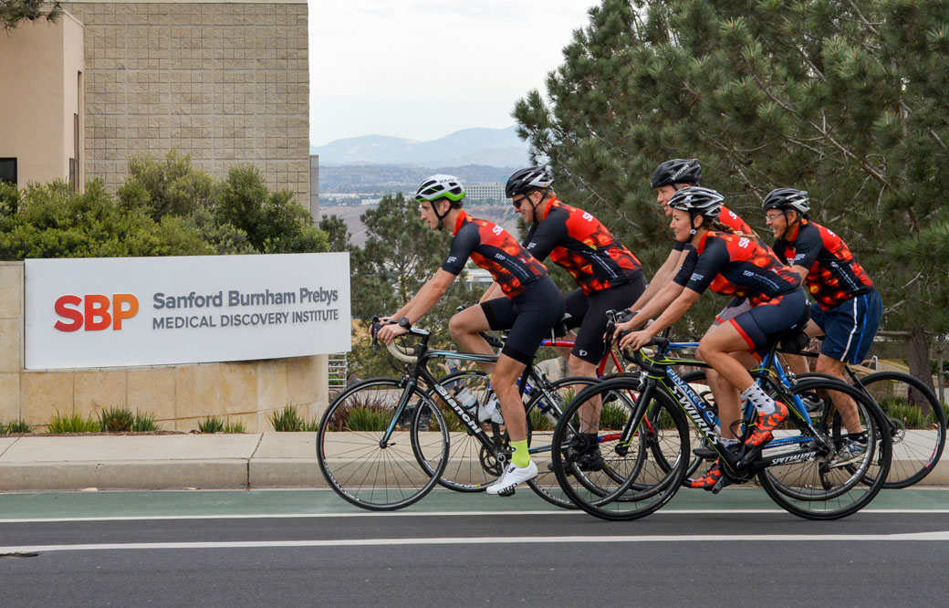
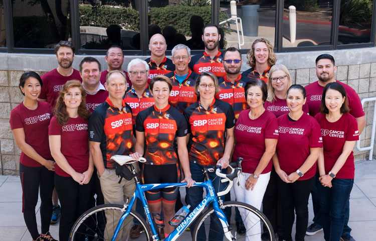
With the goal of improving these statistics, on November 16, 2019, more than 50 scientists, staff and supporters of Sanford Burnham Prebys joined thousands of fellow cancer fighters—including former Padre Tony Gwynn Jr. and San Diego Mayor Kevin Faulconer—at the seventh annual Padres Pedal the Cause (PPTC) fundraiser. Together, team Sanford Burnham Prebys raised more than $30,000 to accelerate collaborative cancer research taking place in San Diego.

Launched in 2013, the annual fundraiser has since expanded from a cycling-only event to include a 5K run or walk and stationary spin classes. To date, the event has raised more than $10 million to accelerate cancer cures, with 100% of the proceeds funding collaborative research taking place at four San Diego research institutes, including Sanford Burnham Prebys. Past PPTC grants have advanced our Institute’s research into cancers of the breast, skin, brain, colon, pancreas and more.

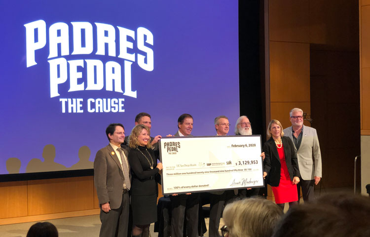
This year’s event had an ambitious goal of raising $3.3 million. Fundraising will continue until December 7, 2019; the final amount raised will be revealed on January 30, 2020, at a special check-presentation ceremony.

Want to help Padres Pedal the Cause meet its fundraising goal? Donate today.

Institute News

Scientists discover new survival strategy for oxygen-starved pancreatic cancer cells

AuthorMonica May
Date

October 23, 2019

Oxygen is essential to life. When fast-growing tumor cells run out of oxygen, they quickly sprout new blood vessels to keep growing, a process called angiogenesis. 

By blocking pancreatic cancer’s oxygen-sensing machinery—the same field of research studied by the winners of the 2019 Nobel Prize in Medicine—Sanford Burnham Prebys scientists have uncovered a new way that tumors turn on angiogenesis in an animal model. The discovery, published in Cancer Research, could lead to a treatment that is given with an anti-angiogenetic medicine, thereby overcoming drug resistance. 

“Treatment resistance is a major challenge for cancer treatments that block blood vessel growth,” says Garth Powis, D.Phil., professor and director of Sanford Burnham Prebys’ National Cancer Institute (NCI)-designated Cancer Center and senior author of the study. “Our research identifies a new way angiogenesis is activated, opening new opportunities to find medicines that might make existing cancer treatments more effective.” 

Many cancer treatments work by blocking angiogenesis, which rarely occurs in healthy tissues. However, these medicines eventually stop working, and the cancer returns, sometimes in as little as two months. Scientists have been researching why this treatment resistance occurs so it can be stopped.

In this study, the scientists focused on pancreatic cancer, which is notoriously desperate for oxygen and also difficult to treat. Fewer than 10% of people diagnosed with pancreatic cancer are alive five years later. 

To see how a pancreatic tumor responds to a disruption in its oxygen supply, the Sanford Burnham Prebys researchers used a mouse model to block an oxygen-sensing protein called HIF1A—which should cripple the tumor’s growth. Instead of dying, however, after about a month the cells multiplied—indicating they had developed a new way to obtain oxygen. 

Further work revealed that the cancer cells were clear and swollen with the nutrient glycogen (a characteristic also seen in some ovarian and kidney cancers). In response to the excess glycogen, special immune system cells were summoned to the tumor, resulting in blood vessel formation and tumor survival. Each of these responses represents a new way scientists could stop pancreatic tumors from evolving resistance to treatment.

“Our team’s next step is to test tumor samples from people with pancreatic cancer to confirm this escape mechanism occurs in a clinical setting,” says Powis. “One day, perhaps we can create a second medicine that keeps anti-angiogenic drugs working and helps more people survive pancreatic cancer.”


Research reported in this press release was supported by the U.S. National Institutes of Health (NIH) (5F31CA203286, CA216424 and P30CA030199). The content is solely the responsibility of the authors and does not necessarily represent the official views of the NIH. The study’s DOI is 10.1158/0008-5472.CAN-18-2994. 

Institute News

Sanford Burnham Prebys scientists win two American Cancer Society awards

AuthorMonica May
Date

October 1, 2019

Innovation and Collaboration of the Year Awards

The San Diego cancer community—including oncologists, oncology nurses, radiologists, cancer researchers and their friends and family—gathered on September 22 to celebrate progress made in reducing cancer deaths and recognize exceptional individuals and institutions at the inaugural American Cancer Society’s Celebration of Cancer Care Champions in San Diego.

More than 40 finalists were selected, including Sanford Burnham Prebys professors Robert Wechsler-Reya, PhD, who received the Innovation of the Year award for his team’s creation of a new model for studying a brain tumor that commonly arises in infants; and Jorge Moscat, PhD, and Maria Diaz-Meco, PhD, who received the Collaboration of the Year award for their partnership with clinicians at Scripps Clinic who uncovered a novel way to potentially identify a deadly form of colorectal cancer.

Nominations were reviewed by an independent review committee composed of representatives from 10 leading healthcare and research institutions, including Celgene, Kaiser Permanente, Rady Children’s Hospital, Scripps MD Anderson Cancer Center, Moores Cancer Center at UC San Diego Health and more. (Note: Members of the review committee did not score nominations for their own institutions.)

Read on to learn more about our award-winning research:

Innovation of the Year: A new model for studying brain tumors that strike infants
Robert Wechsler-Reya, PhD, a professor at Sanford Burnham Prebys and program director of the Joseph Clayes III Research Center for Neuro-Oncology and Genomics at the Rady Children’s Institute for Genomic Medicine, was honored for his development of a novel mouse model of a pediatric brain tumor called choroid plexus carcinoma. This tumor most commonly arises in infants under the age of one who are too young to undergo radiation treatment. Until now, drug development has been hindered by the lack of models that could help researchers better understand the cancer. Wechsler-Reya and his team have already used the model to identify potential drug compounds that may be therapeutically useful.

Collaboration of the Year (tie): Novel biomarkers to help detect a deadly colorectal cancer 
Sanford Burnham Prebys professors Jorge Moscat, PhD, and Maria Diaz-Meco, PhD; and Scripps Clinic clinicians Darren Sigal, MD, and Fei Baio, MD, were recognized for their successful collaboration. Together, the researchers revealed that loss of two genes drives the formation of the deadly serrated form of colorectal cancer—yielding promising biomarkers that could identify the tumor type. This insight could lead to the development of a diagnostic test to identify serrated colorectal cancer, a hurdle that previously limited our understanding of this deadly cancer type and the development of effective treatments. The research also identified a combination treatment that has treated the cancer in mice.

Institute News

5 things to know about immunotherapy and breast cancer

AuthorSusan Gammon
Date

September 30, 2019

If you follow news about medical breakthroughs, you have undoubtedly heard about immunotherapy to treat cancer.

This form of therapy is designed to prime the body’s own immune system to fight the disease head-on. For some cancers, such as melanoma and lung cancer, immunotherapy has helped patients who once had only a life expectancy of months now live for years. But does it work for other cancers?

We sat down with Svasti Haricharan, PhD, assistant professor at Sanford Burnham Prebys and recipient of a Susan G. Komen Career Catalyst Award to discuss where we are with immunotherapy and breast cancer. Here are five things she wants us to know.

  1. As scientists, our job is to understand the biology of why immunotherapy works for some cancers but not others. Our goal is to develop approaches to expand the benefits of immunotherapy to as many patients as possible. With breast cancer, we are still in the early days, but there has been some success. Earlier this year a type of immunotherapy called an “immune checkpoint inhibitor” was approved to treat certain types of metastatic breast cancer. But immunotherapy doesn’t work—yet—for all breast cancers.
  2. No two breast cancers are alike. Even though two women with breast cancer may have the same size tumor, the individual characteristics of the tumor—the receptors, the genetics, even the way the tumor cells gather fuel to grow, can differ. Just as importantly, the way each woman’s body reacts to the growing cancer is predicated by her immune history: her exposure to immunological challenges, the strength of the immune response her body is capable of mounting, and how long she can sustain an immune response. These factors strongly influence the likelihood that a patient will respond to a specific therapy. The more we drill down on breast tumors, and the tricks they use to evade the immune system, the closer we get to outsmarting them.
  3. Today, immunotherapy seems to work best for triple negative breast cancer. Triple negative means three types of receptors—estrogen receptor, progesterone receptor and HER2—are not expressed on the cancer cells. Cancers that express these receptors are easier to treat because these receptors can be targeted directly. We believe part of the reason why immunotherapy is effective for triple negative breast cancer is because these cells can grow rapidly and produce more neoantigens—altered tumor proteins that have not previously been recognized by the immune system. So, these tumors may already have immune cells infiltrating the tumor, and when unleashed via immunotherapy, they can readily attack the cancer. 
  4. Immunotherapy—at least the immune checkpoint agents that are used today—target a protein called PD-1 found on T cells, which are the immune cells that roam the body looking for disease. PD-L1 is another protein found on some normal and some cancer cells. When PD-1 attaches to PD-L1, T cells are queued to leave the cell alone and not attack it. We believe cancer cells use PD-L1 to protect themselves from the immune system, and that cancers with large amounts of PD-L1 are the most likely to respond to checkpoint inhibitors. It’s possible that testing breast tumors for PD-L1 levels will help identify more women likely to benefit from these drugs. 
  5. Collaboration is key. Although we like to think of scientists as having “Eureka” moments, the reality is that much of the progress we make is incremental. We painstakingly plan, control and execute experiments—gathering and analyzing data to open new avenues that can be tested in the clinic. Working alongside professionals who are responsible for patient outcomes is an important part of the research spectrum. Their input provides direction for our goal of achieving cures—and a means to evaluate if what started in the lab will work in the clinic. There are nearly 300 clinical trials currently ongoing that are testing immunotherapeutic approaches for breast cancer. The information we gather from these trials helps guide the future of what we do next in the laboratory. Advances will be made, and progress is on the horizon.
     
Institute News

“We are desperate for new therapies”

AuthorMonica May
Date

September 23, 2019

Experts discuss AML during the Sanford Burnham Prebys community lecture series 

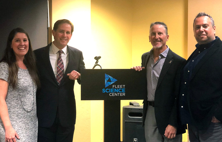
Bill Veljovich had never been sick in his life. “Not even joint pain,” shared the 80-year-old retired engineer at our recent Fleet Science Center discussion about acute myeloid leukemia (AML), a life-threatening type of blood cancer. He was joined by experts from Sanford Burnham Prebys and UC San Diego Health.
 
However, his doctor noticed that his white blood cells counts were off during a routine blood test. He was diagnosed with a blood cancer called myelodysplastic syndrome (MDS), which progressed to AML (this occurs in one out of three people with MDS). Fortunately, Veljovich responded well to a then off-label treatment that only recently was approved for older patients with AML. 

“The truth is, we are desperate for new therapies,” said speaker Rafael Bejar, MD, PhD, a clinician at UC San Diego who specializes in blood cancers. “AML typically occurs in people over the age of 60, who often aren’t able to tolerate intensive chemotherapies.” 

Until two years ago, the treatments for AML remained the same as those used in the 1970s: a chemotherapy combination and perhaps a bone marrow transplant. Only 24% of adults with AML remained alive five years after treatment. 

Now, thanks to foundational research that revealed the underlying genetic drivers of AML, eight new drugs have been approved in the past two years. Several more targeted therapies are nearing potential FDA approval. 

However, AML, which usually arises in cells that turn into white blood cells, is an incredibly complex and fragmented disease. Genome sequencing has revealed that more than 30 genes drive the cancer. Many different treatment types will be needed to truly conquer AML.

Peter Adams, PhD, a professor in Sanford Burnham Prebys’ National Cancer Institute (NCI)-designated Cancer Center, hopes to find a treatment that works for a broader AML population. He focuses on a protein called p53, often called the “guardian of our genome.” This protein senses DNA damage and kills the faulty cell—protecting us from developing cancer. However, to scientists’ surprise, 90% of people with AML have a normal p53 gene. 

“Emerging research suggests that AML inactivates p53 through other means,” said Adams. “My team is working to develop a drug combination that could reactivate the protective powers of p53—and thus fight AML.”

New research advances can’t come soon enough for people living with the cancer. 

“I’ve always taken the approach of learning as much as possible—and then fixing the problem,” said Veljovich, who designed and tested rocket engines before he retired. “I have learned that blood cancers are extremely complex. I wish there was a simple solution, but there isn’t. I’m grateful that we have smart folks like Dr. Bejar and Professor Adams who are working on these tough problems to find better medicines for AML.”

This event was the second of our five-part “Cornering Cancer” series. Join us for discussions on breast cancer (October 20), pancreatic cancer (November 17) and pediatric brain cancer (December 8). Register today.

Institute News

Sanford Burnham Prebys awarded Padres Pedal the Cause grants to advance cancer research

AuthorMonica May
Date

September 13, 2019

Sanford Burnham Prebys scientists have been awarded two collaborative grants with Rady Children’s Hospital and UC San Diego Health from Padres Pedal the Cause (PPTC), an annual fundraiser that aims to accelerate cancer cures. The projects unite the complementary strengths of clinicians and scientists with the hope of uncovering new treatments for colorectal, lung, breast and prostate cancers. 

The grants stem from the record-breaking $2.94 million raised by thousands of participants in the November 2018 event. Launched in 2013, all of the proceeds raised by PPTC stay in San Diego to fund collaborative research that brings us closer to a world without cancer. Past PPTC grants have supported our Institute’s research into cancers of the breast, skin, brain, colon, pancreas and more.

The funded projects are described below:

  • Protecting the gut and halting colon cancer growth (Svasti Haricharan, PhD, and Scott Peterson, PhD, of Sanford Burnham Prebys; Soumita Das, PhD, and Pradipta Ghosh, MD, of UC San Diego Health; Debashis Sahoo, PhD, of UC San Diego Health and Rady Children’s Hospital; and Sherry C. Huang, MD, of Rady Children’s Hospital)

This project will discover and characterize a pathway in the gut that normally protects the gut barrier from microbes—and is lost during the initiation of colon cancers. The researchers aim to uncover a therapeutic target that protects the gut from cancer-causing microbes and halts the formation and progression of colon polyps. The team will also validate biomarkers for detecting polyps in the colon at high risk for progressing to colon cancer.

  • A new pathway to fractioning cancer (Michael Jackson, PhD, of Sanford Burnham Prebys; and Seth Field, MD, PhD, of UC San Diego Health)

To effectively combat cancer, therapies directed at new targets must be developed. A protein called GOLPH3 has been shown to drive the growth of several cancers, including lung, breast, prostate and colorectal cancers. This project aims to find a compound that blocks GOLPH3, which would add a unique approach to the arsenal of cancer treatments.

The seventh annual Padres Pedal the Cause event takes place on November 16, 2019. Participants will cycle, run, walk, spin or volunteer in support of a world without cancer. Join our team or volunteer at our aid station in Mountain Hawk Park in Chula Vista.

Institute News

Cancer’s final frontier: the tumor microenvironment

AuthorMonica May
Date

September 3, 2019

Cancer researchers are setting their sights on a new kind of cancer treatment that targets the tumor’s surrounding environment, called the tumor microenvironment, in contrast to targeting the tumor directly. 

To learn more about this approach, we spoke with cancer experts Jorge Moscat, PhD, director and professor in the Cancer Metabolism and Signaling Networks Program at Sanford Burnham Prebys; and Maria Diaz-Meco, PhD, professor in the Cancer Metabolism and Signaling Networks Program at Sanford Burnham Prebys. Both scientists recently authored a review article centered on a family of cancer-linked proteins that regulate the tumor’s microenvironment. The paper was published in Cancer Cell

What is the tumor microenvironment exactly? 
Moscat: Just like every person is surrounded by a supportive community—their friends, family or teachers—every tumor is surrounded by a microenvironment. This ecosystem includes blood vessels that supply the tumor with nutrients; immune cells that the tumor has inactivated to evade detection; and stroma, glue-like connective tissue that holds the cells together and provides the tumor with nutrients.

Diaz-Meco: These elements are similar to the three legs of a stool. If we remove all three legs, we can deliver a deadly blow to the tumor. FDA-approved drugs exist that target blood vessel growth and reactivate the immune system to destroy the tumor. The final frontier is targeting the stroma.

When did scientists realize it’s important to focus on the tumor’s surroundings—not the tumor itself? 
Diaz-Meco: Scientists have known for more than a century that the tumor’s surroundings are different from normal cells. The tissue surrounding a tumor is inflamed—tumors are often called “wounds that never heal”—and their metabolism is radically different from healthy cells. 

Moscat: The discovery of oncogenes—genes that can lead to cancer—in the 1970s shifted the field’s focus to treatments that target the tumor directly. These targeted treatments work incredibly well, but only for a short time. Cancer researchers are realizing that tumors quickly adapt to this roadblock and become treatment resistant. In addition, many oncogenes are difficult to target, earning the title “undruggable.” As a result, cancer researchers are returning their focus to the tumor microenvironment—especially the stroma. Only a handful of stroma-targeting drugs are in development. None are FDA approved.

Which cancers could benefit most from a stroma-targeting drug? 
Moscat: Pancreatic, colorectal and liver cancers stand to benefit most from a stroma-targeting drug. For example, 90% of a pancreatic tumor consists of stroma—not cancer cells. Combined, these cancers are responsible for more than 20% of all cancer deaths in the U.S. each year. 

What is the focus of your lab’s research? 
Diaz-Meco: Our lab studies the cross talk between tumors and their environment. This conversation is very complex. In addition to “talking” with the tumor, the stroma also “speaks” with the immune system. We are working to map these interactions so we can create drugs that silence this conversation—or change it. For example, we recently showed—in a mouse model that faithfully recapitulates the most aggressive form of human colorectal cancer—that by altering the stroma’s interactions with the immune system, we might make tumors vulnerable to immunotherapy. 

What do new insights into the tumor microenvironment mean for cancer drug development? 
Moscat: It’s likely that the ultimate cancer “cure” won’t be just one drug that kills the tumor cells, but a combination of therapies. I expect this will be a three-part combination treatment that stops blood vessel growth, activates the immune system to attack the tumor and targets the stroma. 

Additionally, this research shows that experimental models of cancer drug development need to take the tumor microenvironment into account. Many current models use mice that lack an immune system—in order to get the tumor to grow—or focus on the tumor in isolation. Based on our knowledge of the tumor microenvironment, this isn’t an accurate representation of human disease. 

Diaz-Meco: In our lab, we have created several animal models of cancers that preserve the immune system and mirror tumor progression. In addition to better modeling human disease, this also allows us to study cancer from its earliest beginnings. This work could lead to early interventions—before the cancer has become large and hard to treat.

Anything else you’d like to add? 
Moscat: We are truly in the golden age of cancer biology. We understand more than we ever have before. New technologies are allowing us to obtain an unprecedented amount of information—we can even map every gene that is “turned on” in a single cancer cell. I am incredibly hopeful for the future. 

Learn more about the future of cancer treatment by attending our next “Conquering Cancer” event at the Fleet Science Center. Details

Institute News

Fleet Science Center cancer series kicks off with lung cancer discussion

AuthorMonica May
Date

August 22, 2019

New lung cancer treatments are making a difference for patients. Pill-based, personalized medicines and immunotherapies are allowing some individuals to survive for years instead of months. Still, lung cancer remains the deadliest cancer—killing more people each year than breast, prostate and colorectal cancer combined. 

To help the public better understand the newly available medicines—and the research advances on the horizon—our Institute teamed up with the Fleet Science Center to host a panel discussion on Sunday, August 18. 

“Many people who live in San Diego aren’t aware of the incredible research advances taking place in their backyard, especially in cancer,” said speaker Garth Powis, D. Phil., professor and director of Sanford Burnham Prebys’ National Cancer Institute (NCI)-designated Cancer Center (on left). “We hope this discussion and future events will help more people understand cancer research and the breakthroughs that might come from their own community.”

Powis was joined by Hatim Husain, MD, a clinician at UC San Diego (on right); and Steven Snyder, PhD, president and CEO of the Fleet Science Center (center), who moderated the discussion. The speakers described how targeted treatments, which are only prescribed if a patient’s tumor has a specific mutation; and immunotherapies, which harness a patient’s immune system to melt the tumor, are extending survival for lung cancer patients. Husain expressed excitement surrounding new blood tests to detect lung cancer—which he hopes will be more commonplace in five to ten years. The speakers also noted that advances made in lung cancer have the potential to extend to other tumor types. 

“Many of the mutations that drive lung cancers are found in other tumors,” said Husain. “Targeted treatments that shrink lung tumors are being studied broadly in patients with a variety of cancers.” 

Powis and Husain also touched on their own collaboration to learn how lung cancer becomes resistant to treatment. Fluid buildup in the pleural space, the area between the lungs and chest wall, is often removed during routine checkups to help patients breathe. Working with Husain, Powis’ team is tracking the cellular and molecular makeup of this pleural fluid over the course of the disease. By regularly analyzing this fluid, they hope to gain insights into how lung cancer becomes treatment resistant and how it can be stopped.

“Scientists are getting close to mapping all of the mutations that drive lung cancer growth,” said Powis. “One day, patients may take one pill that contains all the anti-cancer compounds they need to fight the tumor.”

Upcoming topics in the series include breast, brain, and pancreatic cancer and more. The events will take place from 7:00 p.m. to 8:30 p.m. on select Sundays in the Heikoff Giant Dome Theater at the Fleet Science Center in San Diego. Space is limited. Reserve your ticket today. 

Institute News

Solar power gone awry

AuthorZe’ev Ronai, PhD
Date

July 29, 2019

Are you enjoying the summer? Out grilling, swimming and hiking? Beware: those sunny days may come with a cost. 

When the sun’s rays touch your skin, they don’t stop there. Ultraviolet (UV) light enters your cells, and photons—tiny particles of light—landing on the proteins and DNA in your cells. With just the right amount of activation energy, proteins change their shape and function, and your DNA becomes damaged, or as we say—mutated. Under normal circumstances, cells use special proteins to repair mutated DNA, but when the repair proteins are damaged, DNA mutations become permanent.

Certain DNA segments called genes are more vulnerable to mutations than others. The BRAF gene, which normally makes a protein that controls cell growth, is mutated in more than 50% of melanomas—the most dangerous type of skin cancer. Melanoma appears when BRAF mutations crop up with other mutations in the same skin cell. For patients with these tumors, drugs that target BRAF and related proteins are often successful at slowing or stopping melanoma growth—but only for a while.

Unfortunately, patients who initially respond to such targeted therapy often relapse. Some patients relapse because their tumors generate a new mutation, making it resistant to the drug.  Overall, it may be only a small fraction of cells within the original tumor that develop resistance. So although 99.5% of the cancer cells in a tumor may have a mutated BRAF gene, the other 0.5% can harbor different mutations that either evolved during therapy or were present in the first place, but didn’t drive the initial tumor. For these patients, the bulk of BRAF mutant cancer cells are killed with targeted therapy, but another melanoma can evolve from the remaining 0.5%. This is why combination therapy, where drugs aim for multiple targets, are important.

But targeting every single mutation in a tumor may not be feasible. There will always be a fraction of cells with a different mutation that evolves, making patients vulnerable to a relapse. This is where attacking the tumor from another angle comes into play.  

Checkpoint immunotherapies—which have revolutionized the treatment of melanoma—attack tumors independent of their mutational makeup. They work by loosening the brakes of the immune system—brakes that normally prevent immune cells from attacking our own self. Tumors are very good at hiding from the immune system, but with the brakes released, tumors become exposed and are successfully attacked by the immune system, irrespective of their mutational makeup. 

But not everyone responds to immunotherapy—and we don’t yet know why. Is it the tumor? Is it the patient’s immune system? There is even evidence that the gut microbiome plays a role. Once we understand why some patients respond and or stop responding to immunotherapy, we can improve selection of patients for therapy, the effectiveness of these treatments and the possible combinations that work best. 

So where is skin cancer therapy headed? A combination of checkpoint immunotherapy with targeted therapies, as well as some new tricks we are learning, such as coaching tumor cells to be better recognized by the immune system, are moving the needle.

In my lab at Sanford Burnham Prebys we are dissecting the cell signals that drive cancer. Our studies are guided by data derived from patients’ tumors, coupled with advanced bioinformatics. We seek to understand how physiological processes are modified as cancer develops and how they can be exploited for cancer therapy. For example, we recently demonstrated a connection between the composition of the gut microbiome and the response to immunotherapy, establishing new paradigms, but raising important new questions. Can we better predict who will respond to immunotherapy? Can we enhance the response to immunotherapy by manipulating the gut microbiome? Can we make tumors that don’t initially respond start responding to immunotherapy? The bar is always raised, as one discovery opens so many new avenues to explore and advance our understanding, aspects that members of my lab are working hard on to answer.  

Yes—we are making progress. But preventing the initial sun exposure by using protective gear and sunscreens is needed now as much as ever.

Ze’ev Ronai, PhD, professor in Sanford Burnham Prebys’ Tumor Initiation and Maintenance Program, is a world-renowned cancer research expert and recipient of the Lifetime Achievement Award from the Society of Melanoma Research. The award recognizes his major and impactful contributions to melanoma research over the course of his career.

Institute News

Capturing circulating cancer cell clusters using a new microfluidic device

AuthorMonica May
Date

July 16, 2019

Nearly 90 percent of cancer deaths are a result of metastases, when tumors spread to other vital organs. Researchers are learning that cancer metastases are not due to individual cells but rather distinct clusters of cancer cells that circulate and metastasize to other organs. However, obtaining these clusters to learn more about the metastatic process has proved difficult. 

Now, in a study published in AIP Advances, researchers from Sanford Burnham Prebys, San Diego State University and TumorGen MDx™ have described a new microfluidic device that captures circulating cancer cell clusters. 

“The reason for such little research activity on cancer clusters is the overwhelming difficulty of capturing these extremely rare samples from a patient’s blood sample,” says Peter Teriete, PhD, a study author and a research assistant professor at Sanford Burnham Prebys. “But we realized that if we’re ever going to understand the complex process of cancer metastasis, we’d need to develop a tool to easily find these clusters.”

To do so, the researchers first identified the basic requirements essential to collecting useful information from isolated cancer cell clusters. It involves a sample size large enough to likely contain appreciable numbers of cancer cell clusters (about 10 milliliters of whole blood), as well as using whole blood to preserve rare circulating clusters. Whole blood, however, requires special channel-coating procedures that reduce nonspecific binding properties to prevent biofouling. And the device channel dimensions must be of a suitable size to accommodate single cells and cancer cell clusters of varying diameters.

“Our device’s channel design had to generate microfluidic flow characteristics suitable to facilitate cell capture via antibodies within the coated channels,” Teriete explains. “So we introduced microfeatures—herringbone recesses—to produce the desired functionality. We also developed a unique alginate hydrogel coating that can be readily decorated with antibodies or other biomolecules. By connecting bioengineering with materials science and basic cancer biology, we were able to develop a device and prove that it performs as desired.”

The group’s microfluidic device brings a new therapeutic strategy to the fight against cancer metastasis. Capturing viable circulating cancer stem cell clusters directly from cancer patients is a novel approach for the development of new anti-metastatic drug therapies.

“Drug development that specifically targets distant metastases has been greatly restricted due to the lack of adequate tools that can readily access the metastatic cells responsible for cancer’s dissemination,” says Teriete. “Our microfluidic device will provide cancer researchers with actual human cancer cell clusters so they can begin to understand the critical mechanisms involved with metastasis and develop highly effective drugs that ultimately can save more cancer patients’ lives.”

Story materials courtesy of the American Institute of Physics. Content has been edited for style and length.