melanoma Archives - Sanford Burnham Prebys
Institute News

Perkins Fellow Trains Immune System Against Melanoma

AuthorGreg Calhoun
Date

May 15, 2024

Sanford Burnham Prebys scientist works on new methods to boost the body’s natural defenses against melanoma and other cancers

When she was growing up in India, Sreeja Roy, PhD, looked up to her father as he applied his scientific knowledge to care for the patients in his medical practice.

“He was my inspiration,” says Roy. “I found that I was particularly good at biology, and I liked learning about the mechanisms of how things work. Along the way, I realized I didn’t want to be a medical doctor and began focusing on biomedical research—and I fell in love with it.”

After earning her undergraduate degree in biotechnology from The Australian National University in Canberra, Roy obtained a master’s degree in infection biology from the Universität zu Lübeck in Germany. She returned to The Australian National University for her doctoral degree in immunology with an emphasis on viral vector-based vaccine immunology. After completing graduate school, Roy worked as a postdoctoral researcher at Albany Medical Center in New York before joining Sanford Burnham Prebys in September 2021 as a postdoctoral associate in the Immunity and Pathogenesis Program.

“I had been working on basic science in Albany,” notes Roy. “I really wanted to do translational research so I could work on things that would benefit people much sooner. That is why I chose to move to Sanford Burnham Prebys and focus on cancer immunotherapy.”

Roy’s background in immunology prepared her to enter the emerging field of cancer immunotherapy. This discipline involves developing treatments that enhance the human body’s innate immune response to better locate and dispose of cancer cells. She learned about an opportunity to support her interest in translational immunotherapy through the Jean Perkins Foundation Fellowship.

Roy received one of two prestigious fellowships designed to support postdoctoral researchers in the lab of Carl Ware, PhD, director of the Infectious and Inflammatory Diseases Center and professor in the Immunity and Pathogenesis Program.

“The Jean Perkins Foundation Fellowship has been fantastic,” says Roy. “I can work without the pressure of writing an academic grant, which allows me to focus on the science and be more productive.”

Roy’s project at the Ware lab involves making immunotherapies more effective in treating melanoma, the deadliest form of skin cancer.

“Unfortunately, some tumors never respond to immunotherapy treatments,” explains Roy. “Also, tumors can initially begin to shrink before becoming resistant to a treatment.”

Under Ware’s direction, Roy is testing ways to enhance existing immunotherapies through the lymphotoxin-β receptor, which is found on some types of immune cells.

“When the immune system encounters a foreign substance that may cause an infection, a sample of the invader can be shuttled to the lymph nodes as a way of learning about the threat and generating a better immune response,” explains Roy. “Depending on which tissues are being infected, the lymph nodes cannot always be involved, so an active lymphotoxin-β receptor is able to approximate their effect by organizing immune cells in something akin to training centers so that a better attack can be launched.”

Roy and the Ware lab are developing ways to take advantage of the lymphotoxin-β receptor’s ability to recruit and train immune cells as an approach to making immunotherapies more effective.

“If I can target the lymphotoxin-β receptor signaling against tumors, does that enhance the anti-tumor immunity?” asks Roy. “Do the tumors become more responsive to the treatments now? That is what we’re trying to find out.”

With the help of the Jean Perkins Foundation Fellowship, Roy is determined to continue developing her translational science expertise and find ways to improve the effectiveness of immunotherapies for melanoma and other cancers.

“We’ve made quite a bit of progress,” says Roy. “I look forward to sharing our results and seeing how this project advances from the bench to the bedside.”

Institute News

Melanoma’s mysteries revealed at Sanford Burnham Prebys

AuthorGreg Calhoun
Date

March 26, 2024

Cancer Center open house welcomes San Diego community to learn the latest about melanoma research

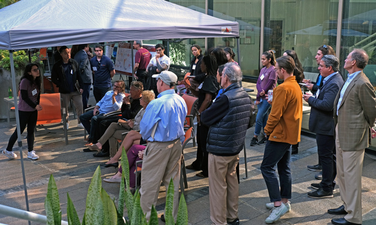
The Institute’s NCI-designated Cancer Center hosted the open house on Wednesday, March 20. It provided an opportunity for community members to meet scientists who seek to better understand melanoma and use this knowledge to improve treatment and prevention.

The event was sponsored by the center’s Community Advisory Board, an eight-member committee that focuses on advocacy, education and community engagement, as well as providing Cancer Center leaders and members with the perspectives of patients, survivors and their loved ones.

Open house participants could select from a variety of activities. Two labs provided brief poster presentations.

Ze’ev Ronai, PhD, director of the Sanford Burnham Prebys Cancer Center and the Jeanne and Gary Herberger Leadership Chair in Cancer Research, and his team discussed several areas of research, including the dissection of microbiota commensals which support the immune system’s fight against melanoma, the studies undertaken to understand melanoma addiction to the metabolic enzyme GCDH, and the development of new drugs to target the molecular machine that translates genetic instructions into proteins, which are known to be hyperactive in cancer cells.

Linda Bradley, PhD, professor in the Cancer Metabolism and Microenvironment Program at Sanford Burnham Prebys, and her group detailed their work on improving the immune system response to viral infections and cancer, including a new potential immune checkpoint therapy and efforts to rejuvenate overstressed immune cells to enhance the effectiveness of immunotherapy.

Attendees also could take tours of two different research facilities. Many participants enjoyed an insider’s view into the field of cryo-electron microscopy (cryo-EM), a technology that garnered three key innovators the 2017 Nobel Prize in Chemistry. The Cryo-EM core facility enables scientists to create 3D images of the cell and all its constituent parts that are accurate to the tiniest detail as it is able to capture individual atoms. Images taken using cryo-EM can be organized sequentially to develop films that show in real time how the cell’s many actors interact, helping scientists map interactions between drugs identified at Sanford Burnham Prebys and their target proteins, thereby advancing novel modalities for the treatment of melanoma and other cancers.

The second tour brought community members to the Conrad Prebys Center for Chemical Genomics. The Prebys Center is the Institute’s comprehensive center for drug discovery and chemical biology. Visitors were able to see the center’s state-of-the-art robots that enable researchers to quickly test the potential effectiveness of hundreds of thousands of compounds to find new prospective treatments. Many scientists at Sanford Burnham Prebys partner with the Prebys Center to conduct drug discovery searches based on new research findings, including those studying melanoma and other cancers.

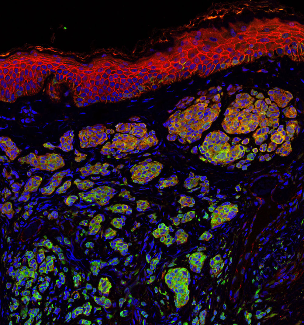
Open House guests conversing in Chairmen's HallMany of the visitors had the opportunity to visit a melanoma research laboratory to learn about research projects in the Ronai lab and view melanoma cells as seen under the microscope.

Following the tours, Ronai shared an overview of the Cancer Center and highlighted recent accomplishments. Attendees interacted with Gregory Daniels, MD, PhD, a medical oncologist and melanoma expert from University of California San Diego and Steven Silverstein, a melanoma survivor, former president of the Melanoma Research Foundation and a melanoma research advocate. The open house concluded with an opportunity for guests to speak with cancer scientists and featured speakers during the evening reception.

“We were honored to provide our valued guests with the opportunity to learn about the research conducted at our Cancer Center, including ongoing melanoma research,” says Ronai. “Our open houses, which focus on different unmet needs in cancer, allow us to welcome and engage with the San Diego community, to share our findings and be inspired by patients and their loved ones.”

Institute News

Our top 10 discoveries of 2020

AuthorMonica May
Date

December 14, 2020

This year required dedication, patience and perseverance as we all adjusted to a new normal—and we’re proud that our scientists more than rose to the occasion.

Despite the challenges presented by staggered-shift work and remote communications, our researchers continued to produce scientific insights that lay the foundation for achieving cures.

Read on to learn more about our top 10 discoveries of the year—which includes progress in the fight against COVID-19, insights into treating deadly cancers, research that may help children born with a rare condition, and more.
 

  1. Nature study identifies 21 existing drugs that could treat COVID-19

    Sumit Chanda, PhD, and his team screened one of the world’s largest drug collections to find compounds that can stop the replication of SARS-CoV-2. This heroic effort was documented by the New York Times, the New York Times Magazine, TIME, NPR and additional outlets—and his team continues to work around the clock to advance these potential treatment options for COVID-19 patients.


  2. Fruit flies reveal new insights into space travel’s effect on the heart

    Wife-and-husband team Karen Ocorr, PhD, and Rolf Bodmer, PhD, shared insights that hold implications for NASA’s plan to build a moon colony by 2024 and send astronauts to Mars.


  3. Personalized drug screens could guide treatment for children with brain cancer

    Robert Wechsler-Reya, PhD, and Jessica Rusert, PhD, demonstrated the power of personalized drug screens for medulloblastoma, the most common malignant brain cancer in children.


  4. Preventing pancreatic cancer metastasis by keeping cells “sheltered in place”

    Cosimo Commisso, PhD, identified druggable targets that hold promise as treatments that stop pancreatic cancer’s deadly spread.


  5. Prebiotics help mice fight melanoma by activating anti-tumor immunity

    Ze’ev Ronai, PhD, showed that two prebiotics, mucin and inulin, slowed the growth of melanoma in mice by boosting the immune system’s ability to fight cancer.


  6. New test for rare disease identifies children who may benefit from a simple supplement

    Hudson Freeze, PhD, helped create a test that determines which children with CAD deficiency—a rare metabolic disease—are likely to benefit from receiving a nutritional supplement that has dramatically improved the lives of other children with the condition.


  7. Drug guides stem cells to desired location, improving their ability to heal

    Evan Snyder, MD, PhD, created the first drug that can lure stem cells to damaged tissue and improve treatment efficacy—a major advance for regenerative medicine.


  8. Scientists identify a new drug target for dry age-related macular degeneration (AMD)

    Francesca Marassi, PhD, showed that the blood protein vitronectin is a promising drug target for dry age-related macular degeneration (AMD), a leading cause of vision loss in Americans 60 years of age and older.


  9. Scientists uncover a novel approach to treating Duchenne muscular dystrophy

    Pier Lorenzo Puri, MD, PhD, collaborated with scientists at Fondazione Santa Lucia IRCCS and Università Cattolica del Sacro Cuore in Rome to show that pharmacological (drug) correction of the content of extracellular vesicles released within dystrophic muscles can restore their ability to regenerate muscle and prevent muscle scarring.


  10. New drug candidate reawakens sleeping HIV in the hopes of a functional cure

    Sumit Chanda, PhD, Nicholas Cosford, PhD, and Lars Pache, PhD, created a next-generation drug called Ciapavir (SBI-0953294) that is effective at reactivating dormant human immunodeficiency virus (HIV)—an approach called “shock and kill.”

Institute News

Solar power gone awry

AuthorZe’ev Ronai, PhD
Date

July 29, 2019

test 2 col list

  • test 2 col list
  • test 2 col list
  • test 2 col list
  • test 2 col list


Are you enjoying the summer? Out grilling, swimming and hiking? Beware: those sunny days may come with a cost. 

When the sun’s rays touch your skin, they don’t stop there. Ultraviolet (UV) light enters your cells, and photons—tiny particles of light—landing on the proteins and DNA in your cells. With just the right amount of activation energy, proteins change their shape and function, and your DNA becomes damaged, or as we say—mutated. Under normal circumstances, cells use special proteins to repair mutated DNA, but when the repair proteins are damaged, DNA mutations become permanent.

Certain DNA segments called genes are more vulnerable to mutations than others. The BRAF gene, which normally makes a protein that controls cell growth, is mutated in more than 50% of melanomas—the most dangerous type of skin cancer. Melanoma appears when BRAF mutations crop up with other mutations in the same skin cell. For patients with these tumors, drugs that target BRAF and related proteins are often successful at slowing or stopping melanoma growth—but only for a while.

Unfortunately, patients who initially respond to such targeted therapy often relapse. Some patients relapse because their tumors generate a new mutation, making it resistant to the drug.  Overall, it may be only a small fraction of cells within the original tumor that develop resistance. So although 99.5% of the cancer cells in a tumor may have a mutated BRAF gene, the other 0.5% can harbor different mutations that either evolved during therapy or were present in the first place, but didn’t drive the initial tumor. For these patients, the bulk of BRAF mutant cancer cells are killed with targeted therapy, but another melanoma can evolve from the remaining 0.5%. This is why combination therapy, where drugs aim for multiple targets, are important.

But targeting every single mutation in a tumor may not be feasible. There will always be a fraction of cells with a different mutation that evolves, making patients vulnerable to a relapse. This is where attacking the tumor from another angle comes into play.  

Checkpoint immunotherapies—which have revolutionized the treatment of melanoma—attack tumors independent of their mutational makeup. They work by loosening the brakes of the immune system—brakes that normally prevent immune cells from attacking our own self. Tumors are very good at hiding from the immune system, but with the brakes released, tumors become exposed and are successfully attacked by the immune system, irrespective of their mutational makeup. 

But not everyone responds to immunotherapy—and we don’t yet know why. Is it the tumor? Is it the patient’s immune system? There is even evidence that the gut microbiome plays a role. Once we understand why some patients respond and or stop responding to immunotherapy, we can improve selection of patients for therapy, the effectiveness of these treatments and the possible combinations that work best. 

So where is skin cancer therapy headed? A combination of checkpoint immunotherapy with targeted therapies, as well as some new tricks we are learning, such as coaching tumor cells to be better recognized by the immune system, are moving the needle.

In my lab at Sanford Burnham Prebys we are dissecting the cell signals that drive cancer. Our studies are guided by data derived from patients’ tumors, coupled with advanced bioinformatics. We seek to understand how physiological processes are modified as cancer develops and how they can be exploited for cancer therapy. For example, we recently demonstrated a connection between the composition of the gut microbiome and the response to immunotherapy, establishing new paradigms, but raising important new questions. Can we better predict who will respond to immunotherapy? Can we enhance the response to immunotherapy by manipulating the gut microbiome? Can we make tumors that don’t initially respond start responding to immunotherapy? The bar is always raised, as one discovery opens so many new avenues to explore and advance our understanding, aspects that members of my lab are working hard on to answer.  

Yes—we are making progress. But preventing the initial sun exposure by using protective gear and sunscreens is needed now as much as ever.

Ze’ev Ronai, PhD, professor in Sanford Burnham Prebys’ Tumor Initiation and Maintenance Program, is a world-renowned cancer research expert and recipient of the Lifetime Achievement Award from the Society of Melanoma Research. The award recognizes his major and impactful contributions to melanoma research over the course of his career.

Institute News

Taking out a microRNA to thwart melanoma

AuthorSusan Gammon
Date

August 6, 2018

Melanoma is a deadly disease with limited treatment options. However, even when those therapies are initially successful, the cancer often comes back. Researchers continue to hunt for new approaches to make the disease more vulnerable.

Ranjan Perera, PhD, an adjunct professor at SBP’s Lake Nona campus, has been studying melanoma for many years, looking for mechanisms that can help control the disease. These efforts helped his lab discover miR-211, a molecule found in melanocytes, the cells that sometimes go awry and become cancerous. Not surprisingly, miR-211 is sometimes overexpressed in melanoma. 

Ranjan Perara, PhD
     Ranjan Perera, PhD 

Because miR-211 is a microRNA—a small molecule that interferes with the cellular machinery that produces proteins—it can have a big impact on gene expression, and the gene it impacts is pretty interesting.

“MicroRNA-211 is targeted to a gene called PDK4, which is important for mitochondrial energy metabolism,” says Perera. 

Scientists have known for more than 100 years that tumors restructure their metabolisms to compensate for their out-of-control growth. If miR-211 is part of that process, taking it out could make cancer cells more treatable.

To better understand what miR-211 is doing, researchers in Perera’s lab used CRISPR/Cas9 gene editing tools to eliminate it from cancer cell lines. They found that removing the molecule impacted mitochondria, the cells’ energy plants, and made them metabolically vulnerable. In addition, miR-211 loss dampened pathways that drive melanoma growth—so there was a double benefit. In animal models, cells without miR-211 had trouble forming tumors. These results were recently published in the Journal of Investigative Dermatology.

Perera and colleagues were also curious whether removing the microRNA might affect how cancer cells respond to the drug Vemurafenib—a therapy used for the treatment of late-stage melanoma. While the drug is effective in certain patients, tumors often develop resistance after several months. Further study showed that eliminating miR-211 made the melanoma cells much more sensitive to Vemurafenib.

These findings add to the body of evidence that helped Perera and SBP get a patent covering approaches using miR-211 to detect and treat melanoma. Perera’s team will continue to study this molecule, as well as the genes it impacts, to gain more insights and potentially transform these findings into new melanoma diagnostics and treatments.

Though it’s still early, these findings make miR-211 an interesting potential drug target, and Perera believes further investigation is definitely warranted.

“Given that miR-211 loss has a dual anti-cancer effect, by inhibiting both critical growth-promoting cell signaling pathways and rendering cells metabolically vulnerable, it is an extremely attractive candidate for combinatorial therapeutics,” says Perera. “This is especially true if, like here, miR-211 is upregulated in Vemurafenib-resistant melanomas in the clinic, since it provides both a highly specific target.”

Interested in keeping up with SBP’s latest discoveries, upcoming events and more? Subscribe to our monthly newsletter, Discoveries.

Institute News

What SBP Scientists are Researching to Battle Skin Cancer

AuthorHelen I. Hwang
Date

May 16, 2017

Skin cancer is one of the most common of all cancers, and melanoma accounts for about 1 percent of skin cancers. However, melanoma causes a large majority of deaths from that particular type of cancer. Alarmingly, rates of skin cancer have been on the rise in the last 30 years. Here in Southern California, our everlasting summer comes with a price. Exposure to sun increases our risk to melanoma. 

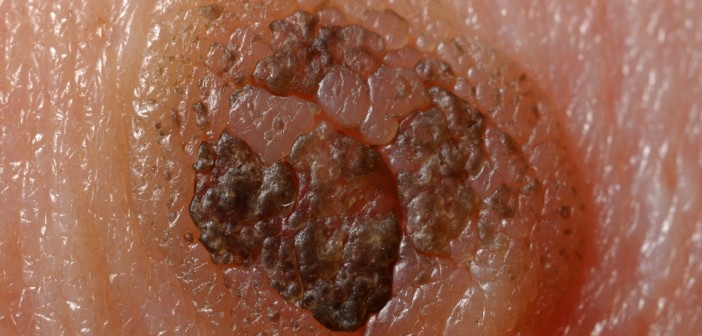
Melanoma occurs when the pigment-producing cells that give color to the skin become cancerous. Symptoms might include a new, unusual growth or a change in an existing mole. Melanomas can occur anywhere on the body. 

At Sanford Burnham Prebys Medical Discovery Institute (SBP), we have several researchers working on the causes of melanoma and discovering new ways to treat this deadly disease.


Here is a roundup of SBP’s latest research

Key findings show how melanoma develops in order to identify potential therapeutic targets

Ze’ev Ronai, PhD
Professor and SBP Chief Scientific Advisor

Ronai’s laboratory has been studying how rewired signaling networks can underlie melanoma development, including resistance to therapy and metastatic propensity. One player in that rewiring is a protein called ATF-2, which can switch from its usual tumor-preventive function to become a tumor promoter when combined with a mutation in the human gene called BRAF.

Ronai’s work on a protein, ubiquitin ligases, led to the identification of RNF125 as an important regulator of melanoma resistance to a common chemotherapy drug. RNF125 impacts melanoma resistance by its regulation of JAK2, an important protein kinase which could play an important role in melanoma resistance to therapy. 

Work on the ubiquitin ligase Siah2 identified its important role in melanoma growth and metastasis, and its contribution to melanomagenesis. Melanoma is believed to be a multi-step process (melanomagenesis) of genetic mutations that increase cell proliferation, differentiation, and death. 

Work in the lab also concern novel metabolic pathways that are exploited by melanoma for their survival, with the goal of identifying combination drug therapies to combat the spread of melanoma. Earlier work on the enzyme PDK1 showed how it can be a potential therapeutic target for melanoma treatment.
 

Immunotherapy discovery has led to partnership with Eli Lilly

Linda Bradley, PhD
Professor, Immunity and Pathogenesis Program, Infectious and Inflammatory Diseases Center

Bradley’s group is focused on understanding how anti-tumor T cells can be optimized to kill melanoma tumors. They discovered an important molecule (PSGL-1) that puts the “break” on killer T cells, allowing melanoma tumors to survive and grow. Using animal models, they removed this “break” and T cells were able to destroy melanoma tumors. They have extended their studies and found that in melanoma tumors from patients, T cells also have this PSGL-1 “break”. Bradley’s lab has partnered with Eli Lilly to discover drugs that can modulate PSGL-1 activity in human disease that may offer new therapies for patients.

Knocking out a specific protein can slow melanoma growth 

William Stallcup, PhD
Professor, Tumor Microenvironment and Cancer Immunology Program

The danger of melanomas is their metastasis to organs, such as the brain, in which surgical removal is not effective. By injecting melanoma cells into the brains of mice, we have shown that the NG2 protein found in host tissues makes the brain a much “friendlier” environment for melanoma growth. 

Specifically, NG2 is found on blood vessel cells called pericytes and on immune cells called macrophages. The presence of NG2 on both cell types improves the formation of blood vessels in brain melanomas, contributing to delivery of nutrients and thus to accelerated tumor growth. Genetically knocking out NG2 in either pericytes or macrophages greatly impairs blood vessel development and slows melanoma growth.


Mysterious molecule’s function in skin cancer identified

Ranjan Perera, PhD
Associate Professor, Integrative Metabolism Program

Ranjan’s research uncovered the workings of a mysterious molecule called SPRIGHTLY that has been previously implicated in colorectal cancer, breast cancer and melanoma. These findings bolster the case for exploring SPRIGHTLY as a potential therapeutic target or a biological marker that identifies cancer or predicts disease prognosis.


Drug discovery to help babies has led to a clinical trial at a children’s hospital

Peter D. Adams, PhD
Professor, Tumor Initiation and Maintenance Program

Approximately 1 in 4 cases of melanoma begins with a mole, or nevus. Genetic mutations can cause cells to grow uncontrollably. By investigating how this occurs, we can understand why melanoma develops from some moles, but not others.

Babies born with a giant nevus that covers a large part of the body have especially high risk of melanoma, and the nevus cells can spread into their spine and brain. Adams’ research identified a drug that deters the cells from growing. The drug identified will be used in a clinical trial at Great Ormond Street Children’s Hospital in London, England that may help babies with this debilitating disease.  


Discovery of a receptor mutation correlates with longer patient survival

Elena Pasquale, PhD
Professor, Tumor Initiation and Maintenance Program

Pasquale’s work has included whether mutations in the Eph receptor, tyrosine kinases, play a role in melanoma malignancy. Eph receptor mutations occur in approximately half of metastatic melanomas. We found that some melanoma mutations can drastically affect the signaling ability of Eph receptors, but could not detect any obvious effects of the mutations on melanoma cell malignancy. 

Bioinformatic analysis of metastatic melanoma samples showed that Eph receptor mutations correlate with longer overall patient survival. In contrast, high expression of some Eph receptors correlates with decreased overall patient survival, suggesting that Eph receptor signaling can promote malignancy. 

Institute News

Super-oncogenic protein that promotes development of melanoma

AuthorJessica Moore
Date

May 19, 2016

An international collaborative study led by scientists at the Sanford Burnham Prebys Medical Discovery Institute (SBP) has identified a malicious form of a protein that drives the formation of melanoma. The findings, published in Cell Reports, reveal unexpected insight into how this lethal skin cancer develops and progresses, and may help understand and develop novel therapies against these aggressive tumors.

“We found that an inactive version of a protein called activating transcription factor 2 (ATF2) elicits a tumor-promoting effect in a way not seen before,” said Ze’ev Ronai, PhD, chief scientific advisor of SBP and professor of its NCI-designated Cancer Center. “We have known for years that the active version of ATF2 promotes melanoma, but this result was a surprise because we thought ATF2 transcriptional activity was essential to activate cancer-related genes.”

Ronai’s team has been studying ATF2’s role in melanoma for two decades. Their past work led to the view that it’s dangerous when it’s in the nucleus because it controls cancer-enabling genes, but benign when it’s not.

In the current study, researchers looked at the oncogenic potential of a ‘dead’ form of ATF2 in mice with mutations in BRAF, a kinase that transmits signals promoting cell division and is often mutated in pigmented skin cells. The same mutation is found in about half of all human melanomas.

“Inactive ATF2, in mice with mutant BRAF, resulted in the formation of pigmented lesions and later, melanoma tumors,” said Ronai, senior author of the study.

“What makes this discovery relevant to human melanoma is that we identified a structurally similar form of inactive ATF2 in human melanoma samples that has the same effects on cancer cells,” added Ronai. “Inactive ATF2 could be an indicator of tumor aggressiveness in patients with BRAF mutations, and maybe other types of cancer as well.”

“Unlike models with more complex genetic changes, like the inactivation of PTEN and p16 combined with BRAF mutations that result in rapid tumorigenesis (within a few weeks), the inactive ATF2 caused BRAF mutant mice to develop melanoma much slower, more similar to the timescale seen in patients,” commented Ronai. “This improves our ability to monitor the development of melanoma and efficacy of possible interventions.”

“We’re now investigating why inactive ATF2 so potently promotes BRAF-mutant melanoma, and looking for other types of cancer where it acts the same way,” Ronai said. “Our findings may guide precision therapies for tumors with mutant ATF2.”

The paper is available online here.

Institute News

Scientists identify promising new melanoma drug

Authorsgammon
Date

November 25, 2015

A new drug discovered by scientists at Sanford Burnham Prebys Medical Discovery Institute (SBP) may show promise for treating skin cancers that are resistant or unresponsive to today’s leading therapies.

In the United States, 5 million people are treated annually for skin cancer, and 9,000 people die from the deadliest form—melanoma—according to the US Department of Health and Human Services.

The new compound, named SBI-756, targets a specific molecular machine known as the translation initiation complex. These structures are in every cell and play the critical role of translating mRNA into proteins. In cancer cells the complex is impaired, producing extra protein and providing a growth advantage to tumors. SBI-756 causes the translation complex to dissociate, and was shown to inhibit melanoma cell growth in the study, published today in Cancer Research.

“The unique target of SBI-756 makes it especially promising for use in combination therapy,” said Ze’ev Ronai, senior author and scientific director of SBP’s La Jolla campus. “A major issue limiting the effectiveness of current melanoma therapies is that tumors become resistant to treatment. Combining drugs that come at a melanoma from different angles may help overcome the problem of drug resistance.”

About 50% of melanomas are caused by mutations in a specific gene called BRAF. Patients with these tumors are commonly prescribed vemurafenib, a BRAF inhibitor that shrinks tumors. However, many patients experience a relapse within weeks, months, or even years because tumors evolve and become resistant to the drug. A similar phenomenon is seen in mice, where treatment of BRAF melanomas results in an initial response, but 3-4 weeks later the tumors return.

The team found that if SBI-756 is co-administered with vemurafenib, the tumors disappeared and most importantly, they did not reoccur. Even in mice with advanced/late stage BRAF driven cancer, the reappearance of . These data suggests that SBI-756 provides a significant advantage in overcoming tumor resistance.

“The ability of this compound to delay or eliminate the formation of resistant melanomas is very exciting,” said Ronai.

In other forms of melanoma, caused by mutations in the genes NRAS and NF1—which are known as unresponsive to BRAF drugs—administering SBI-756 alone significantly the scientists found. The team is now testing whether combining SBI-756 with existing drugs used for treating these types of melanomas can make the tumors disappear.

Drugs that target the translation initiation complex have been intensely pursued in the past few years, not just for melanoma, but for a wide array of cancers. SBI-756 is considered a first-in-class drug because it is the first successful attempt to target a specific part of the complex called eIF4G1.

In fact, SBI-756 is the culmination of seven years of work in Ronai’s group—testing and tweaking the drug’s features to help it bind to the target more readily and to make it easier to formulate. The resulting compound is a significant improvement over the initial version.

“It appears that the dose we need to administer is very low. Even in the experiments where the drug was administered to mice with tumors over a significant period of time, we have not found any toxicity,” Ronai said.

“The finding of SBI-756 is also exciting for the possible treatment of diseases other than cancer, such as neurodegenerative diseases, where the activity of the translation initiation complex is reported to be higher,” said professor Nahum Sonenberg of McGill University, a world renowned leader in the field of protein translation.

“We hope that we’re going to come up with the next generation of the compound that can go into clinical trials—first in melanoma but likely in other tumors,” Ronai said.

The study was performed in collaboration with the Conrad Prebys Center for Chemical Genomics at SBP, Lady Davis Institute for Medical Research, Sir Mortimer B. Davis-Jewish General Hospital, McGill University (Canada), the National Cancer Institute, MD Anderson Cancer Center, and Yale University.

Institute News

Sanford-Burnham researchers identify a new target for treating drug-resistant melanoma

Authorsgammon
Date

May 28, 2015

A new collaborative study led by researchers at Sanford-Burnham, published today in Cell Reports, provides new insight into the molecular changes that lead to resistance to a commonly prescribed group of drugs called BRAF inhibitors. The findings suggest that targeting newly discovered pathways could be an effective approach to improving the clinical outcome of patients with BRAF inhibitor-resistant melanoma tumors. Continue reading “Sanford-Burnham researchers identify a new target for treating drug-resistant melanoma”